

 PDF
PDF ePub
ePub Citation
 Print
Print


INTRODUCTION
Angiogenesis is a highly regulated process balanced by inhibitors and stimulators of endothelial cell proliferation, endothelial cell migration, and capillary formation molecules.1-3 Angiogenesis plays a critical role in solid tumor development and metastasis. Vascular endothelial growth factor (VEGF) and basic fibroblast growth factor (bFGF), produced by a number of neoplastic and non-neoplastic cell types, are major stimulators of tumor-related angiogenesis.3-5 Recently, it has been suggested that angiogenesis plays a role in the pathophysiology of hematologic malignancies as well.2,3,6
In children with acute lymphocytic leukemia (ALL), elevated levels of urine bFGF have been associated with increased density of bone marrow (BM) vessels.7 A recent study found significantly higher peripheral blood mononuclear cell VEGF expression in recurrent ALL compared to newly diagnosed ALL and that VEGF levels in newly diagnosed ALL showed prognostic value.8 It has also been shown that microvessel densities (MVD) at presentation are significantly increased in patients with ALL compared to controls and that MVD drops towards normal in remission. However, there was no significant difference in MVD at presentation or remission between patients found to have poor prognostic factors and those who subsequently relapsed.9 In contrast, Faderl et al.10 reported that higher levels of angiogenic factors in adult ALL patients were predictive of longer survival.
Currently, the role of angiogenic factors in childhood ALL is not fully understood. To study this, we examined diagnostic BM plasma aspirates and retrospectively evaluated and compared VEGF and bFGF levels among patients with standard-, high-risk, and relapsed ALL.
MATERIALS AND METHODS
From May 2003 to May 2004, 33 children with ALL were consecutively and retrospectively enrolled. All patients were enrolled and had samples drawn at time of initial diagnosis. Control patients were retrospectively selected from patients with more than two-lineage cytopenia on peripheral blood, but normal light microscopy BM findings. Patients received a 2 year treatment course, according to initial risk group classification of standard or high risk. For analysis, patients were retrospectively divided into three groups, standard-, high-risk, and relapse ALL. National Cancer Institute criteria were used to classify those without relapse into standard or high risk ALL. The third group was defined by patients who experienced relapse(s).
BM aspiration samples (5mL) were collected using sterile ethylenediamine tetraacetic acid (EDTA) tubes. Each marrow sample contained greater than 90% blasts. Separated plasmas were processed by refrigerated centrifuge at 1500g for 10 minutes, and stored at -70℃. Control plasma values for VEGF and bFGF were determined from samples of seven patients with normal BM.
Commercially available kits (R&D Systems) were used to measure VEGF and bFGF levels following manufacturer protocols. Briefly, 100µL and 200µL of plasma, for VEGF and bFGF respectively, were added to separate microplates. Plates contained specific monoclonal antibody for either VEGF or bFGF. Mixtures were incubated for 2 hours at room temperature, and then washed three times to remove unbound substance. Enzyme-linked polyclonal antibodies specific for each protein were added, mixtures were again incubated for 2 hours at room temperature, and washed. Substrate solution to develop color was added. Color intensity was measured at 450-nm wavelength, and a standard curve was used to determine the proportionate VEGF or bFGF bound. Concentration was expressed in pg/mL.
Results were presented as average BM plasma VEGF and bFGF concentrations with standard error of the mean (SEM). SEM was calculated as standard deviation divided by square root of the number of samples.
VEGF and bFGF levels were compared among each group, including the control group. Pearson correlations were performed to assess the relationship between angiogenic factors and prognostic factors, including WBC count and age at diagnosis. Additionally, the relationship between platelet count at time of aspiration and VEGF was examined with a Pearson correlation.
Analysis was performed using SPSS (Windows program package version 11.5). VEGF and bFGF levels between groups were compared by one way analysis of variance (ANOVA). Differences were evaluated by the Scheffe test. P values < 0.05 were considered significant.
RESULTS
Median age for patients with ALL was 5.9 (range, 1.8-13.9) years. Twenty three males and ten females participated in the study. There were 9, 17, and 7 patients in the standard-, high-risk, and relapse ALL group, respectively.
Median age for the 7 control subjects was 11 (range, 0.1-13.6) years. Three were males, and four were females.
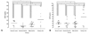
Average VEGF was higher in relapse ALL (N=7, 216.6±79.9pg/mL, range; 12.2-577.1pg/mL) compared to standard (N=9, 36.8±12.1pg/mL, range; 0-117.4pg/mL) (p=0.013) or high risk ALL (N=17, 80.0±12.2pg/mL, range; 15.2-166.0pg/mL) (p=0.023). However, compared to controls, it was not significantly lower (N=7, 119.3±29.4 pg/mL, range; 13.3-190.7pg/mL) (p=0.269). Average VGEF was lower in standard risk ALL compared to controls. There was no significant difference between high risk ALL and controls (Fig. 1A).
bFGF levels were also significantly higher in relapse than standard-, or high-risk ALL patients (relapse ALL; 48.6±15.4pg/mL, standard risk ALL; 18.9±5.5pg/mL, high risk ALL; 19.0±3.5 pg/mL, normal control; 18.6±4.0pg/mL) (p=0.003). Average bFGF level was similar between standard risk ALL and controls. There was no significant difference between high risk ALL compared to controls (Fig. 1B).
Three patients with refractory relapse and death had much higher VEGF and bFGF values (VEGF; 420.0±81.6pg/mL, bFGF; 85.6±3.2pg/mL).
There was no significant correlation between WBC count at diagnosis and angiogenic factors (VEGF and bFGF). R2 values for WBC vs. VEGF and bFGF were 0.01 (p=0.68) and 0.04 (p=0.27), respectively. Further, there was no correlation between age and either VEGF or bFGF (R2=0.02, p=0.40 and R2=0.00, p=0.84 for VEGF and bFGF, respectively).
There was no significant correlation between VEGF and platelet count at sampling time (R2=0.10, p=0.07).
The Pearson correlation coefficient between VEGF and bFGF was 0.775 (p<0.01). The ANOVA revealed that VEGF and bFGF values different significantly between groups, with p values of 0.006 and 0.003 for VEGF and bFGF, respectively.
DISCUSSION
During the past few years, it has been proposed that angiogenesis may play a role in not only solid tumors, but also in hematologic malignancies. Previous studies of hematologic malignancies have demonstrated a close relationship between BM and angiogenesis.7,10,11 In the current study, we detected VEGF and bFGF BM plasma levels in childhood ALL and control cases. We showed that VEGF and bFGF levels in relapsed patients were higher than in other risk groups and controls.
The prognostic significance of neovascularization has been demonstrated for solid tumors, but few reports are available for hematologic malignancies, and results are controversial. Aguayo et al.12 reported that increasing bFGF levels were associated with shorter overall and disease-free survival. However, bFGF levels did not correlate with established prognostic factors of WBC count, age, and cytogenetic abnormalities at diagnosis. In this study, VEGF and bFGF levels were not statistically different between standard and high risk ALL, classified at time of diagnosis with traditional risk factors of WBC count and age. No correlation between WBC count and age with angiogenic factors was found, which supports this result. Furthermore, this study found no significant difference in angiogenic factor levels between ALL patients and controls (Fig. 1). This differs from a previous study that reported different MVD density in the two groups.7
Peres-Atayde et al.7 found that MVD in BM of children with ALL was significantly higher than in controls. These findings suggested that leukemia cells induce angiogenesis in the BM and that leukemia can be dependent on angiogenesis. Other studies have found no significant difference in MVD at presentation or remission between children with poor prognostic factors and those who had relapse.9 Interestingly, Schneider et al.13 reported that ALL patients with poor outcomes had significantly lower bFGF levels and that VEGF value was not significantly higher in ALL patients compared to control subjects, or in relapse compared to non-relapse patients. Furthermore, Faderl et al.10 reported that ALL patients with high levels of VEGF and bFGF demonstrated shorter survival. Koomagi et al.8 showed that VEGF levels were significantly higher in recurrent ALL compared to newly diagnosed ALL. In this study, VEGF and bFGF correlated closely (Pearson correlation coefficient=0.775), and both angiogenic factors were elevated in patients with relapse compared to all groups. Of note, the 3 patients who died due to refractory disease and relapse were found to have much higher VEGF and bFGF value at diagnosis. Therefore, both VEGF and bFGF may affect the pathophysiology of childhood ALL, and elevated levels may be associated with dismal outcome.
Interestingly, average VEGF in standard risk patients was lower than controls (Fig. 1A). This result is similar to previous findings.14 Average levels in the high risk group were similar to controls. Most importantly, the relapse group had higher values than controls. Therefore, VEGF may have an important role in more aggressive childhood ALL.
In this study, BM plasma VEGF values were not correlated with platelet counts. Previous reports presented that high amounts of soluble VEGF have been detected in platelets.15 Therefore, release of VEGF from platelets may contribute to levels in cancer patients. However, Gunsilius et al.16 showed that serum VEGF was correlated with platelet count but that plasma VEGF was not. Therefore, plasma VEGF values are a more precise marker for cancer patients than serum levels.
We showed that VEGF and bFGF was related with relapse in childhood ALL. However, the small sample size and short follow-up period are limitations of this study. Therefore, a prospective study with a larger patient cohort and longer follow up should be followed.
Our data suggested that VEGF and bFGF at diagnosis may play an important role in progression of childhood ALL. Even though BM plasma VEGF and bFGF were not associated with traditional risk factors of age and WBC count at diagnosis, they were associated with disease progression and response to treatment. In conclusion, levels of BM plasma VEGF and bFGF at diagnosis may be a useful predictor of relapse in childhood ALL patients.
 XML Download