

 PDF
PDF ePub
ePub Citation
 Print
Print


INTRODUCTION
The parasitological diagnosis of Clonorchis sinensis (CS) is based on the presence of characteristic eggs in the feces, duodenal fluid, or bile. Although a number of techniques have been developed for diagnosing CS infection, the most reliable technique has been stool examination.1 However, in light infections or in the presence of a biliary obstruction, the eggs might escape detection by this method, and some practical problems such as difficulties in stool collections may be encountered.2 On the other hand, in view of collecting the eggs as near as possible to where they were laid and before they become diluted in the feces, an examination of the bile collected by endoscopic or percutaneous biliary drainage is believed to be the most accurate diagnostic technique.3 The acquisition of bile is not always easy in normal situations and the technique of bile collection can sometimes result in serious complications. Therefore, the diagnosis of clonorchiasis from bile is believed to be feasible only in certain circumstances, particularly when a biliary tract evaluation or intervention is absolutely required.
The aim of this study was to determine the infection state of CS based on a bile examination in patients with biliary tract diseases in Ulsan, Korea, which was known to be an endemic area of CS infections in the 1980s.4
MATERIALS AND METHODS
About Ulsan
Ulsan Metropolitan city is located at the south-eastern edge of the Korean Peninsula and is one of the 7 largest cities in Korea, with a gross area of 1,056 km2 and a population of more than one million. Further, 74.6% of the population has an independent financial status. In addition, Ulsan is a representative port city. The Taehwa River flows across Ulsan city and consecutive ports, including the ports of Ulsan, Onsan, and Bangeojin are arranged along the Gulf of Ulsan. Utilizing these ports, Ulsan has provided a gateway into Korea from East Asia. Before industrialization, the CS infection rate in Ulsan was extremely high. The first survey of clonorchiaisis in the Taewha River crossing Ulsan was carried out by Joo in 1980.4 He examined stool samples from 1,723 residents in the vicinity of the river using the formalin-ether sedimentation technique and found that 22.2% were positive for CS. Therefore, Ulsan was designated as a representative endemic area for clonorchiasis in Korea.
Survey of CS infections and statistics
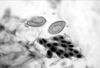
From January 2001 to August 2003, 238 patients with biliary tract diseases were enrolled in this study. All patients had undergone endoscopic biliary drainage as a result of biliary obstruction with/without cholangitis. The patients' bile samples were obtained from the nasobiliary drainage tube the day after the endoscopic biliary drainage procedures in order to exclude contamination from the previous injected dyes. All the biliary drainage procedures were carried out by one physician. The analysis of the bile to detect CS eggs was also performed by one expert technician (Fig. 1). The patients' ages ranged from 25 to 91 years and the mean age was 59.4 years (SD 13.8). The male to female ratio was 130:108. Disease distribution of the patients was as follows: 182 gallstone diseases (52 with gallbladder stones, 92 with extrahepatic bile duct stones, and 38 with intrahepatic bile duct stones), 43 bile duct cancers, and 13 gallbladder cancers. Both bile duct and gallbladder cancers were diagnosed by either radiographic studies or by pathological findings of biopsy specimens and/or by surgical findings with/without pathological examination of the resected tumors. Statistical analysis was performed using a chi-square test to analyze differences in the CS infection states according to age, gender, and disease type. A p-value <0.05 was considered significant. Statistical analysis was performed using the SPSS/PC+ version 11.0 software.
RESULTS
The overall egg positive rate of CS was 28.2% (67/238). The prevalence was significantly higher in males (35.4%, 46/130), than in females (19.4%, 21/108) (p<0.05). The egg positive rate was not significantly different according to age group: 26.7% (4/15) in 30-39 years, 25.0% (12/48) in 40-49 years, 24.4% (11/45) in 50-59 years, 30.2% (19/63) in 60-69 years, 35.3% (18/51) in 70-79 years, and 25.0% (3/12) in 80 years of age and over. The egg positive rate in males was similar in each age group: 40.0% in 30-39 years, 42.3% in 40-49 years, 28.6% in 50-59 years, 25.0% in 60-69 years, 48.0% in 70-79 years, and 40.0% in 80 years of age and over. However, in females, the majority of egg positive cases were in patients over 50 years of age: none in 30-39 years, 4.5% in 40-49 years, 17.6% in 50-59 years, 37.0% in 60-69 years, 23.1% in 70-79 years, and 14.3% in 80 years of age and over.
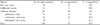
Table 1 shows the prevalence of clonorchiasis among the disease groups. The CS egg positive rate according to the disease group was not statistically different: 32.6% in patients with bile duct cancer, 38.5% in those with gallbladder cancer, and 26.4% in those with gallstone diseases. The egg positive rate according to gallstone locations was also not statistically different: 32.7% in gallbladder, 28.3% in extrahepatic bile duct, and 13.2% in the intrahepatic bile duct.
Of the 52 patients with gallbladder stones, only 40 had normal cholangiographic findings and 12 showed the characteristic findings of clonorchiasis. The former was 20% (8/40) of the CS egg positive rates, and the latter was 75% (9/12) of those that underwent bile examination.
DISCUSSION
Clonorchiasis is a snail-transmitted trematodiasis caused by the Chinese liver fluke. It is generally acquired by eating raw, inadequately cooked or pickled flesh of fresh water fish.1 The geographic distribution of clonorchiasis is largely confined to southeast Asia, including Korea, China, Hong Kong, Japan, Taiwan, and Vietnam.1-11 With the increasing popularity of travel to these countries, as well as the global migration of Asians, physicians need to be aware of this condition. In Korea, the infection rates of soil-transmitted helminthiases have reduced remarkably due to the high level of economic growth. However, a recent survey revealed that snail-transmitted trematodiasis, particularly CS, remains highly prevalent, especially in the riverside areas of Korea.1 According to national surveys, the overall egg-positive rates of CS were: 4.6% in 1971, 1.8% in 1976, 2.6% in 1981, 2.7% in 1986, 2.2% in 1992, and 1.4% in 1997.12 This means that the infection status of CS has remained high despite the development of effective anthelmintics such as praziquantel. The Korean dietary habits of eating raw freshwater fish, underestimating the pathogenicity of the liver fluke, and overestimating the efficacy of praziquantel might contribute to the failure of control and re-infection after treatment. Furthermore, considering that the CS egg detection rate differs according to the intensity of the infection of the subject, the introduction of praziquantel made most clonorchiasis cases light infections, thereby making the diagnosis problematic.
The parasitological diagnosis of CS is based on the presence of characteristic eggs in the stool, duodenal fluid, or bile samples. Until now, stool examination was the preferred method. Other supplementary diagnostic methods have been applied including an intradermal test, ELISA, immunoblotting, sonography, CT scan, and cholangiography.1,13-16 However, all of these methods have their limitations with regard to sensitivity, specificity, or applicability and none of the methods surpass stool examinations for detection of the eggs. In addition to stool examination, bile or duodenal fluid examination is another technique for detecting eggs or adult worms when making a diagnosis of clonorchiasis. In these parasitological diagnostic methods, an examination of the bile collected directly from the bile duct is believed to be the most precise diagnostic method for clonorchiasis, particularly in cases of light infections.3 However, this technique is invasive and can be inconvenient to the patient. Therefore, it is difficult to apply this method routinely for a diagnosis of clonorchiasis.
In this study, the overall CS egg positive rate was higher than usual. Although the study area is one of the representative endemic areas of clonorchiasis in Korea4 and all the enrolled patients suffered from various biliary tract diseases, the high prevalence rate is concerning. This high rate may have been caused, in part, by applying an unusual diagnostic method for detecting CS eggs: a bile examination. Bile appears to be the most adequate material for detecting CS eggs because CS live, grow, and shed their characteristic eggs in the bile duct. Therefore, bile examination is believed to be the most sensitive method for detecting CS eggs, even in light infections. Furthermore, compared with the stool or duodenal fluid, bile is very clear, so CS eggs can be easily detected.
In this study, the egg positive rates, according to age and disease type, were similar and evenly distributed. Although there was a somewhat higher incidence of clonorchiasis among patients with hepatobiliary diseases than in those without, this phenomenon is believed to be caused by a widespread infection of CS in this study area. Meanwhile, the prevalence of clonorchiasis in males was found to be significantly higher than in females. This difference appears to be caused by the fact that males are more likely to ingest raw fresh water fish and by a disregard of the infections rather than by ignorance of the possibility of infection.
Clonorchiasis may lead to cholangitis. During an infection by the fluke, the bile duct is severely dilated, and the ductal wall is thickened as a result of mucosal hyperplasia and periductal fibrosis. This produces an easily recognizable radiological image. In particular, the cholangiographic findings consist of characteristic filling defects and several changes in the intrahepatic and extrahepatic ducts.16,17 As a result, this study also evaluated the diagnostic accuracy of cholangiography for clonorchiasis based on a bile examination. For a precise evaluation, 52 patients with gallbladder stones were enrolled. The remainder of the patients, including those with bile duct stones, bile duct cancer, and gallbladder cancer, which can adversely affect an accurate evaluation of the bile duct tract, were excluded. The sensitivity and specificity of the cholangiography for diagnosing CS infections were 75 and 80%, respectively. The egg positive rate in the patients with normal cholangiographic findings was 20%. This suggests that many cases had a light or early infection, and the bile duct changes had not been developed.
The present study had several limitations. First, the results of bile examination were not compared with those of a stool examination, which is known to be a reliable method for diagnosing clonorchiasis. Therefore, it could not be verified that bile examination is a superior diagnostic method for detecting CS eggs in the feces. Second, the enrolled cases were limited to patients with biliary tract diseases. Therefore, the results are not absolutely representative of the study area. Third, a bile examination is a good method for detecting CS eggs but it is difficult to use in clinical practice due to the invasiveness of the acquisition technique.
Nevertheless, the data demonstrated that clonorchiasis is still prevalent around Ulsan, even though Ulsan has become one of the 7 biggest cities in the Korea and represents a symbol of industrial and economic growth in the country. For the control of the disease, health education programs along with praziquantel treatment are recommended. Therefore, an easily available and accurate diagnostic tool for detecting CS infections, particularly in cases with light worm infections, should be developed.
 XML Download