

 PDF
PDF ePub
ePub Citation
 Print
Print


INTRODUCTION
Although the hand is generally exposed to all kinds of injuries, the proximal phalangeal neck is particularly vulnerable to trauma, especially in children.1-3 These fractures are likely to be missed, unless they are confirmed by careful physical examination and radiographic study.4 If the appropriate treatment plan is not selected, phalangeal neck fracture in children may result in malunion of the fracture, deformity of the hand, stiffness of the affected joint, pain, and accelerated degenerative changes.5-8 Displacement of a fracture fragment in a phalangeal neck fracture is usually more severe in the proximal phalanx than in the middle phalanx. However, there are few reports on the results of treatment in proximal phalangeal neck fractures in children.6 This paper presents data from a retrospective study on proximal phalangeal neck fracture in children to identify the types of fractures, their causes, operative treatments, complications, and functional outcomes.
MATERIALS AND METHODS
We reviewed the records of 24 children who were treated for proximal phalangeal neck fracture from January 1990 to January 1998. Patients were followed up until they had complete bony union and recovery of proximal interphalangeal movement, for a mean of 14 months (range: 12-30 months). Unicondylar or bicondylar head fractures were excluded. Anteroposterior (AP), true lateral, and oblique radiographies were reviewed. The fracture angle was measured on AP and lateral films. On the AP view, we drew a line through the center of the shaft of the proximal phalanx. Next, the radial and ulnar margins of the distal articular surface of the proximal phalanx were connected and a second line was drawn perpendicular to that. The angle formed by those two lines was determined as the radial and ulnar angulation. Dorsal and palmar angulations were determined on the true lateral view. These angulations were also measured as the angle formed by two lines. One line bisected the distal articular surface and ran parallel to the palmar or dorsal cortex of the distal fragment, and the other was drawn through the center of the shaft. The fractures were classified into three types. Type I fractures were not displaced and without finger deformity. In Type II fractures, the distal fragment was displaced but maintained some bone to bone contact with the proximal fragment. In type III fractures, there was no bony contact between the proximal and distal fragment.9 Open reduction was indicated in patients with fractures that were not reduced by an initial treatment of closed reduction, unstable fractures which were not maintained after reduction, malunited fractures because of failure of conservative treatment and gross rotational deformity of the nail plane in full grip. The average length of immobilization with a finger splint and pin removal was 3.5 weeks (range: 3-5 weeks). Distal interphalangeal joint motion was allowed immediately after the operation, and proximal interphalangeal joint motion was started after pin removal. For the follow-up study, the following data were recorded: deviation of the finger during active maximum extension, degree of flexion of finger grip, relief of pain, and the subjective cosmetic result. Objective data on the functional positions of the hand needed for performing daily activities were analyzed using standard techniques to assess active and functional ranges of motion (ROM) of the proximal interphalangeal joint. The results were rated as follows: "excellent" indicates fracture union plus full ROM of the proximal interphalangeal joint, no clinical deformity, less than 5° angulation in any plane, no rotation, no pain, and no infection; "good" indicates fracture union and one criterion missing; "fair" indicates fracture union with two criteria missing; and "poor" indicates fracture union with three criteria missing based on this grading system.
RESULTS
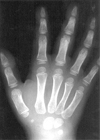
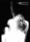
The age of the patients ranged from 2 to 14 years (average: 4.8 years) at the time of surgery. There were 14 boys and 10 girls. The dominant hand was involved in 15 of the 24 patients. The little finger was the most frequently involved finger (10 patients), followed by the index finger (4 patients). The most common cause of injury was sports-related (12 patients). Two cases were caused by slipping and falling and 6 were crushing injuries. Four patients had door- slam injuries. Ulnar angulation was noted in six patients (Fig. 1) and radial angulation was seen in two patients. On the lateral view, dorsal or volar displacement of the distal fragment was seen in 16 patients. Dorsal displacement with volar angulation greater than 45° was present in 12 patients (Fig. 2), volar displacement with dorsal angulation greater than 45° was present in two patients, and mild volar displacement was present in two patients. Three of 12 patients with dorsal displacement of the distal fragment, with volar angulation, had 180° rotation of the head. There were 4 patients with Type I fractures, 17 with Type II, and 3 with Type III fractures.
There was one open fracture. Four Type I fracture patients had closed reduction and percutaneous pinning. Open reduction and internal fixation was done in Type II and Type III patients. The interval between trauma and operation ranged from 2 days to 2 months, with a mean of 18 days. Seven patients had closed reduction and splint immobilization treatment at private clinics when initially injured. Six patients were transferred to the author's institution because of delayed recognition of a displaced and rotated fracture fragment. Four patients also reported feeling instability in the proximal interphalangeal joint.
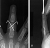
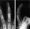
The operative technique was as follows. A skin incision was made in the dorsal midline over the fracture site. A parasagittal incision through the transverse retinacular ligament with mobilization of the central tendon was needed in six of 14 patients in whom midline dorsal incisions were made (Fig. 5). Open reduction and internal fixation with 0.035-inch Kirschner (K) wires (8 patients), 23-gauge needles (11 patients) or pull-out steel wire with K wire (1 patient) was done. Criss-cross fixation was performed in 14 patients (Fig. 3, A and B). Single K wire fixation was used for the others. Osteoclasis was performed in six patients with malunited fractures. No bone graft was performed for malunions. Twelve patients (all Type I & 8 Type II) had excellent results, six patients (Type II) had good results, four patients (3 Type II & 1 Type III) had fair results, and two patients (Type III) had poor results. Complete ROM, grip, and pinch strength were restored in 16 of the 24 patients. After the operation, the radiographs revealed no avascular necrosis or traumatic joint damage around the injury site (Fig. 4, A and B). Complications developed in 2 patients. One mild buttonhole deformity occurred, but there was no functional disturbance. Volar angular deformity greater than 15° was present in one patient with a severe dorsal communited fracture one week after K wire removal. Callus overgrowth and proximal interphalangeal joint swelling was present in four patients, but none had any functional disturbance. No pin site infections or osteonecrosis of the phalangeal head occurred.
DISCUSSION
Neck fracture of the proximal phalanx of the finger is a relatively uncommon injury and occurs almost exclusively in children. Leonard and Dubrovnik,10 in a review of 263 finger fractures in children, found that 38 (15%) were fractures of the phalangeal neck. Of 38 patients, nine required open reduction and internal fixation, either for dorsal displacement with volar angulation of the head or for 180° proximal rotation of the head. The current authors have seen 248 finger fractures in children. Of those fractures, 27 were proximal phalangeal neck fractures (10.9%). Surgery was performed in 24 patients. In the current study, closed reduction and pinning with splint immobilization was used only for the patients who had less than 5° angulation deformity after closed reduction, without rotational deformity. Advantageously, stabilization of the fracture by percutaneous pinning allows the patient to move the small joints of the injured digit during fracture healing to prevent stiffness. Therefore, closed reduction and internal fixation are recommended as the initial treatment. Closed reduction may be difficult if there is displacement with some degree of rotation of the fractured fragment (Type II & III). Dixon and Moon11 reported that, in children, the volar plate could roll on itself, causing 90° rotation of the condylar fragment which then is entrapped in the capsule and collateral ligaments. This fracture may easily be overlooked because of obscure radiographic findings and a lack of finger deformity. The fractured distal fragment is usually cartilaginous and may be radiolucent. The ossified portion of the distal fragment is usually small. Because the phalangeal neck fractures have a tendency to rotate dorsally, with the intact collateral ligaments acting as the pivot point, the proximal and distal fragments can overlap each other and be missed on normal AP radiographs. Thus, the true lateral view is more helpful in diagnosing phalangeal neck fractures.
In the current study, seven patients who visited private clinics when initially injured received closed reduction and splint immobilization. Six patients were transferred to the author's institution because of late recognition of fracture fragment displacement or rotation. Two patients' parents did not recognize the finger deformity or fracture at the time of initial injury. On average, the patients visited the author's institution 5 weeks after the trauma. The initial finger radiographs showed shortened phalangeal length with minimal AP displacement and 180° rotation of the head on lateral projection. Hastings and Simmons6 reported that operative treatment was needed for patients with displacement greater than 5° angulation, displacement of more than 2 mm radiographically, or any clinical evidence of malrotation or angulation. They argued that the most common causes of malunion were reliance on inadequate radiographic views, failure to critically evaluate for rotational alignment with the digit in full flexion, inadequate immobilization of the fractures, or an assumption that the fracture would always remodel. In the hand, growth plates normally exist only in the proximal end of phalanges, the proximal end of thumb metacarpal, and the distal end of the finger metacarpals. The literature reports a limited potential for remodeling after fractures of the neck of the proximal phalanx because of the distance of the fracture from the physis. If treated unreduced, these fractures may heal with angular deformity and a bony block to flexion.7,9,12 To prevent such deformity, we performed surgical treatment for anatomical reduction. Simmons and Peters12treated three children with such malunions using the volar approach with incision of the accessory collateral ligaments to provide exposure. A power burr was used to extensively resect the bony block, with average gain of 42 degrees of flexion at the proximal interphalangeal joint for their patients. Early open reduction or closed reduction and percutaneous pinning of the fracture generally yield better results than attempts to correct the malunion later.13 We proposed that the indications for open reduction are the failure of closed reduction as an initial treatment, an unstable fracture that was not maintained after reduction, or a malunited fracture due to the failure of conservative treatment and gross rotational deformity of the nail plane in full grip. In open reduction for malunited cases, osteoclasis must be performed to obtain acceptable range of motion 6 weeks after the initial injury. In our study, malunion of the fracture, due to late recognition of fracture fragment displacement and rotation, was found in 6 cases, which were treated between 4 and 6 weeks after the initial injury. All of them required open reduction and internal fixation, including osteoclasis or osteotomy. In some of the children, the affected phalanges were so small that Kirschner's wire was sometimes inadequate for fixation.14 In those cases, we used 23-gauge needles as fixation material. Criss-cross needle fixation was adequate for the union.
The initial X-ray, and especially the true lateral view, must be carefully examined. We recommend performing the dorsal midline incision and occasionally the parasagittal incision through the transverse retinacular ligament for accurate rotational alignment. Twenty-three gauge, criss-cross fixation needles can be best used for younger children or small fracture fragments. The best results can only be obtained with early active motion exercise and restoration of the plane of motion and the axis of rotation.
 XML Download