

 PDF
PDF ePub
ePub Citation
 Print
Print


INTRODUCTION
Due to anatomical location, nasopharyngeal cancers are considered unresectable, and radiation has been the conventional treatment approach.1,2 Because of high rates of both local and distant failures from radiation alone, recently more advanced radiotherapy and combined chemoradiotherapy have been actively used.3,4
The tumor stage, histopathologic type, involvement of lymph nodes in the lower neck, and intracranial extension have been well established as prognostic factors of nasopharyngeal carcinoma.5-7 In addition to these factors, tumor bulk has been recognized as an important prognostic factor in the treatment of malignancy. Fletcher8 first suggested that tumor volume indicated the number of tumor clonogen that should be removed. An increasing tumor bulk relates to an increasing number of tumor clonogen requiring sterilization.9-12 In nonoperative therapy of certain malignancies like lung cancer, the largest dimensional and bi-dimensional product, which can be easily assessed clinically, were reported as a reliable tool in evaluating response. This technique is similar to the more difficult method of measuring tumor volume.
The main purpose of any staging system is to segregate patients into subgroups with different prognosis, in order for the appropriate treatment strategy to be employed. The Ho's and AJCC staging systems13,14 have been widely used for nasopharyngeal carcinoma. Both staging systems classify the T stage according to the anatomic site involved and intracranial extension, not to the three-dimensional tumor volume or tumor diameter. Due to the limitation of the current staging system for prognosis, a refined staging system is needed. This study is aimed at delineating the relationship between tumor volumetric measurement and treatment results in nasopharyngeal carcinoma, and finding more accurate prognostic factors in staging nasopharyngeal carcinoma.
MATERIALS AND METHODS
Patient population
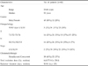
Sixty patients with newly diagnosed nasopharyngeal carcinoma treated with chemoradiotherapy in Chonnam National University Hospital from January 1991 to September 2001 were analyzed retrospectively. All the patients were staged according to 1997 AJCC14 and had pretreatment computed tomography (CT) scans for their tumor volume measurement. There were 48 male (80.0%) and 12 female (20.0%) patients, with a median age of 53 (range, 19-68 years). All the patients received chemotherapy and radiation therapy. The patients received one to three cycles of chemotherapy consisting of cisplatin and 5-fluorouracil (5-FU). Thirty-eight patients before 1998 received neoadjuvant chemotherapy (cisplatin 100 mg/m2 and 5-FU 1000 mg/m2) and 22 patients after 1998 received neoadjuvant and concurrent chemoradiotherapy (cisplatin 60-80 mg/m2 and 5-FU 600-800 mg/m2). All patients received conventional radiation therapy with a fraction size of 180 or 200 cGy once a day, and 5 times a week by a 6MV photon linear accelerator. The total delivered dose was 64.8-77.0 Gy (median: 70.0 Gy) for the primary tumor and 45.0 Gy was prescribed for the elective neck irradiation. The follow-up period ranged from 11 to 144 months with a median of 51 months (Table 1).
Volume determination
A pretreatment contrast CT scan was performed in all patients with contiguous axial scans of 3-5 mm slice thickness. Patients were staged according to the AJCC stage classification system for nasopharyngeal carcinoma. The primary tumor and nodes were outlined separately. Neck node involvement was diagnosed based on the criteria recommended by Mancuso et al.15 and Som16: 1) a discrete mass more than 1 cm in diameter, not enhancing to the extent expected of vessels in the lymph node bearing region of the neck or 2) presence of central necrosis of suspected nodal mass or 3) grouping of three or more contiguous nodes, each 8-15 mm in diameter. Nodes that were embedded within the primary tumor (e.g., retropharyngeal nodes) were included as part of the tumor.
All CT scans were digitalized using a film scanner. The contour of the primary tumor and lymph nodes was outlined using the computer image analysis software Image-Proplus 4.0 (Media Cybernetics, USA) and the area calculated. The maximal perimeter was calculated in the primary tumor. In the lymph nodes, the maximal perimeter could not be calculated due to a large error in measuring multiple lymph nodes of varying contiguity. The primary tumor volume (PTV) and nodal tumor volume (NTV) were then calculated by a summation-of-areas technique. The total tumor volume (TTV) was outlined by adding the PTV and NTV. The PTV, NTV, TTV, and MPP were divided into two groups with the cut-off values decided by a stepwise pattern.
Statistical analysis
The correlations between T stage and PTV, N stage and NTV, staging and TTV were analyzed. For the survival analysis, each parameter was divided into two groups as follows: T stage (T1/T2 vs. T3/T4), N stage (N0/N1 vs. N2/N3), MPP (≤ 18 cm vs. > 18 cm), PTV (≤ 30 cm3 vs. > 30 cm3), NTV (≤ 5 cm3 vs. > 5 cm3), TTV (≤ 35 cm3 vs. > 35 cm3), chemoradiotherapy (neoadjuvant vs. concurrent), histopathologic type (WHO type I vs. II vs. III), intracranial extension, cranial nerve involvement, and skull base erosion.
The product-limit method of Kaplan and Meier and log-rank test were used for the univariate analysis of prognostic significance of the parameters on treatment results (local control rate, nodal control rate and disease-specific survival). Multivariate analysis was also performed using the Cox proportional hazard model. A p-value less than 0.05 was considered to be statistically significant.
RESULTS
Volumetric analysis of primary tumor and metastatic lymphadenopathies
The PTV, NTV, and TTV were 0.4 to 77.5 cm3 (median, 12.2 cm3), 2.7-121.0 cm3 (median, 6.6 cm3), and 0.4 to 184.8 cm3 (median, 27.8 cm3), respectively, and the MPP was 2.5 to 27.1 cm with a median of 15.6 cm.
Relation between tumor volume and stage
The PTV (≤ 30 cm3, n=46 vs. > 30 cm3, n=14), NTV (≤ 5 cm3, n=27 vs. > 5 cm3, n=33), and TTV (≤ 35 cm3, n=37 vs. > 35 cm3, n=23) were correlated with T stage (p=0.001), N stage (p < 0.001), and stage group (p=0.028), respectively, although there were variations such as small volume and high stage, or vice versa.
Treatment outcome
Survival analysis showed the 5-year local control rate to be 75.5%, the 5-year nodal control rate to be 74.6%, and the 5-year disease-specific survival rate to be 60.2%.
Univariate analysis of tumor volume and treatment outcome
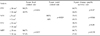
Large PTV (> 30 cm3) was associated with a significantly lower local control (p=0.004). Large NTV (> 5 cm3) was associated with a lower nodal control (p=0.019) and lower disease-specific survival rate (p=0.046) with statistical significance. Although a lower disease-specific survival rate was also observed in patients with a large PTV or TTV, there was no statistical significance. The MPP was associated with a local control rate with statistical significance (p=0.017) (Table 2).
Multivariate analysis of prognostic factors in nasopharyngeal carcinoma
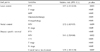
The results are summarized in Table 3. Only PTV was found to be an independent predictor of local control, with T stage or MPP no longer a significant factor. In nodal control, only NTV was found to be an independent prognostic factor, N stage no longer being a significant factor. In disease-specific survival, the NTV and cranial nerve involvement were found to be independent prognostic factors.
DISCUSSION
Tumor bulk has been well recognized as one of the major prognostic factors in the treatment of malignancy, as increasing tumor bulk relates to an increasing number of tumor clonogen needing to be sterilized.5-9 The prognostic significance of tumor bulk has been recognized and adopted in the staging systems of most malignancies, which often employs a crude measurement of tumor diameter and assessment of tumor extent. Such methods of evaluating tumor bulk may be less applicable in tumors that tend to be infiltrative and irregularly shaped, especially if the tumor is difficult to assess and measure clinically.
Nasopharyngeal carcinoma represents a tumor with a highly infiltrative growth pattern, with a propensity to spread along parapharyngeal space as well as to the skull base and foramina.17 In addition, there is a variation of the anatomic structure of the nasopharynx between individuals. The tumor volume cannot be easily assessed clinically, and even with the help of imaging, the irregularly shaped tumor would make a crude measurement difficult and have limited accuracy. Accurate measurement of tumor volume in nasopharyngeal carcinoma therefore requires a detailed outlining of the tumor extent from imaging, and a calculation of tumor volume from a three-dimensional perspective.
Recently, tumor volume has been actively studied in head and neck malignancies. The ability of tumor volume to predict local control in supraglottic and glottic carcinoma,18,19 and in oropharyngeal and hypopharyngeal carcinoma20,21 was reported. Furthermore, Chua et al.22 suggested that measurement of the primary tumor volume in nasopharyngeal carcinoma offered a more important prognostic value in predicting local control than either Ho's or AJCC staging systems. Both the Ho's and AJCC staging systems for nasopharyngeal carcinoma classify the T stage according to the anatomic sites and not to the tumor bulk. The AJCC staging system classifies the N stage according to bilaterality, the greatest dimension, and extension to supraclavicular fossa. The main purpose of any staging system is to segregate patients into subgroups with different prognosis so that an appropriate treatment strategy can be employed. Due to the limitation of the current staging systems to predict prognosis, there has been an effort to refine the staging system.
Although large tumor volume was more commonly observed in the higher stages (T stage, N stage, stage group) of disease in this study, there was substantial variation of tumor volume in all stages. This finding means that similar values of tumor volume could be classified in different stages according to the direction of tumor extension.
Chua et al. reported that patients with large PTV > 60 cm3 had a poor chance of control of nasopharyngeal carcinoma22 and that 15 cm3 was the cutoff point that predicted the prognosis for early T1 and T2 nasopharyngeal carcinoma.23 Willner et al.24 reported that patients with PTV > 64 cm3 had a lower local control treated by radiotherapy. Sze et al.25 suggested that patients with primary tumor-retropharyngeal lymph node volumes over 15 cm3 had lower local control and that the local control rate increased by 1% as the primary tumor volume increased by 1 cm3. Our study showed that patients with PTV over 30 cm3 had lower local control and PTV was found to be a significant factor in predicting local failure. The T stage of the AJCC staging system failed to predict local control in multivariate analysis.
Johnson et al.26 reported that patients with TTV over 35 cm3 had lower local control. In our study, TTV was divided into PTV and NTV. According to analysis, PTV was significantly related to local control rate and NTV was significantly related to nodal control rate. Furthermore, NTV was significantly related to disease-specific survival rate, as patients with large NTV over 5 cm3 had a statistically lower survival rate. This differs from the report of Chua et al.23 that a large NTV over 4 cm3 was related to a high distant failure rate without any relationship to disease-specific survival. This can be explained by the finding that the relatively small tumor volume of lymph nodes in early nasopharyngeal carcinoma was included in their study. Although there was a report that disease-specific survival varied according to the N stage,23 it was not an independent prognostic factor of survival in this multivariate analysis.
Tumor perimeter has not yet been studied for treatment outcome. However, it was included in this study due to its indication of irregularity of tumor extension and its usage as the two-dimensional data of tumor size. Although the MPP was a significant parameter that predicted local control in the univariate analysis, it was not significant in the multivariate analysis where the tumor volume was included. This suggests that nasopharyngeal carcinoma tends to have an infiltrative growth pattern, often with a highly irregular tumor contour. The tumor perimeter is not as significant as tumor volume due to its limitation as a two-dimensional factor, even though it reflects the irregularity of each tumor relatively well.
Cranial nerve involvement was recognized as a prognostic factor of local control and survival, irrespective of skull base erosion.27 Intracranial extension was reported to have an inverse correlation with survival.4 In our study, cranial nerve involvement was found to be related to a lower disease-specific survival rate. This may be explained by the extension of the tumor along cranial nerves to other sites out of the port of radiation.
Other factors contributing to apparent tumor radioresistance must be considered. Although this study demonstrated an inverse correlation between tumor control and disease volume, there were several failures in small tumors and cures in cases with massive disease. This suggests that other factors besides those studied here contribute to radiation response. Cellular factors such as repopulation, intrinsic radioresistance, and/or reoxygenation and redistribution are suggested as important variables for tumor control.
In conclusion, we suggest that in order to better refine the staging system, volumetric analysis of the primary tumor and lymph nodes, anatomic sites involved, and intracranial extension should be included in the current TNM staging system for nasopharyngeal carcinoma.
 XML Download