

 PDF
PDF ePub
ePub Citation
 Print
Print


INTRODUCTION
Although a recent clinical trial found no benefit of carotid artery stenting (CAS) relative to carotid endarterectomy (CEA), CAS may still represent a potential alternative treatment for patients with a high surgical risk (1).
Recently, the CAS procedure has been optimized through refinement of technical devices including stents and distal protection devices. Various different stent designs (such as open-cell and closed-cell) and materials exist; however, the safety of the stent itself is rarely taken into consideration. Chang et al. (2) reported that stent deformation is not uncommon after CAS; among 116 CAS cases studied, stent fracture and deformation occurred in 27%. However, the clinical impact of stent fracture or deformation is unclear.
Herein, we report a case of delayed cerebral infarction caused by longitudinal folding deformation and in-stent thrombosis following CAS using a self-expandable stent with an open-cell design.
This report had been approved by the Institutional Review Board, with informed consent given by the patient.
CASE REPORT
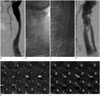
A 72-year-old man presented with motor aphasia and transient right hemiparesis. He had a history of hypertension. Cerebral angiography showed more than 80% stenosis of the left common carotid artery (CCA) (Fig. 1A): the diameter of the stenotic segment was less than 1.5 mm whereas unaffected proximal and distal segments of the CCA measured 9 and 8 mm in diameter, respectively (Fig. 1E).
Carotid artery stenting of the left CCA was performed percutaneously via the right femoral artery with a self-expandable stent with an open-cell design (Protégé® RX Carotid Stent System, EV3, Plymouth, MN, USA; 10 × 60 mm). This lesion was pre-dilated and then post-dilated following placement of the stent. The stent appeared completely expanded in the left CCA after procedure (Fig. 1B). Neither periprocedural complications nor neurological symptoms developed after the procedure. Aspirin (100 mg/day) and clopidogrel (75 mg/day) were administered daily for 8 months following stent implantation, and the latter was stopped thereafter.
After 4 years, a sudden neurological episode occurred, characterized by the right hand weakness. MRI revealed an embolic infarction in the left precentral gyrus. Computed tomography angiography (CTA) revealed the deformation to comprise longitudinal folding and divide into two separated lumens along the whole segment of the implanted stent (Fig. 1F). The left CCA angiography confirmed extensive in-stent thrombosis with longitudinal folding deformation of the stent (Fig. 1C, D), and clopidogrel (75 mg/day) was administered immediately. The patient remained asymptomatic during 12 months of follow-up after discharge.
DISCUSSION
Carotid stent fracture has been reported with markedly disparate incidences from 2% to 29%, and stent deformation without fracture is also likely to occur frequently with the incidence of 23% (2, 3, 4, 5). However the clinical impact of stent fracture or deformation is still unclear. To our knowledge, this is the first case report for the longitudinal folding deformation of the carotid stent combined with delayed cerebral infarction. In CREATE trial of the Protégé stent (6), they did not demonstrate delayed ischemic complication due to stent folding deformation among the 408 CAS cases studied.
In this case, we used the self-expandable nitinol stent with an open-cell design. Generally, the cell size in open-cell stents differs between the ends and the body of a stent. Relative to stents of the same design, the used stent is known to have a wider cell size in both ends, which may weaken the radial force from the stent ends, thereby resulting in stent folding deformation. Sfyroeras et al. (7) analyzed carotid stent fracture that occurred mainly in self-expandable nitinol stents, and Chang et al. (2) observed that stent deformation was significantly more common in open cell stents than closed cell stents.
This stent was placed in the left CCA, which is located in a highly active part of the human body. The carotid artery is recently considered as a possible region of stent fracture because of various neck motion and high-pressure pulsatile flows. Carotid stents are subject to external mechanical forces of axial and crush deformation by complex head and neck movement (8). Vos et al. (9) demonstrated that carotid artery could lose its natural flexion, extension, and rotational flexibility after CAS. Therefore, the stented segment of the artery may be a rigid unit in all head positions, thus resulting in kinking or compression at the junctions between the stent and artery. Boehm et al. (10) reported head rotation initiated neurologic symptoms in patients with carotid stent fracture.
Other possible causes of stent deformation are vessel angulation of more than 45° and presence of heavy calcification around the stented artery (2, 5). However, in our case, no calcification along the left CCA occurred, and the stented artery was a straight configuration without curvature (Fig. 1E).
We experienced a delayed ischemic complication due to such a stent deformation, which was an uncommon phenomenon. Some authors described case reports of late stroke related to stent fractures but not stent deformations (10, 11). However, delayed neurological symptoms are known to develop rarely in cases of stent fractures or deformations. Although stent fractures were often associated with restenosis, stent fractures or deformations were independent of late stroke (2, 5, 7).
No established method of managing deformation and collapse of a carotid stent, which may precipitate cerebral infarction through in-stent thrombus and restenosis, exists. In symptomatic cases of stent fracture, various therapeutic interventions including anticoagulant treatment, restenting, stent removal by open surgery, endarterectomy, or a bypass graft may be considered (7). In this case, we employed a conservative approach to management with dual antiplatelet therapy; the patient was successfully treated and remained asymptomatic during 12 months of follow-up after discharge.
Conclusion
Delayed cerebral infarction due to longitudinal folding deformation of the carotid stent is a very rare complication of CAS. Although the causes of such a complication remain unclear, attending clinicians should be aware of the possibility of this complication following CAS and should inform patients of the prospective risk prior to treatment.
 XML Download