

 PDF
PDF ePub
ePub Citation
 Print
Print


Endosalpingiosis is a non-neoplastic process that's characterized by a proliferation of ectopic tubal epithelium, and this malady is most often diagnosed incidentally or it may present with chronic pelvic pain. There has been a report on the florid form of endosalpingiosis with cystification that mimicked a neoplastic process (1). To the best of our knowledge, the MR imaging appearance of florid cystic endosalpingiosis infiltrating both the uterus and the surrounding tissue and that looked like a gynecologic malignancy has not been previously described in the medical literature. We report here on the MR imaging appearance of a case of florid cystic endosalpingiosis that involved the uterine, the cervical serosa and the adjoining pelvic tissue, and this all resembled a malignancy.
CASE REPORT
A 40-year-old woman presented with chronic pelvic pain and dysfunctional uterine bleeding. The patient had a history of undergoing uterine ablation twice and this had been done due to dysfunctional uterine bleeding about nine and six months prior to the current presentation. The CT scan that had been done elsewhere revealed an irregular soft tissue mass in the pelvis and the mass was indenting the posterolateral aspect of the uterus and cervix. The manual vaginal examination that was performed at presentation revealed a large soft tender mass that was felt in the left fornix. In addition, a pedunculated cervical polyp was also noted; this was removed and the histology showed it to be an endometroid polyp with simple hyperplasia and it was without atypia or focal squamous metaplasia.
A subsequent MRI of the pelvis was done on a 1.5T AVANTO (Siemens, Erlangan, Germany) MRI scanner with the T1 weighted (T1W) high resolution sequence, the T2 weighted (T2W) sequence and the short tau inversion recovery (STIR) sequence in the axial plane and the T1W and T2W fast spin echo sequence in the sagittal plane. The post-contrast T1W images with and without fat suppression were acquired in the axial and sagittal planes. The pre-contrast T1W images were digitally subtracted from the post-contrast T1W images for evaluation.
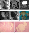
The MRI findings revealed a heterogeneously enhancing, complex cystic mass involving the posterior myometrium at the uterocervical junction with an extraserosal extension infiltrating into the pouch of Douglas and a thickening that extended posterolaterally to the upper left to the pararectal region (Fig. 1A-C). A discrete well circumscribed cystic lesion was also noted in the posterior cervical stroma with no appreciable post-contrast enhancement (Fig. 1D). Focal hyperintensity was noted to be interspersed in the discrete well circumscribed cystic lesion, and this focal hyperintensity was presumed to be subacute blood, but there was no evidence of any hemorrhage in the histopathology specimen and this possibly represented mucinous contents (Fig. 1E). There were no enlarged retroperitoneal/pelvic nodes. Our imaging differential diagnosis was endometriosis versus cystic neoplasm. Total abdominal hysterectomy was done together with bilateral salpingo-oopherectomy. The operative findings revealed nodule in the pouch of Douglas with no apparent cystic mass (possibly collapsed) and there was a fibrotic thickening along the left uterosacral ligament extending to the left pararectal area.
Gross examination of the resected specimen revealed irregular grayish white tissue adhered to the posterior cervical wall (Fig. 1F). Both ovaries and both fallopian tubes were unremarkable except for a few simple cysts. Microscopic examination revealed cystic structures lined by the tubal type of epithelium. On examination of the multiple sections from the lesion, these dilated tubal structures were devoid of any endometrial stromal tissue, and this excluded a diagnosis of endometriosis (Fig. 1G, H). Similar histological findings were noted at the corpus uterine serosa, the left cervical serosa and in the pouch of Douglas. The final diagnosis was florid cystic endosalpingiosis.
DISCUSSION
Endosalpingiosis is non-neoplastic proliferation of ectopic tubal epithelium (2). According to Zinsser and Wheeler (3), the frequency of endosalpingiosis is up to 12.5%, on the basis of the surgically removed omentum that was histologically examined. These patients are usually asymptomatic and their condition is usually incidentally diagnosed at the time of operation or by microscopic examination of the resected/biopsied specimen. The symptomatic cases may present with chronic pelvic pain (4), as in our case.
Cystic endosalpingiosis has been described on MRI as a well circumscribed, intramural serous fluid-filled unilocular cystic mass in the uterine fundus and as a simple cyst in the right ovary with no hemorrhagic component (5). Multiple disseminated pelvic calcifications in endosalpingiosis have been described on CT (6). Yet we found no evidence of any calcifications on the retrospective review of the CT study that was done prior to MRI. Tumor-like foci of endosalpingiosis have only rarely been described in the urinary bladder (7) and in the vermiform appendix (8).
Florid cystic endosalpingiosis is rare. The pathogenesis of florid cystic endosalpingiosis is largely unknown; however, mullerianosis wherein the coelomic epithelium lining of the peritoneal cavity might undergo a change towards primary mullerian epithelium, including tubal, endometrial and endocervical epithelium, has been described (9). Clement and Young (1), in their series of four cases of endosalpingiosis, described the clinical and histopathology findings in a case of florid cystic endosalpingiosis that had tumor-like masses, which were characterized by a polypoidal mass composed of multiple cysts lined by tubular type epithelium, hyperplastic smooth muscle tissue and a myofibromatous stroma. Atypical endosalpingiosis has been reported by the same authors with marked cellular stratification and a varying degree of cellular atypia. No cellular atypia was noted in our case.
The morphology of the lesion in our case resembled a multicystic mass with intrusion into the cervical stroma. Various differential diagnoses have been proposed for cystic cervical masses (10), and these include deep nabothian cysts of the cervix, florid deep glands of the uterine cervix, endometriosis, cystic adenocarcinoma and adenoma malignum. Of the tumor like lesions, florid deep glands of the uterine cervix can infiltrate the cervical stroma with no evidence of any atypia, as in our case. Moreover, in our case, a multicystic mass was seen in the pouch of Douglas on imaging. Tubal and tuboendometroid metaplasia may penetrate deeply into the cervical stroma, but it should originate from the endocervical canal, rather than arising from or beneath the serosa (10), as in our case.
We describe this case to demonstrate the MRI appearance of florid cystic endosalpingiosis, which can clinically and radiologically mimic neoplasia. Awareness of the imaging appearance of this malady may broaden differential diagnosis of cystic pelvic masses and help prevent over diagnosis and over treatment.
 XML Download