

 PDF
PDF ePub
ePub Citation
 Print
Print


A bronchobiliary fistula (BBF), defined as a passageway between the biliary and bronchial trees, is a very rare condition. In Western countries, trauma, postoperative biliary stenosis and biliary lithiasis are the predominant causative factors of a BBF (1, 2). For the treatment of patients with a BBF, endoscopic or transhepatic biliary drainage has been successful and avoids the need for surgical exploration (1, 3, 4).
A BBF is also an extremely rare complication of radiofrequency ablation (RFA). To date, only seven cases of BBF, including one fatality, have been reported after RFA of hepatic tumors (5-10). In one case, a patient was managed with the use of an endoscopic sphincterotomy that resulted in complete closure of the fistulae (5). Four patients underwent both endoscopic and percutaneous drainage for successful management of a BBF and/or an associated biloma or abscess (6, 7, 10). Another patient was managed with the use of an external drainage catheter; however, the fistula was still persistent two months later, although symptomatic improvement was achieved (8). We present the first case of a BBF as a complication of RFA that was eliminated by catheter placement for external drainage.
CASE REPORT
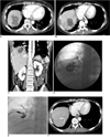
A 43-year-old woman positive for hepatitis B surface antigen was diagnosed as having hepatocellular carcinoma (HCC) with two intrahepatic tumor nodules, one nodule 2 cm in diameter in the left lateral segment of the liver and the other nodule 3.5 cm in diameter in the dome. The patient underwent one session of transarterial chemoembolization (TACE), followed by percutaneous ethanol injection targeting the area of the residual viable tumor in the liver dome. However, two months later, a viable portion was found to be present at the margin of the nodule in the hepatic dome and the patient was referred to our center. At that time, the laboratory test results included an aspartate transaminase (AST) level of 86 IU/L (reference, 0-40 IU/L), alanine transferase (ALT) level of 88 IU/L (reference, 0-40 IU/L), total bilirubin level of 1.0 mg/dL (reference, 0.2-1.2 mg/dL) and alpha-fetoprotein level of 1,460 ng/mL (reference, 0-20 ng/mL). RFA was performed percutaneously under ultrasonographic guidance, with the use of local anesthesia and conscious sedation. A single, 17-G, internally cooled electrode (ValleyLab, Burlington, MA) was used and radiofrequency current was emitted for 12 minutes with a 200 W generator set to deliver maximum power with the automatic impedance control method. Two ablations were performed with the use of the same method. A follow-up CT scan obtained immediately after RFA showed complete ablation of the HCC without direct evidence of diaphragmatic injury except for a small amount of reactive pleural effusion (Fig. 1A). The patient reported pain at the right upper quadrant of the abdomen and right shoulder after RFA and the pain gradually improved over five days with the administration of analgesics. The patient developed a 38.0℃ fever on the next day after RFA, which subsided after one day. Two months after RFA, the patient revisited our hospital with a complaint of a productive cough with large amounts of green-yellow sputum (up to 200 mL per day), accompanied with a 38.4℃ fever with chills and right upper quadrant abdominal pain. Decreases in breath sounds on the right lower lung field and slight tenderness to percussion of the right upper quadrant of the abdomen were noted on a physical examination. Icteric sclera or hepatosplenomegaly was not noted. Plain chest radiograph demonstrated the presence of an ill-defined consolidation in the right lower lobe with right-sided pleural effusion. An abdominal CT scan showed the presence of a liver abscess at the RFA site and pneumonia, with a small abscess in the right lower lung, suggestive of a focal diaphragmatic defect with communication between the lung and liver abscesses (Fig. 1B, C). Ultrasonography-guided percutaneous drainage of the liver abscess with a 10.2 Fr pigtail catheter was performed and 10 ml of thick dark-yellowish pus mixed with necrotic debris was drained. Contrast material injection via the needle demonstrated the presence of opacified cavities of the lung and the bronchial tree communicating with the biliary tree, which was consistent with a BBF (Fig. 1D). The patient coughed vigorously when the above contrast study was performed. No organism other than Bacillus spp, which is a common contaminant, was isolated from a culture of the drainage material. Following catheter insertion and treatment with antibiotics, symptoms including fever and bilioptysis were markedly improved. Bile mixed with pus (20 to 100 ml daily) was drained initially, but the rate of drainage gradually decreased to less than 10 ml per day over a month. Follow-up abdominal CT scans obtained five weeks after catheter insertion showed the presence of a small residual lesion with low attenuation in the hepatic dome around the catheter, and repeated cholangiography confirmed no leakage of contrast material through the fistula. The pigtail catheter was therefore removed and antibiotic treatment was discontinued. However, fever and productive cough with sputum containing bile resumed the day after removal of the pigtail catheter. A new cavitary lesion with low attenuation and parenchymal consolidation in the right basal lung were observed on CT images, suggesting reopening of the BBF. A pigtail catheter was reinserted into the remnant abscess in the liver through the previous drainage tract. Percutaneous tubography showed filling of the contrast material in the right lower lobe of the lung, with contrast material leakage into the right subphrenic space, indicating the presence of a BBF although there was no definite communication between the biliary and bronchial systems (Fig. 1E). Thereafter, the amount of percutaneous drainage decreased gradually and the lesions in the liver and lung shrank significantly, as shown on follow-up CT scans three weeks after reinsertion of the pigtail catheter (Fig. 1F). As there was no drainage for a week and the patient did not complain of any fistula-related symptoms, the pigtail catheter was removed. Four weeks after removal, there was no evidence of a BBF or other related lesions on CT images. Two months later, the patient underwent a repeat session of RFA for a recurrent HCC in segment 7 without any treatment-related complications, including a BBF.
DISCUSSION
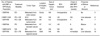
Radiofrequency ablation has been increasingly used for the treatment of primary and metastatic hepatic tumors as an alternative or adjunct to surgical resection. Although RFA is a safe modality overall, RFA is associated with minor and major complication rates of 9% (9). Biliary complications, including stricture and leakage, are relatively uncommon, with a prevalence of 1% (9). A BBF as a complication of RFA is extremely rare, with only seven cases reported worldwide (Table 1) (5-10). To the best of our knowledge, no patient with a BBF that occurred after RFA has undergone successful percutaneous drainage leading to complete blockage of the fistula. Thus, the present case is the first report of BBF due to RFA that was successfully managed by percutaneous drainage and safe removal of the catheter.
A BBF due to RFA may be caused by rupture of a growing biloma into a diaphragmatic defect caused by thermal injury, a finding supported by our imaging results. A BBF that occurs after RFA may also depend on tumor location and size. The proximity of a tumor to the lung base could cause thermal injury to the diaphragm following RFA, and symptomatic biloma formation may develop in patients with large tumors owing to more severe bile duct damage (8). The ablated tumor in our patient was located in the hepatic dome adjacent to the right diaphragm, although the tumor was not large.
As described in the present case, clinical findings for a BBF include pneumonitis, fever, chest pain, right upper abdominal pain and cough productive of bile-stained sputum, known as bilioptysis (8). Plain chest radiographs often show abnormal findings and may demonstrate right pleural effusion, right basilar atelectasis or the presence of a lung abscess. A CT scan can be used to identify both hepatic abscesses and air within the biliary tree, but images will not delineate a fistulous tract. Endoscopic retrograde cholangiography or percutaneous transhepatic cholangiography will adequately demonstrate the presence of a BBF (3, 4). In our patient, we were able to identify the fistula tract with tubography after percutaneous drainage.
To date, there has been no consensus on the standard treatment for a BBF. Traditionally, the definitive treatment for a BBF has involved extensive surgery, with or without simple drainage of the subdiaphragmatic abscess and resection of the fistulous tract (2). The conservative approach that has been reported most frequently has involved the use of endoscopic sphincterotomy with removal of any stones or sludge, followed by stent placement in the common bile duct (3, 5). Reduction of the pressure gradient between the common bile duct and the duodenum may lead to preferential drainage of bile through the stent, which then has a lower pressure than the fistula. The resultant decrease in fistula output allows the tract to close and heal.
Percutaneous drainage for a BBF not related to RFA has been previously described (4). In patients with a BBF complicated with liver abscess, as in our patient, external drainage of an abscess or biloma can facilitate both alleviation of hepatic inflammation and the pressure gradient across the biliary tract and bronchial tree. In all of the previous cases, patients with a BBF following RFA complicated by a biloma or liver abscess underwent external drainage with or without endoscopic drainage (6, 8, 10). We found that an initial five weeks of percutaneous drainage plus antibiotic therapy was insufficient to clear the fistula tract. Hence, long-term drainage maintenance with antibiotics after symptomatic improvement is likely to be required for complete remission of a BBF.
In summary, although RFA is a safe and effective modality for the treatment of HCC, the procedure should be performed with caution to prevent BBF formation in patients with liver dome tumors. We report the first case of a BBF as a complication of RFA in a patient with HCC in the liver dome adjacent to the right diaphragm, which was successfully eliminated by percutaneous drainage and antibiotic treatment.
 XML Download