

 PDF
PDF ePub
ePub Citation
 Print
Print


Foreign body ingestion is a common problem in children due to their inherent natural curiosity to put things in their mouth. The button battery represents less than 2% of pediatric ingested foreign bodies (1). However, the relative frequency of ingestion has been on the rise over the last two decades due to increased accessibility to electronic gadgets and toys (1). An impacted button battery in the esophagus could be associated with such complications as ulceration, perforation, fistulization, or even death (2-6). Spondylodiscitis is a recognized complication of the penetrating esophageal foreign bodies. However, to the best of our knowledge, it is not described in the English literature as a complication following the ingestion of a button battery.
CASE REPORT
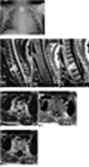
A one and-a-half year old boy was presented to the emergency department with a four day history of coughing, repeated vomiting, irritability and refusal of food. His symptoms began after returning from a picnic. The boy had no prior history of breathing difficulties or cyanosis. A medical examination revealed that the boy had a low grade fever and a few crepitations in the left lower chest. A chest radiograph (Fig. 1A) revealed a rounded, coin-like foreign body at the cervicothoracic junction. An endoscopy performed under general anaesthesia revealed a foreign body in the upper esophagus at 15 cm from the incisors. The button battery was partially embedded in the esophageal wall, which was edematous and ulcerated with charred tissues around the foreign body. During manipulation, the patient's blood oxygen saturation level dropped, which suggested that signs of pneumothorax were present and this prompted the insertion of bilateral chest tubes. As a result, the patient's oxygen saturation level improved and the bilateral air entry was confirmed by a chest radiograph. Following a difficult manipulation, a lithium button battery was removed from the patient. The battery shell remained intact without signs of leakage. Regardless, the patient was kept on nasogastric tube feeds and broad spectrum antibiotic coverage. Ten days after the procedure, a gastrograffin esophagogram was performed, which did not reveal any evidence of a leak. Consequently, the patient was started on oral feeds and was discharged two days later, however was readmitted almost six weeks later, with complaints of neck pain, stiffness, restricted neck movement and intermittent fever since the previous two weeks. The MR imaging (Fig. 1B-D) showed evidence of spondylodiscitis involving the T1-2 disc with enhancing endplates and extension of the enhancing granulation tissues into the prevertebral region. In addition, narrowing of the tracheal lumen was also noted at this level. Also, there was diffuse enhancement in the prevertebral soft tissues around the esophagus, trachea and mediastinal planes, which suggested mediastinitis (Fig. 1E-G). A gastrograffin esophagogram had not revealed any obvious leakage; however, the patient did improve with antibiotics and was afebrile, free of pain and had improved neck movement at the time of discharge.
DISCUSSION
The ingestion of foreign bodies, in the form of button batteries by infants, is on the rise due to their ubiquity in electronic devices and toys. The peak age of incidence is between six months and thee years of age (7). Button batteries contain heavy metals such as mercury, silver, lithium or a strong alkali (7). Possible mechanisms of injury as a result of their ingestion include electrolyte leakage from the batteries, alkali production from external currents, mercury toxicity and pressure necrosis (8).
Although most cases of ingestion are without complication, one in a 1,000 cases cause serious problems, especially when impacted in the esophagus (4, 7). A variety of complications have been described with esophageal impaction; the most common being ulceration (7). Other serious complications include perforation, stricture, and tracheo-esophageal fistula formation (2, 3, 5, 6). Chang et al. (4) reported one case of death following the development of pneumothorax and pneumoperitoneum following the ingestion of a button battery.
Spondylodiscitis is a recognized complication of sharp esophageal foreign bodies. Wadie et al. (9) reported a case of spondylodiscitis in an adolescent girl following the accidental ingestion of a sewing pin. Fonga-Djimi et al. (10) described the esophageal perforation, mediastinitis, and C6-7 spondylodiscitis resulting from a radiolucent foreign body (a rigid plastic gear wheel) in a 6-year-old boy. Spondylodiscitis secondary to phonatory prosthesis insertion or endotracheal intubation have also been reported (11, 12). However, to the best of our knowledge, spondylodiscitis as a result of an impacted button battery in the esophagus has not been reported in the literature. An external current flowing from a battery through adjacent tissue causes the hydrolysis of tissue fluids and local generation of alkali. Furthermore, associated pressure necrosis may cause direct extension of infection, from the esophagus across the pre-vertebral fascia to the spine.
The advocated rest time following esophageal surgery is six to seven days and about 10 days for caustic ingestion injuries of the esophagus before starting oral feeds (13, 14). Oral feeds were started as early as 48 hrs and were delayed up to nine days in various case reports of esophageal injury, following button battery ingestion, depending on the degree of the esophageal burns (7, 15). Oral feeds should not be started until perforation has been ruled out by a contrast esophagogram (13). Baum et al. (16) advocated the algorithm, which states initiating "nothing by mouth" for at least one week, and nasogastric tube feeds and broad spectrum antibiotics for one to two weeks for esophageal perforations. In our case, oral feeds were started 11 days after confirming the absence of a leak on the gastogarffin esophagogram; however, this still led to the exterior spread of infection.
A high index of suspicion is important in diagnosing esophageal foreign bodies. Any battery lodged in the esophagus should be urgently removed by endoscopy. Since button battery ingestion can be hazardous, prevention should be directed at making them inaccessible to children by proper child proofing the appliances containing these batteries and proper safe disposal of used batteries.
In conclusion, we report a case of spondylodiscitis secondary to an esophageal injury following the removal of an accidentally ingested and impacted button battery at the cervicothoracic esophagus in a one-and-a-half year old child. The patient developed mediastinitis and spondylodiscitis even after commencing the oral feeds with an adequate esophageal rest time and after confirming absence of leak on a gastrograffin esophagogram. Hence, we recommend that a gastrograffin esophagogram be followed by an esophagogram with thin barium before starting the oral feeds in such cases in order to increase the sensitivity of detecting leaks, as well as broad spectrum antibiotic coverage for two weeks and a follow-up image if there is no clinical improvement.
 XML Download