

 PDF
PDF ePub
ePub Citation
 Print
Print


Penile Mondor's disease is a rare disease that's characterized by thrombosis in the dorsal vein of the penis. Doppler ultrasonography (US) clearly visualizes dorsal vein thrombosis and the associated hemodynamic alterations. Previous studies have demonstrated the typical color Doppler US findings of superficial dorsal vein thrombosis without the flow signals in this area, yet this is insufficient to understand the hemodynamics in penile Mondor's disease. We report here for the first time a cavernosal artery flow signal pattern in a penile Mondor's disease patient, in addition to its previously reported classic US findings.
CASE REPORT
A 38-year-old male presented with complaints of penile pain during sexual intercourse, and he had experienced this pain for one week. There was no evidence of penile discharge, hematuria, dysuria, fever or erectile dysfunction, and he denied any recent vigorous sexual activity or a history of any type of trauma. His medical history was unremarkable, and he denied ever being infected with a sexually transmitted disease. On the physical examination, a cord-like induration (0.5 cm in diameter and 3 cm in length) was palpated in the dorsal aspect of the proximal penis, and this was associated with tenderness and overlying skin erythema. There were no palpable lymph nodes in his groin and his blood cell count showed leukocytosis (WBC 12 × 103 µL), but the routine biochemical evaluations (blood sugar, LFTs and renal function tests) were normal.
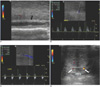
Gray scale ultrasonography revealed internal echogenicity in the superficial dorsal vein of the penis, but color Doppler sonography failed to detect the venous flow signals in this region (Fig. 1A). Pulsed wave Doppler US without an intracavernosal vasoactive agent showed weak flow with a low peak systolic velocity and a high-resistance pattern. The right side peak systolic velocity (PSV) of the cavernosal artery was 24.9 cm/sec with a resistance index (RI) of 1.0 (Fig. 1B); the corresponding left-side values were 16 cm/sec and 1.0, respectively (Fig. 1C). Thirty minutes after an injection of an intracavernosal vasoactive agent, he showed a full erection with unbending rigidity. Doppler US indicated that an enlarged superficial dorsal vein was compressing the deep dorsal vein, and no flow signals were evident in either the superficial dorsal vein or the deep dorsal vein. Bilateral dorsal arterial flow signals were noted (Fig. 1D). The PSV of the cavernosal artery and its RI were unchanged by injection of the intracavernosal vasoactive agent.
He was treated with nonsteroidal anti-inflammatory drugs for six weeks and his symptom were much improved.
DISCUSSION
Mondor's disease of the penis is an uncommon disease that usually involves the superficial dorsal veins. In 1939, Henri Mondor first described a sclerosing thrombophlebitis of the subcutaneous veins of the anterior chest wall (1), and in 1955, Braun-Falco described phlebitis of the dorsal veins of the penis within the context of generalized phlebitis (2). Isolated penile Monor's disease was first described in 1958 by Helm and Hodge (3).
Mondor's disease is a benign and usually a self-limited process. Patients complain of a cord-like induration, which is often painful, in the dorsal aspect of the penis, and this pain can be constant or episodic. The etiology of this condition is usually unknown, but various causative factors have been reported, e.g., penile trauma, excessive sexual activity, prolonged sexual abstinence, infection, pelvic tumors and the constrictive elements used during certain sexual practices; of these, the trauma caused by sexual intercourse appears to be the main etiologic factor (4, 5). For most patients, their symptoms completely resolve after 6 to 8 weeks of conservative management with anti-inflammatory drugs and antibiotics. However, surgery is indicated when such symptoms persist after conservative management, although the long-term results after conservative or surgical treatment of superficial penile vein thrombosis have not been reported (6).
In the present case, Doppler sonography showed a high flow resistance pattern, which resembled that of venous thrombosis after pancreatic or kidney transplantation. The Doppler sonographic findings of venous thrombosis in transplant graft recipients have revealed an absence of venous flow with a high resistance arterial waveform (7-9).
Low flow priapism is a pathology that can produce a low-flow, high resistance flow pattern in the corpus cavernosal artery, and this presumably develops due to a disturbed venous outflow. On Doppler US, low flow priapism usually presents as the absence of cavernosal artery blood flow or as a very high-resistance cavernosal artery flow pattern. We consider that superficial dorsal vein thrombosis may result from a venous outflow disturbance and so it shows low-flow, priapism-like Doppler US findings.
The primary venous drainage of the corpora cavernosa is into the deep dorsal vein (only the most distal portion of the corpora cavernosa, skin and glans drain into the superficial dorsal vein); thus, it appears that superficial dorsal vein thrombosis does not significantly affect the cavernosal venous outflow. However, the enlargement of the superficial dorsal vein and the adjacent soft tissue edema that was caused by thrombophlebitis may cause compression and venous outflow disturbance of the deep dorsal vein. Nevertheless, this is insufficient to explain the high RI value observed in the present case. We suggests that further analysis of Doppler US findings in a larger number of cases needs to be done.
In conclusion, the Doppler US findings of thrombus without blood flow in the superficial dorsal vein and the low-flow, high resistance in the cavernosal artery may be suggestive of penile Mondor's disease.
 XML Download