

 PDF
PDF ePub
ePub Citation
 Print
Print


Mazabraud's syndrome is a rare benign disease, characterized by the association of intramuscular myxoma and fibrous dysplasia of the bone. The majority of patients with Mazabraud's syndrome have multiple myxomas and a polyostotic form of fibrous dysplasia (1). However, we recently encountered a case of Mazabraud's syndrome with the monostotic form of fibrous dysplasia. The patient also presented with a uterine tumor resembling an ovarian sex cord tumor (UTROSCT). These tumors are rare and have been classified as group II ovarian sex cord like neoplasms of the uterus by Clement and Scully (2). Group II tumors are composed almost completely of epithelial-like cells arranged in cords, trabecula, nests or solid sheets resembling ovarian sex-cord elements (3). To the best of our knowledge, the coexistence of the two different entities has not been reported in the literature.
CASE REPORT
A 65-year-old woman had noted a slowly growing painless mass in the left gluteal area for 10 years. As the mass was not troublesome, the patient never consulted a physician. When it was noticed that the mass had slowly increased in size over a period of four months, the patient underwent a pre-employment physical. The large mass localized in the left gluteal area was noted. The patient was referred to our university hospital orthopedic surgery department for evaluation of the soft tissue mass in the left gluteal region. There was no history of trauma.
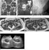
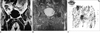
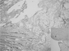
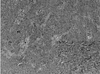
A physical examination demonstrated a large, painless and mobile mass at the area of concern. Magnetic resonance imaging (MRI) scans of the left hip and gluteal area including axial and coronal T1 weighted spin-echo (SE) images (TR: 582 ms, TE: 16 ms) and turbo short tau inversion recovery (STIR, TR 3000 ms, TE 18 ms, TI 100) images were obtained on a 1-T unit with a body coil. In addition axial T2 weighted turbo-spin-echo (TSE) imaging (TR: 3000 ms, TE: 112 ms) was included. These studies demonstrated at least four lobulated soft tissue masses located within the left gluteal musculature, a soft tissue mass within the anterolateral musculature of the left thigh and a solitary bone lesion involving the left ilium. The soft tissue masses were markedly hypointense compared with that of skeletal muscle on T1-weighted images and hyperintense on T2 weighted and STIR images. The bone lesion showed decreased signal intensity on T1-weighted images and increased signal intensity on T2 weighted and STIR images (Figs. 1A-D). Radiography of the pelvis demonstrated a solitary, osteolytic lesion with thin sclerotic margins within the left ilium, which is consistent with the presence of fibrous dysplasia (Fig. 1E). Ultrasonography of the symptomatic and the largest left gluteal mass showed a 4.5 × 4 × 4 cm lobulated, solid, heterogeneous, hypoechoic tumor with multiple small-sized fluid filled cystic areas (Fig. 1F). A differential diagnosis at the time included soft tissue sarcomas and soft tissue metastases. Magnetic resonance imaging also revealed an enlarged uterus and endometrial cavity completely occupied by a large mass that measured approximately 11 cm in the transverse diameter. The mass showed intermediate signal intensity on STIR and low signal intensity on T1 weighted images (Figs. 2A, B). In laboratory studies, the CA 125 level was higher than normal. FDG-PET showed accumulation of FDG in the lesion located in the uterus. No accumulation was observed in the solitary bone lesion located within the left ilium and soft tissue masses (Fig. 2C). The patient underwent excisional biopsy of the symptomatic and the largest left gluteal mass. At the time of surgery, the tumor was found to be located within the gluteus maximus muscle. The lesion was completely excised, including a small cuff of normal appearing musculature. Grossly, the tumor measured 5 × 4 × 4 cm in dimension and appeared as a lobulated ovoid cystic mass that was partially covered by and adherent to skeletal muscle. The cut surface of the tumor revealed many cystic spaces (size ranged from 1 to 2.5 cm), which were filled with mucoid material, and solid gelatinous areas. A histological evaluation showed an intramuscular myxoma, with no sign of malignancy (Fig. 3). An abnormal left ilium and soft tissue myxomas indicated a typical case of Mazabraud's syndrome. Later, a total abdominal hysterectomy and bilateral salpingo-oophorectomy were performed because of the tumor of the uterus. Macroscopically, the tumor grew as a nodular mass within the myometrium and was 8.5 cm in diameter. On a histological examination, the tumor was composed entirely of epithelioid cells displaying sex cord-like structures as cords, anastomosing trabecula, and solid sheets of cells. Tumor cells had plump vesicular nuclei with prominent nucleoli. The stroma was scanty, and was seen as fibrous or hyalinized bands between cell groups. Nine mitoses/10 high power fields were determined (Fig. 4).
DISCUSSION
Intramuscular myxoma is a relatively uncommon benign soft tissue tumor (1, 4). Its association with fibrous dysplasia of bone represents a rare syndrome described by Mazabraud and Girard in 1957 (5). The relationship between fibrous dysplasia and myxoma remains unclear. A common histogenesis has been proposed for both lesions. Wirth et al. has suggested a basic metabolic error of both tissues during the initial growth period, restricted to the region of bone involvement (6). Myxomas may appear at any age, but have a predilection for older individuals, occurring most commonly during the sixth and seventh decades of life (4). They are often located in the large muscles of the thigh, shoulder and buttocks. The majority of intramuscular myxomas are solitary (1). Cross-sectional techniques are essential in the preoperative planning of excision of soft tissue tumors. The ability to evaluate soft tissue myxomas is best accomplished with MR imaging (4). Myxomas typically demonstrate the following MR features: very sharply defined contour and homogeneous signal intensity (7). In particular, the lesions are significantly low in signal intensity on T1-weighted images and high in signal intensity on T2-weighted images. In the patient of this case, the MR appearance was in agreement with previously reported cases. Ultrasonography usually shows hypoechoic tumors with multiple small fluid-filled clefts or cystic areas of various sizes (1). The US findings of our patient were consistent with previous descriptions.
Fibrous dysplasia of the bone is a rare benign condition of unknown origin, with fibro-osseous metaplasia of one (monostotic) or multiple (polyostotic) bones. Any bone may be affected; however, the long bones, ribs, and skull are the most common sites of occurrence. The monostotic form of fibrous dysplasia is much more common than its counterparts (1).
The association of intramuscular myxoma and fibrous dysplasia has been previously reported (1, 4, 6). The key to accurate diagnosis of myxomas within the spectrum of Mazabraud's syndrome lies in awareness of their association with fibrous dysplasia. In the presence of fibrous dysplasia, a soft tissue mass should suggest the possibility of a myxoma, although histological proof surely will still be required (4). Although myxomas tend to occur singly, 81% of patients with Mazabraud's syndrome had multiple myxomas. The myxomas appear to be located in the lower extremities near the bone most affected by fibrous dysplasia (8). The syndrome is more common with the polyostotic form of fibrous dysplasia, but has been reported with monostotic involvement as in our case (4). Some of the findings displayed by the patient in this case are consistent with the features in other cases namely, the myxomas were multiple and occurred principally in the lower extremity. In general, fibrous dysplasia antedates the appearance of an intramuscular myxoma and the soft tissue tumors become apparent many years later (6), although this was not the case in our patient. Unlike most reported cases, the intramuscular myxoma located in the left thigh was detected without an osseous lesion in our patient.
The uterine tumor resembling an ovarian sex cord tumor has similarity with an ovarian sex cord tumor, particularly with a granulosa cell tumor. The histogenesis of these tumors is unclear. Most are benign but some have recurred and/or metastasized. The tumors with non-infiltrative growth and low mitosis behave as benign lesions. Malignant forms behave as low-grade stromal sarcomas (3). Our case showed a high mitotic activity and cellular atypia and therapy was planned as for a low-grade endometrial stromal sarcoma. There are few reports concerning the imaging diagnosis of this rare tumor. Suzuki et al. used MRI in a case of UTROSCT that was cervical in its location (9). In that case, MRI demonstrated a round mass with hypointensity on T1 weighted images and intermediate signal intensity with small areas of hyperintensity on T2 weighted images (9). MRI was used in another case of an UTROSCT located in the myometrium, and the tumor was described as slightly hyperintense on T2 and isointense to the myometrium on T1 weighted images (10). The MRI findings in our patient were consistent with these previous descriptions.
The present case is of interest as the patient presented with both Mazabraud's syndrome and a uterine UTROSCT. Coexistence of Mazabraud's syndrome with a UTROSCT remains unclear; the UTROSCT was most probably not related to the syndrome.
 XML Download