

 PDF
PDF ePub
ePub Citation
 Print
Print


Ever since inflamed appendix was initially demonstrated by ultrasonography (US) in 1981 (1), graded-compression ultrasonography by Puylaert (2) has been widely used for the past two decades as an aid to clinically diagnose acute appendicitis. The use of US for evaluating the patient clinically suspected of having appendicitis has been extensively reported on in the literature (3-24). However, there has been deal of great variability in the reported performance of US for the diagnosis of appendicitis. While the range of reported accuracy (82%-96%) for US in children has been acceptable (3, 5, 9, 11, 19), the sensitivity (44% to 100%) and the specificity (47% to 99%) have varied considerably (4, 6-8, 10, 13-15, 18, 21-22). Also, the visualization rates vary widely in the published literature, from a low of 22% to a high of 98% (13, 15). Various factors might be considered as the causes of these variations. Because US is highly user-dependent, operator skill may be an important factor in the diagnostic accuracy of appendicitis (14, 21, 25). Also, patient age or sex-based differences in the diagnosis of appendicitis with using some clinical presentations alone may be observed in the elder or female patients because of the broad overlap of the symptoms for acute abdominal conditions (including gynecologic abnormalities) (26-27). Especially because of the inability to compress the right lower quadrant (RLQ), particularly in obese patients, or because of a retrocecal location of the appendix, US could not appropriately visualize the appendix. Thus, most of the false-negative diagnoses with using US result from non-visualization of the appendix or from inflammation limited to the appendiceal tip (7, 28-30).
Meta-analysis is the critical review and statistical combination and evaluation of the results of previous research, and this is potentially useful for assessing diagnostic accuracy (31-32). Some meta-analyses related to the diagnostic methods used for appendicitis have been conducted (33-37). Among these, 3 studies have addressed the clinical outcomes of US for the diagnosis of appendicitis (33, 35, 37). In a landmark 1995 article by Orr et al. (33), an overall sensitivity of 84.7% and a specificity of 92.1% were reported for US by using meta-analysis of the previous pediatric and adult studies (17 studies, 3,358 patients), that were published between 1986 and 1994. In that metaanalysis, the accuracy and usefulness of US were related to the likelihood of appendicitis. However, no systematic quantitative overview about the diagnostic accuracy of US, according to the researchers' and patients' factors, has been undertaken to date. Therefore, the objective of this study was to determine the usefulness of US for evaluating abdominal pain patients in Korea who had possible acute appendicitis, and to evaluate the diagnostic performance of US according to the patients' and researchers' characteristics with using meta-analysis method.
MATERIALS AND METHODS
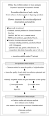
The flow chart of the meta-analysis in this study is shown in Fig. 1. This process consisted of the retrieval of the relevant literature according to the goals that were set, and then performing qualitative1 and quantitative2 meta-analysis (38-40).
Gathering of Data
The first search step was to examine not only the journal database sites such as the Medical Research Information Center (http://www.medric.or.kr/) and the Research Information Center for Health (http://www.richis.org), but also the journal search window of homepages such as the Korean Radiological Society (http://www.radiology.or.kr/), the Korean Society of Medical Ultrasound (http://www.ultrasound.or.kr/), and the Korean Surgical Society (http://www.surgery.or.kr/) from 1985 to 2003. The second step involved a manual search of the contents and the bibliographies cited in each of the retrieved study. The medical subject headings used for this search were acute abdomen, appendix, appendicitis, diagnosis and ultrasound or ultrasonography (or US).
Qualitative Meta-analysis
A total of 73 Korean articles that contained information on ultrasonography for the diagnosis of acute appendicitis were selected. A diagnostic radiologist and a meta-analyst independently extracted the outcome variables of the individual articles onto a data sheet; disagreements were resolved by discussion or by cross-checking with the other co-workers. A systemic review team consisted of a diagnostic radiologist (for data extraction and clinical interpretation of the study results), a surgeon (for study selection and the clinical interpretation of the study results), a biostatistician (for data synthesis and analysis), and two meta-analysts (for the study design, the assessment of study quality and the statistical interpretation of the study results). A systematic literature review was conducted based on the previously suggested meta-analysis evaluation guidelines (40). The criteria for quality evaluation were as follows. 1) Only original articles were included. 2) Patients must have the predominant clinical findings for acute appendicitis. These clinical findings were mainly RLQ abdominal pain and RLQ tenderness or RLQ rebound tenderness. 3) The disease positive group within the studies must certainly include the histopathologic findings as a reference standard to confirm appendicitis, but the disease negative group could be confirmed by the surgical results or the clinical follow-up. The inflamed appendix was assessed by high-resolution, real time US according to the graded-compression method (2). The US criteria (41-42) for the diagnosis of appendicitis were an appendiceal diameter greater than 6 mm, a lack of compressibility, inflammation, echogenic periappendiceal fat, appendicolith, adjacent fluid collections (and hyperemia on color Doppler imaging). This study included the articles that presented over three of the US criteria in the materials and methods section of each study. Also, sufficient or available numeric information such as a 2×2 contingency table for data or the patient outcome data (sensitivity and/or specificity with the absolute numbers of positive and negative findings or the standard errors) of the US testing were contained in our inclusion criteria. Of the 33 articles evaluated at the final stage, 22 studies that had extensively used US for the diagnosis of acute appendicitis met these inclusion criteria; thus, all these were selected for the quantitative meta-analysis.
Quantitative Meta-analysis
The Hasselblad method with the SAS program was utilized for analyzing the contingency tables in this quantitative meta-analysis (43-44). The estimate of đ and the 95% confidence intervals (CIs) were estimated using the sensitivity and specificity for each of the study's outcome data. The đ measure used in this study is analogous to the effect-size measure described for continuous-outcome measures as a more simple calculation. Homogeneity testing was done to test whether the effect size parameters were reasonably constant across the studies (43). Because there was evidence of heterogeneity (Q = 111.913, p-value < 0.001), a random effects model was used instead of a fixed effects model.
Subgroup analyses (39, 45) were performed to provide further insight into the heterogeneity. Additionally we calculated the likelihood ratio for a positive US result3 (46). According to the literature review (18, 25, 26, 33, 35, 37, 47-50) and the results of the qualitative meta-analysis about the factors related to US accuracy for the diagnosis of appendicitis, we classified the subgroup criteria as the characteristics of the patients or the researcher. The patient characteristics included age, gender and the clinical status. Among these, the age and gender groups were classified as three age groups (young, adult and older) and two gender (male and female) dominant groups according to the weight (the percentage points). This was done by consensus of the systemic review team because of secondary aggregating data and the insufficient information concerning these variables in each study. The researcher characteristics included the sonographic examiner, the type of US and the diagnostic method. On the basis of the clinical presentations before the imaging test, we categorized the diagnostic method into two groups: the clinical examination (by the initial physical examination, the diagnostic scoring system4 or leukocytosis) and the US examination.
RESULTS
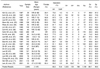
The general characteristics of the 22 studies (51-72) used for this meta-analysis are summarized in Table 1. A total of 2,643 patients with right lower quadrant abdominal pain underwent graded-compression US for the diagnosis of acute appendicitis. Of the 2,643 patients, 1,717 (65.0%) were treated by operational appendectomy (OA), and clinical follow-up without any surgical intervention was performed on 926 patients (35.0%). Among the OA patients, 1,411 patients (82.2%) with a wide range (59.7%-100.0%), according to the individual study had been pathologically diagnosed as having acute appendicitis with/without periappendiceal abscess or perforated appendicitis. One hundred eighty-three patients (10.6%) with a limited range (0.0%-33.6%), had a normal appendix (negative appendectomy rate). The other patients (7.2%) had right ovarian cyst, endosalpingosis, ectopic pregnancy, ascending colon cancer and acute peritonitis, etc. The age range of patients was 1-87 years. One study (61) was conducted upon children only, and the other studies involved all age groups. The proportion of females ranged from 42.3% to 78.9%. Most of the first authors (among the coauthors) were diagnostic radiologists (in 12 studies), 7 studies were conducted by surgeons and the other studies (n = 3) were conducted by pediatricians or emergency physicians. Among those, 8 studies (51-53, 56, 59-61, 68) also had diagnostic radiologists, surgeons and pediatricians or internal physicians involved in them. Also, the sonographic examiners5 were clearly described in 12 studies (58-59, 62-65, 67-72). It should be noted that diagnostic radiologists were not coauthors, but the sonographic examiners were in 4 studies (58, 62-63, 72).
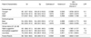
The sensitivity and specificity of the graded-compression US tests that were used for the diagnosis of acute appendicitis in each of the 22 studies are listed in Table 1. When the US appendiceal (a diameter enlarged to 6 mm or greater, intraluminal fluid and lack of compressibility) and periappendiceal (periileal inflammatory changes, cecal wall thickening, periileal lymph nodes and peritoneal fluid) evaluations were included as diagnostic criteria, the overall sensitivity was 86.7% (95% CI: 85.4, 88.0) with a range from 51.3% to 100.0%. Also, the overall specificity was 90.0% (95% CI: 88.9, 91.2) with a range from 60.0% to 100.0%, and each parameter was widely scattered. Thus, on a quantitative meta-analysis using the Hasselblad method (43), the estimate of đ6 for the US was 2.0054 (95% CI: 1.8553, 2.1554), so using the graded-compression US for the diagnosis of acute appendicitis was judged to be effective (Tables 2, 3).
The results of the subgroup meta-analysis by the patients characteristics are shown in Table 2. For the studies of the younger-age dominant group, the overall sensitivity and specificity (95% CI) of graded-compression US were 90.1% (87.7 to 92.5) and 93.6% (91.6 to 95.6), respectively. The likelihood ratio (LR) for a positive US result in younger-age dominant groups was 14.1 and it was higher than those (7.1 or 3.3) in adult or older-age dominant groups. For the studies of the male dominant group, the overall sensitivity and specificity (95% CI) of graded-compression US were 94.4% (92.4 to 96.5) and 94.4% (92.3 to 96.5), respectively. The LR for a positive US result in the male dominant groups was 16.9 and it was higher than that (6.4) in the female dominant groups. Also, when the studies included the highly clinical suggestive groups (57, 62, 68), the overall sensitivity and specificity (95% CI) of the graded-compression US were 93.1% (89.2 to 97.1) and 92.3% (88.1 to 96.5), respectively. The LR for a positive US result in the highly clinical suggestive group was 12.1. These đ estimates of the younger age, male and high-clinical status dominant groups for the gradedcompression US were 2.2388 (95% CI: 1.8758, 2.6019), 2.7131 (95% CI: 2.2493, 3.1770), and 2.4582 (95% CI: 1.7387, 3.1777), respectively. Thus, the graded-compression US effectively influences the diagnosis of acute appendicitis when the above-mentioned factors are present in the patients' characteristics.
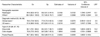
According to the results of the subgroup meta-analysis by the study researchers' characteristics, if sonographic examiners were the only diagnostic radiologists groups (10 studies), the overall sensitivity and specificity (95% CI) of graded-compression US were 84.9% (82.9 to 87.0) and 93.0% (91.5 to 94.4), respectively. When there are other groups (including surgeons or emergency physicians) in the 2 studies, the overall sensitivity and specificity (95% CI) of graded-compression US were 98.4% (96.4 to 100.0) and 72.7% (65.8 to 79.7), respectively. Therefore the LR for a positive US result for the diagnostic radiologist groups was higher (12.1) than that (3.6) for the other groups. On the other hand, when the usefulness of the diagnostic method was simultaneously compared within only 3 studies (53, 66, 68), the overall sensitivity and specificity (95% CI) of the ultrasonographic method were 91.8% (88.9 to 94.8) and 87.9% (84.4 to 91.4), respectively. The LR for a positive result for the US examination groups was 7.6 and it was higher than that (3.5) for the clinical examination groups. If the type of US was the gray-scale group (64, 67, 70), the overall sensitivity and specificity (95% CI) of US were 82.1% (78.1 to 86.2) and 94.2% (91.7 to 96.7), respectively. The LR for a positive result for the gray-scale US groups was 14.2 and it was higher than that (10.6) for the color-doppler US. The estimates of đ by the diagnostic radiologists, the US examination groups and the gray-scale group for US were 2.0195 (95% CI: 1.7942, 2.2447), 2.3216 (95% CI: 1.9167, 2.7266), and 2.2630 (95% CI: 1.8444, 2.6815), respectively. So, some factors in the researchers characteristics were judged to be effective in the diagnosis of acute appendicitis with using US (Table 3).
DISCUSSION
As a diagnostic tool of acute appendicitis, US has been popularly used in Korea since the mid-1980s (73). In this meta-analysis study, estimates of the diagnostic accuracy of graded-compression US were conducted with the method of pooling sensitivity and specificity measurments. We defined the pooled estimate for sensitivity (86.7%) and specificity (90.0%) of graded-compression US in this study. Because the estimated values of đ were moderately large (> 2.0) and the estimates of specificity was higher than that (86.0%) for a given level of sensitivity (86.0%) and đ (2.0), we concluded that using graded-compression US for the diagnosis of acute appendicitis in symptomatic Koreans was effective. That is, an estimated đ of over 2.0 would suggest good discrimination by the diagnostic tool for the detection of patients with suspected disease (43).
Our result that covered 22 studies concurs with the result of meta-analysis study covering 17 studies by Orr and colleagues in the USA (33), but our study has a little bit higher value than that obtained with a quantitatively systemic review by other systemic review teams (35, 37). Obermaier et al. in Germany (35) performed a systemic literature research with using 69 articles, and the results of single-center studies (sensitivity 81.6%, specificity 89.8%) or the results of studies that had less than 10 investigators (sensitivity 84.3%, specificity 86.8%) showed better diagnostic values of appendicitis than those of the multicenter studies or the studies with 10 or more investigators. Also, Terasawa et al. (37) reported that US had an overall sensitivity of 86%, a specificity of 81%, a positive likelihood ratio of 5.8, and a negative likelihood ratio of 0.19. According to the recent study by Kessler et al. (74), the most accurate appendiceal finding for appendicitis was the presence of a 6 mm or larger diameter appendix. Using these diagnostic criteria, US showed 98% for the sensitivity, specificity, positive predictive value, and negative predictive value. Thus, US is more useful for those patients who have an indeterminate probability of appendicitis after the initial evaluation.
If the US finding is positive, patients should have an operation, otherwise, they should be observed without performing an operation. Such a strategy reduces the unnecessary appendectomy rate. In our results, the negative appendectomy rate when using graded-compression US was 10.6%. In approximately 10%-20% of all the cases in several studies (26, 27, 75-79), a misdiagnosis was made and patients underwent operations without them having acute appendicitis at all. Therefore, the clinician s goals are to minimize the negative appendectomy rate and to approach 100% sensitivity for the diagnosis. Achieving these goals requires various diagnostic technologies such as taking a comprehensive clinical history and complete physical examination, a scoring system computer analysis technique, measuring the inflammatory markers (C-reactive protein and the leucocyte count), laparoscopy, computer tomography scan (CT), magnetic resonance imaging (MRI), scintigraphy, US and etc. (40, 80). According to the result of an analysis that used United States Census Bureau data by Flum et al. (78), the population-based incidence (15.5%) of unnecessary appendectomies did not change significantly over time (1987-1998) and it increased yearly for women of productive age or for patients older than 65 years even with the introduction of computed tomography, US and laparoscopy.
The choice of statistical method for pooling the results of different studies depends on the summary statistics, the source of heterogeneity and notably variation in diagnostic thresholds. First of all, in this study, the sensitivity and specificity were combined directly. Diagnostic odds ratios and summary receiver operating characteristic curves with using more complex formulas may also be synthesized (43, 81). We have also carefully looked into several major outcomes and subgroup meta-analyses for combining specific subgroup data across the different studies through the stratification of the study variables by the patients or researchers characteristics (Tables 2, 3). Thus, these important results were essentially the same as those seen in the analyses from a total of 22 articles. When a patient predominately belonged to younger age, male or clinically highly suggestive group, the graded-compression US method could diagnose appendicitis more accurately. The overall sensitivity and specificity of graded-compression US for the younger age dominant group (in which the age distribution below 19 years was over 32%) were 90.1% and 93.6%, respectively; those for US in the male dominant group (the male percentage was over 55%) were 94.4%, and those for US in the clinically highly suggestive group (probability > 75%) were 93.1% and 92.3%, respectively. Also, the likelihood ratio (LR) for a positive US result for the younger age group, the male dominant group and highly suggestive group was 14.1, 16.9 and 12.1, respectively. This result means that the US results of these sorts are about 14 times, 17 times, and 12 times as likely to come from patients with acute appendicitis as from patients without acute appendicitis, according to each group. Our result in the clinically highly suggestive group is similar to those results of Rettenbacher et al. (18) and Orr et al. (33). However, the use of graded-compression US is known to be restricted for pediatric patients with unclear clinical findings or for female patients of childbearing age and/or with gynecologic diseases, or for obese adolescents (23, 30, 75-77).
Ultrasonography has recently been performed in emergency rooms by surgeons or emergency physicians with appropriate instrumentation and training (47, 48). When the sonographic examiners (despite of their specialty or there was no description by the study author) were diagnostic radiologists, they differentiated appendicitis from other acute abdominal conditions more accurately. The overall sensitivity and specificity for US for the diagnostic radiologist group were 84.9% and 93.0%, respectively; those for US for the others group were 98.4% and 72.7%, respectively. The LR for a positive US result for diagnostic radiologist group (12.1) was higher than that for the others group (3.6). Such discrepancies might be influenced by the number of studies (10 versus 2 studies). However, Obermaier et al. (35) reported there were no distinct differences between the investigating departments (the overall sensitivity, specificity, and accuracy for radiologists were 83.1%, 88.1% and 83.5%; for surgeons they were 78.9%, 88.9% and 86.0%, respectively). So clinicians have to thoroughly apply the utilization guide of US for the diagnosis of appendicitis considering the technologic advances of the US facilities or in-depth radiologic experience in their training (25, 74). Also, appendicitis was identified more accurately by US than by only clinical examination (the initial physical examination, the diagnostic score or the presence of leukocytosis, etc.). The overall sensitivity and specificity for US testing groups were 91.8% and 87.9%, respectively. This result also concurs with the result of Kessler et al. (74). Therefore, US was superior to a clinical (or laboratory) examination solely for affirming or excluding appendicitis.
Some limitations of our study must be considered. First, because the outcome data used in this study were based on retrospective observational studies, there was considerable variation in the results of US as the primary imaging modality by the study. Particularly, there was considered disparities between each study (including diversity of the sonographic examiners, the inter-examiner skill, the US facilities and study periods). It was indirectly proven that 22 studies were heterogeneous with a statistical significance (Q = 111.913, p-value < 0.001) for the graded-compression US criteria. This variation may be caused by chance alone (small sample sizes), but it can also reflect true heterogeneity.
Second, verification bias may have occurred when the reference standard was assessed on patients sampled differentially in the categories of test results (32, 82). To eliminate these variations in study quality on the meta-analysis for diagnostic tests, the Cochrane Methods Working Group on Screening and Diagnostic Tests have suggested the comprehensive validity checklist for the primary studies include the target population, method of patient selection (selection bias), method of verification (differential reference standard bias), method of interpretation of tests, and method to avoid residual confounding (40, 83, 84). This meta-analysis used only the studies that met the inclusion criteria for quality evaluation. Thus we excluded the studies with the lack of biopsy results as a reference standard or without the evidence of utilization of graded-compression US or without the evidence on sonographic signs of appendicitis (85-94).
Third, because we were restricted to Korean-language studies for the study selection, so there may be considerable language bias as a kind of publication bias (95). However, this result could directly or indirectly compare the results of meta-analyses using non-Korean-language literature (33, 35, 37).
Four, because of the insufficient information extracted in each study, the classification criteria of the subgroups (like dominant age or gender groups) may be arbitrary. Thus, we may consider the spectrum effect, which reflects the inherent variation in test performance among population subgroups (96). So then, our results for subgroup analyses should be interpreted with caution.
In this analysis, the medical cost of US utilization was not considered. Thus, future investigations should analyze the cost-effectiveness of the US method. Also, future studies are needed to compare the usefulness of this methodology by the type of US facility (67) as well as comparing it with computed tomography, when considering the breakthroughs of imaging technique (37). Currently, according to the study protocol of Bachmann et al. (40), systemic reviews of diagnostic literature (including MEDLINE, EMBASE, DARE, Cochrane Database of Systemic Reviews, conference proceedings, MEDION, SCISEARCH, BIOSIS) for prediction of acute appendicitis will allow us to assess the quality of the available evidence and to identify the value of the specific diagnostic tests (including the history, physical examination and ultrasonography tests, etc.). Although this meta-analysis provides a statistically robust outcome despite of some limitation of study method, randomized clinical trials or well-designed prospective studies for adopting a new diagnostic modality will be continuously required in clinical outcome research. To our knowledge, this study is a cornerstone of the estimate formula of for assessing the accuracy of a new diagnostic test and it provides an evidence-based clinical outcome for medical education and health insurance policy.
In conclusion, US may be suggested as a useful diagnostic method for acute appendicitis, especially when the symptomatic patients are younger age, male and have clear clinical suggestions of disease. This procedure is evidently user-dependent and it has to be performed by a well-trained physician prior to the decision-making regarding an appendectomy.
 XML Download