

 PDF
PDF ePub
ePub Citation
 Print
Print


Malignant mesenchymoma is a rare malignant neoplasm, usually containing two or more malignant mesenchymal cellular types (1). Despite the publication of several cases reports involving a small series (2-5), the varying composition of tumors and the small number of reported cases has led to a lack of widespread awareness of the related radiological findings. We report a rare case of primary retroperitoneal malignant mesenchymoma showing distinctive radiological findings.
CASE REPORT
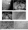
A 47-year-old man was admitted to hospital due to a palpable mass in the left flank. His laboratory data were normal, but plain radiography of the abdomen showed that in the left upper quadrant, a huge mass with exuberant calcifications was present (Fig. 1A). Abdominal CT revealed that the peritoneum contained a large heterogeneous soft tissue mass composed of several nodules with different components. The upper portion of the mass showed dense or stippled calcifications (Fig. 1B), whereas in the lower portion there were several non-calcified soft tissue nodules with strong peripheral enhancement and a central necrotic area (Fig. 1C). The HU of the low-density area was 42, and this was attributed to the presence of non-fatty tissue. The mass had invaded the renal capsule, pancreas, and adrenal gland (though not the left-side colon and spleen, which strongly adhered to it), and surgical resection was thus required, together with distal pancreatectomy, splenectomy, left nephrectomy and left hemicolectomy. The resected mass measured about 18 cm in its longest diameter, and was composed of several soft tissue nodules. In its upper portion, a hard bone-like structure was present. Microscopically, the nodule around the dense calcification was consistent with osteosarcoma (Fig. 1D), and the soft tissue nodules in the lower part corresponded to leiomyosarcoma (Fig. 1E), liposarcoma (Fig. 1F), and fibrosarcoma.
Fourteen months after surgery, follow-up abdominal CT revealed a recurrent retroperitoneal mass in the left paraaortic area at the level of the inferior pole of the kidney, without evidence of calcification. The mass was surgically excised, and a resected specimen contained a mainly fibrosarcomatous component. During the subsequent 15-month period, the patient was in good health, with no evidence of recurrence.
DISCUSSION
Malignant mesenchymoma, first described by Stout in 1948, is a malignant soft tissue tumor consisting of two or more distinct mesenchymal components, either or any of which might in itself be viewed as a primary malignant neoplasm (1). Nowadays strict diagnostic criteria for malignant mesenchymoma require that each component is sufficiently differentiated histogenetically (6, 7). For this purpose, fibrosarcoma, hemangiopericytoma, malignant fibrous histiocytoma, myxosarcoma, and malignant peripheral nerve sheath tumor are not considered as separate malignant tumor components; those that normally fall into this category are liposarcoma, leiomyosarcoma, rhabdomyosarcoma, osteosarcoma, chondrosarcoma, and angiosarcoma (6).
The previously reported radiologic features of malignant mesenchymoma vary (2-4). Although none were common to all the cases they described, Suzuki et al. (5) suggested that the typical findings of malignant mesenchymoma were large tumor size, a sharp margin, heterogeneous make-up, and massive calcification. The tumor in our case was also a huge heterogeneous mass with massive intratumorous calcification, but a feature different from those previously noted was also present. Earlier reports described a large mass with various intermingled components, whereas in our case the components were discrete. The massive calcification we found corresponded to an osteosarcomatous component, while the non-calcified enhancing nodules were, respectively, consistent with leiomyosarcoma, liposarcoma, and fibrosarcoma. A well-differentiated liposarcomatous component can have a fatty component (5), though in our case none was apparent at CT. The composition of a malignant mesenchymoma - whether or not it contains calcification or ossification, a fatty component, or necrotic soft tissue - seems to determine its radiological features.
A malignant mesenchymoma is generally considered to be highly malignant, usually with a poor prognosis (7), though the findings of this same study, based on clinicopatholgic analysis and long-term follow-up, suggest a more favorable prognosis. In our case, the clinical course was rather indolent; although the mass recurred locally 14 months after initial surgery, there was no evidence of metastasis during the 29 months following initial presentation, and this seems to reaffirm a more favorable prognosis.
For the proper diagnosis of malignant mesenchyoma, the radiologist must be aware of its nature; if any of the sarcomatous components are overlooked, it might be diagnosed as single-type or predominant sarcoma with divergent differentiation.
In summary, depending on its composition, malignant mesenchymoma may demonstrate various radiologic features. If a large retroperitoneal soft tissue mass is present, together with several different sarcomatous features such as prominent calcification/ossification or a fatty component, the differential diagnosis should include malignant mesenchymoma.
 XML Download