

 PDF
PDF ePub
ePub Citation
 Print
Print


To relieve mechanical ileus, a stent is commonly used for the palliative treatment of colorectal obstruction secondary to colorectal neoplasm. For the palliative and preoperative treatment of colorectal carcinoma, a bare expandable or covered metallic stent is used (1-6). Complications after insertion include rectal bleeding, abdominal pain, malpositioning, pseudo-obstruction, tumor ingrowth, migration and perforation (1, 3, 5). Delayed perforation due to a bare rectal stent has rarely been reported, however. We report a case of delayed colonic perforation occurring one month after the insertion of a bare rectal stent used for the palliative treatment of rectal carcinoma.
CASE REPORT
A 70-year-old female was admitted to hospital due to abdominal pain and tenesmus lasting for one month and followed by mechanical ileus caused by rectal carcinoma. Abdominal and pelvic computed tomography revealed rectal carcinoma, with multiple liver and intraperitoneal metastases. The patient also had a history of gastric ulcer, unstable angina and hyperlipidemia, and was diabetic. The rectal carcinoma, which was at stage IV, was inoperable. Sigmoidoscopy showed marked luminal narrowing and a huge mass in the rectosigmoid area, and biopsy revealed adenocarcinoma of the rectosigmoid colon.
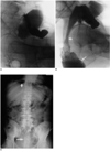
Because of the advanced state of malignancy and the patient's poor general health, the option of palliative treatment using a rectal stent was chosen. The procedure used has been described elsewhere (1-6). After lubrication of the anal sphincter, a Foley catheter was inserted, the end-hole being made by cutting the tip. An angled 0.038-inch stiff hydrophilic guide wire (Radiofocus; Terumo, Tokyo, Japan) and a 7-Fr Headhunter catheter (Cook, Bloomington, Ind.) were assembled, and the latter was inserted through the Foley catheter. The guide wire and catheter were advanced with fluoroscopic guidance to pass through the area of obstruction (Fig. 1A). To define the stenotic area and to rule out colonic perforation, nonionic contrast material was flushed through the catheter. Once the hydrophilic guide wire and catheter had been advanced through the stenosis, the former was replaced by a 260-cm-long Amplatz 0.038-inch stiff guide wire (Meditech/Boston Scientific, Watertown, Mass.) in order to straighten the tortuous rectosigmoid region. It was then easy, with fluoroscopic guidance, to introduce the delivery system over the stiff guide wire and thus assess wheter the stent, 30 mm in diameter and 10 cm in length (Stentech, Seoul, Korea), was correctly positioned. After stent insertion, the patient expelled both gas and contrast media (Fig. 1B).
Post procedurally, the patient made very good progress and the symptoms of obstruction were relieved. One month later, however, she complained of spontaneous abdominal pain. Chest PA and plain radiographs of the abdomen revealed the presence of huge amounts of free air in subphrenic areas, thus suggesting perforation of the hollow viscus (Fig. 1C). The patient underwent emergency surgery, and this revealed colonic perforation caused by wires at the proximal end of the stent. This was carefully removed and a colostomy was performed. The patient's vital signs were subsequently stable, and a normal regular diet was tolerated, but one week later she died due to uncontrolled hypotension probably caused by sepsis.
DISCUSSION
Since it can immediately relieve mechanical ileus in almost all patients, the use of self-expandable metallic stents in the palliative treatment of rectosigmoid colon carcinoma is a valuable alternative to colostomy, and both palliatively and preoperatively, the use of bare stents in such cases has achieved good clinical results (2-5). The advantage of a bare stent is its small deployment system, which makes for an easy, safe and comfortable procedure. The clinical and radiographic findings in cases involving bowel obstruction show that in patients requiring preoperative treatment, the obstruction was resolved within 24 hours of stent placement in over 90% of cases, and that in those requiring palliation, success was achieved within four days of treatment in a similar proportion of cases (3, 5). Minor complications reported in 13% of patients, included rectal bleeding, abdominal pain, malpositioning, pseudo-obstructive episodes due to fecal impaction, and occlusive tumor ingrowth into the stent lumen (1, 3, 5). According to Mainar's study (5), colonic perforation was a major complication. One patient underwent immediate surgery to resolve a perforation caused by wires at the ends of the stent; two other focal perforations were discovered during surgical resection, but no symptoms were reported. For preoperative decompression, the mean time between stent placement and surgery was 8.6 days (5). The stent immediately resolved mechanical ileus and improved the patients' condition prior to surgery nine days later (5).
The technical success rate of preoperative and palliative treatment with a covered stent is above 90% (1). In 75% of cases, symptoms of obstruction were resolved within 24 hours. The mean time between stent placement and surgery was 5-7 days. The main complication in cases involving use of the covered stent was stent migration, occurring in 50% of cases, and the type of stent employed was thus changed: instead of the completely covered type, a stent with two-thirds of its proximal part uncovered was used (1).
In generally, immediate colonic perforation after stent placement may have been caused by technical problems. Placement may have pushed the delivery system too much, thus suddenly expelling the stent, leading to perforation of the bowel wall. Delayed colonic perforation after stent placement may be caused by the weakening of junction areas between normal bowel wall and the tumor or by tumoral perforation due to mechanical tumor necrosis arising from stent and wire problems. In our case, the wires at the ends of the proximal portion of the stent penetrated the colon wall. Due to its continuous movement, the bowel wall came in contact with the wire, which caused erosion, and possibly led to bowel perforation. The covered stent, the end of which is protectively wrapped, did not pose problems in this respect, though migration was one of the major complications. The end of the bare stent takes the form of an inverted "v", and this may lead to wire problems, as in our case, in which the bare stent was inserted because the tumor had a long stenotic segment and severe angulation deformity. The bare stent has its advantages: a very easy delivery system, good shape in cases involving curved lesions, and no stent migration.
In conclusion, in the palliative treatment of colorectal carcinoma, caution must be exercised when using a bare stent, and close follow-up is required. In addition, the possibility of bowel wall perforation must be borne closely in mind.
 XML Download