

 PDF
PDF ePub
ePub Citation
 Print
Print


INTRODUCTION
Many congenital anatomic variations of the pancreatic duct have been described in the literature, such as complete or incomplete pancreatic divisum, annular pancreas, and ansa pancreatica. Among various types of pancreas ductal anomalies, pancreas divisum is the most frequent anatomic variation, whereas ansa pancreatica is a rare anatomic variation, with a reported prevalence of 1.1% (12).
Ansa pancreatica is characterized by focal accessory duct atrophy and an additional curved duct linking main and accessory ducts replacing atrophied duct. Ansa pancreatica is considered as a predisposing factor for recurrent pancreatitis (1). Adibelli et al. (3) reported that patients with recurrent pancreatitis had a higher frequency of ansa pancreatica than the general population (11.1% vs. 0.85%). The mechanisms of ansa pancreatica causing acute pancreatitis have not been clear yet. However, recent evidence consistently have suggested that the curved duct cause impaired pancreatic juice drainage, mainly from pancreas head and uncinate process, resulting in recurrent focal pancreatitis (13).
There have only a few articles exploring the ansa pancreatica and recurrent/chronic pancreatitis, thus ansa pancreatica is an under-reported disease entity (1345678). As comprehensive evaluation of the pancreatic duct variation by imaging such as computed tomography (CT) or magnetic resonance cholangiopancreatography (MRCP) is extremely important in order to correctly guide the next management steps, radiologists should be aware of this rare disease entity. Thus, we intend to comprehensively analyze the imaging findings with three cases of recurrent pancreatitis or localized chronic pancreatitis in patients with ansa pancreatica.
CASE REPORT
We reported the following three cases of recurrent pancreatitis with underlying ansa pancreatica ductal anomaly, as described in Fig. 1. Interestingly, the pancreatitis was localized in the pancreas head and uncinate process in all three cases.
CASE 1
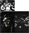
A 24-year-old female patient presented with recurrent acute pancreatitis. The first episode of acute pancreatitis was 17 years ago and the most recent prior event was 3 years ago. At the time of hospital administration, serum amylase level was 145 U/L (normal range, 30–110 U/L) and lipase level was 136 U/L (normal range, 13–60 U/L), which were moderately higher than the upper normal limits.
Abdominal CT and MRCP were performed to evaluate recurrent pancreatitis and the underlying predisposing cause. Abdominal CT revealed clustered pancreatic parenchymal calcification in the pancreas head, suggesting localized chronic pancreatitis. Also several pancreaticoliths were impacted within the dilated duct of Santorini continuing to the minor papillae (Fig. 2A). The MRCP revealed a normal appearing duct of Wirsung draining to major papillae, a dilated duct of Santorini, and another curved duct between the former two ducts, suggesting ansa pancreatica type anatomic variation (Fig. 2B).
CASE 2
A 62-year-old male patient presented with an incidental pancreas lesion on routine check-up. He denied any symptoms of abdominal discomfort at the time of visit. Indeed, he experienced several episodes of acute abdominal pain during his whole life without knowing the exact cause of pain. The patient had rarely consumed alcohol before. At the time of visit, laboratory examination was not remarkable except for hyperlipidemia.
Abdominal CT showed a 2-cm size nodular lesion with multiple calcification at the pancreas uncinate process, suggesting localized chronic pancreatitis (Fig. 3A). Pancreatic ductal dilatation and pancreaticolith were not obvious on the CT scan. The patient underwent MRCP for further characterization of the pancreas lesion. MRCP showed normal appearance of the duct of Wirsung and another hooked duct arising from the flexion point of the pancreas duct draining to minor papillae, suggesting ansa pancreatica type ductal variation (Fig. 3B).
CASE 3
A 29-year-old male patient was referred from other hospital to evaluate the cause of recurrent acute pancreatitis. He had not consumed alcohol before and he had no biliary stone disease. At the time of visit, laboratory examination was not remarkable. However, he had suffered from frequent episodes of abdominal pain and the most recent prior event was 2 months ago.
Abdominal CT revealed mild pancreas swelling localized in the pancreas head. Pancreas duct dilatation and panceaticolith were not obvious on the CT scan. Follow-up MRI and MRCP were performed 2 months after the CT scan. MRI showed improvement of pancreas head swelling. MRCP revealed arched duct linking the main pancreatic duct draining to the minor papillae, which was compatible with ansa pancreatica type anatomic variation.
DISCUSSION
Here, we reported three cases of recurrent pancreatitis localized in the pancreas head and uncinate process with underlying ansa pancreatica ductal anomaly. Ansa pancreatica is a rare type of pancreas ductal variation first reported by Dawson and Langman in 1961 (4). The reported prevalence of this type ductal variation is 1.1% (3). The ansa pancreatica is a ductal variation related with the developmental process of the pancreas.
The pancreatic duct is constituted of a dorsal duct (or a duct of Santorini) and a ventral duct (or duct of Wirsung). The ventral duct or duct of Wirsung arises from the main pancreatic duct, which empties through the major duodenal papillae. The dorsal duct or duct of Santorini forms the accessory pancreatic duct, draining to the minor duodenal papilla. These two ducts fuse in the pancreas head portion as a result of asymmetric duodenal rotation during gestational age 6 to 8 weeks. The ventral pancreatic bud rotates 180° counterclockwise, arriving at the dorsal pancreatic bud. During the development of the pancreatic duct, a variable degree of accessory duct atrophy occurs. Ansa pancreatica is characterized by focal accessory duct atrophy around its junctions to the main pancreatic duct, which is replaced by an additional curved duct linking main and accessory ducts. This curved additional duct is formed from the proximal portion of the dorsal duct and inferior branches of both dorsal and ventral ducts. Thus, in ansa pancreatica, the accessory duct arises from the main pancreatic duct and runs a hooked course anteriorly to the main duct ending in or around the minor papilla (579).
Ansa pancreatica type ductal anatomic variation might be a predisposing factor for recurrent or chronic pancreatitis. Few observations reported coexistence of acute idiopathic pancreatitis and underlying ansa pancreatica anatomic variation. But it is unclear whether the coexistence of theses two conditions is a simple coincidence or causal relationship (567). Ishii et al. (10) reported that approximately 7% of the patients with ansa pancreatica presented with acute pancreatitis. In ansa pancreatica ductal variation, the arched additional duct meets the main duct at on oblique angle, whereas the other tributaries of the main pancreatic duct join at a right angle. Based on this ductal variation, the pancreas area served by the additional arched duct has poor pancreatic juice drainage, resulting in recurrent pancreatitis (1). When condition predisposing pancreatitis such as heavy alcoholism or functional stenosis of the sphincter of Oddi, this anatomic arrangement makes patients more vulnerable to the development of pancreatitis.
Summarizing, ansa pancreatica is a predisposing factor of recurrent pancreatitis localized in pancreas head and uncinate process, because drainage of pancreatic juice from the pancreas head and uncinate process might be impaired due to the hooked duct linking main and accessory ducts. Through our experience and current consistent evidence, we would propose that if there is recurrent pancreatitis, especially localized in the pancreas head and uncinate process, underlying ductal anomaly such as ansa pancreatica should be considered in the diagnosis and management of these patients.


 XML Download