Journal List > J Korean Soc Radiol > v.79(1) > 1098286

Lim, Ha, Youn, Yoon, Baek, Do, Choi, Choi, Lee, and Na: Guidelines for Primary Imaging Test and Biopsy Methods in the Diagnosis of Thyroid Nodules: Joint Report by the Korean Society of Radiology and National Evidence-Based Healthcare Collaborating Agency

Abstract

The Korean Society of Radiology and the National Evidence-based Healthcare Collaborating Agency developed the guideline for primary imaging and biopsy methods in the diagnosis of thyroid nodules. The development committee, the working committee, and the advisory committee were formed to develop the recommendation. The development committee mainly plays a role of methodological consulting and overall planning and management of the advisory development stage. The working committee was composed of experts recommended by the Korean Society of Thyroid Radiology and conducted a practical adaptation process from the selection of core questions to the final recommendation. The Advisory Committee consisted of clinical experts recommended by the Korean Thyroid Association and reviewed core questions and draft recommendations and participated in the Expert Panel Survey. This guideline recommends cervical ultrasound as the first imaging modality for diagnosis of suspected thyroid nodules and recommends ultrasound-guided fine needle aspiration for histologic diagnosis of thyroid nodules. This guideline is expected to be of significant benefit to clinicians treating thyroid nodules.

REFERENCES

1.Frates MC., Benson CB., Charboneau JW., Cibas ES., Clark OH., Coleman BG, et al. Management of thyroid nodules detected at US: Society of Radiologists in Ultrasound consensus conference statement. Radiology. 2005. 237:794–800.
crossref
2.Guth S., Theune U., Aberle J., Galach A., Bamberger CM. Very high prevalence of thyroid nodules detected by high frequency (13 MHz) ultrasound examination. Eur J Clin Invest. 2009. 39:699–706.
3.Tan GH., Gharib H. Thyroid incidentalomas: management approaches to nonpalpable nodules discovered incidentally on thyroid imaging. Ann Intern Med. 1997. 126:226–231.
crossref
4.Moon WJ., Baek JH., Jung SL., Kim DW., Kim EK., Kim JY, et al. Ultrasonography and the ultrasound-based management of thyroid nodules: consensus statement and recommendations. Korean J Radiol. 2011. 12:1–14.
crossref
5.Statistics of the Ministry of Health and Welfare. Available at. https://www.data.go.kr/dataset/15003005/fileDa-ta.do. Published 2017. Accessed Nov 13. 2017.
6.Choi SJ., Jeong WK., Jo AJ., Choi JA., Kim MJ., Lee M, et al. Methodology for developing evidence-based clinical imaging guidelines: joint recommendations by Korean Society of Radiology and National Evidence-Based Healthcare Collaborating Agency. Korean J Radiol. 2017. 18:208–216.
crossref
7.Gharib H., Papini E., Garber JR., Duick DS., Harrell RM., Hegedüs L, et al. American Association of Clinical Endocrinologists, American College of Endocrinology, and Associazione Medici Endocrinologi Medical Guidelines for clinical practice for the diagnosis and management of thyroid nodules—2016 update. Endocr Pract. 2016. 22:622–639.
8.Haugen BR., Alexander EK., Bible KC., Doherty GM., Mandel SJ., Nikiforov YE, et al. 2015 American Thyroid Association management guidelines for adult patients with thyroid nodules and differentiated thyroid cancer: The American Thyroid Association Guidelines Task Force on thyroid nodules and differentiated thyroid cancer. Thyroid. 2016. 26:1–133.
crossref
9.Perros P., Boelaert K., Colley S., Evans C., Evans RM., Gerrard Ba G, et al. Guidelines for the management of thyroid cancer. Clin Endocrinol (Oxf). 2014. 81(Suppl 1):1–122.
crossref
10.National Comprehensive Cancer Network (NCCN). NCCN Guidelines for Thyroid Carcinoma. Available at.https://www.nccn.org/professionals/physician_gls/pdf/thyroid.pdf. Published 2015. Accessed Aug 14. 2015.
11.Smith-Bindman R., Lebda P., Feldstein VA., Sellami D., Goldstein RB., Brasic N, et al. Risk of thyroid cancer based on thyroid ultrasound imaging characteristics: results of a population-based study. JAMA Intern Med. 2013. 173:1788–1796.
12.Brito JP., Gionfriddo MR., Al Nofal A., Boehmer KR., Leppin AL., Reading C, et al. The accuracy of thyroid nodule ultrasound to predict thyroid cancer: systematic review and meta-analysis. J Clin Endocrinol Metab. 2014. 99:1253–1263.
crossref
13.Cesur M., Corapcioglu D., Bulut S., Gursoy A., Yilmaz AE., Erdo-gan N, et al. Comparison of palpation-guided fine-needle aspiration biopsy to ultrasound-guided fine-needle aspiration biopsy in the evaluation of thyroid nodules. Thyroid. 2006. 16:555–561.
crossref
14.Hambly NM., Gonen M., Gerst SR., Li D., Jia X., Mironov S, et al. Implementation of evidence-based guidelines for thyroid nodule biopsy: a model for establishment of practice standards. AJR Am J Roentgenol. 2011. 196:655–660.
crossref
15.Solbiati L., Osti V., Cova L., Tonolini M. Ultrasound of thyroid, parathyroid glands and neck lymph nodes. Eur Radiol. 2001. 11:2411–2424.
crossref
16.Lee YH., Kim DW., In HS., Park JS., Kim SH., Eom JW, et al. Differentiation between benign and malignant solid thyroid nodules using an US classification system. Korean J Radiol. 2011. 12:559–567.
crossref
17.Danese D., Sciacchitano S., Farsetti A., Andreoli M., Pontecor-vi A. Diagnostic accuracy of conventional versus sonography-guided fine-needle aspiration biopsy of thyroid nodules. Thyroid. 1998. 8:15–21.
crossref
18.Carmeci C., Jeffrey RB., McDougall IR., Nowels KW., Weigel RJ. Ultrasound-guided fine-needle aspiration biopsy of thyroid masses. Thyroid. 1998. 8:283–289.
crossref
19.Gharib H., Papini E. Thyroid nodules: clinical importance, assessment, and treatment. Endocrinol Metab Clin North Am. 2007. 36:707–35. vi.
crossref
20.Wu HH., Jones JN., Osman J. Fine-needle aspiration cytology of the thyroid: ten years experience in a community teaching hospital. Diagn Cytopathol. 2006. 34:93–96.
crossref
21.Yang J., Schnadig V., Logrono R., Wasserman PG. Fine-needle aspiration of thyroid nodules: a study of 4703 patients with histologic and clinical correlations. Cancer. 2007. 111:306–315.
crossref
22.Deandrea M., Mormile A., Veglio M., Motta M., Pellerito R., Gal-lone G, et al. Fine-needle aspiration biopsy of the thyroid: comparison between thyroid palpation and ultrasonography. Endocr Pract. 2002. 8:282–286.
crossref
23.Can AS., Peker K. Comparison of palpation-versus ultrasound-guided fine-needle aspiration biopsies in the evaluation of thyroid nodules. BMC Res Notes. 2008. 1:12.
crossref

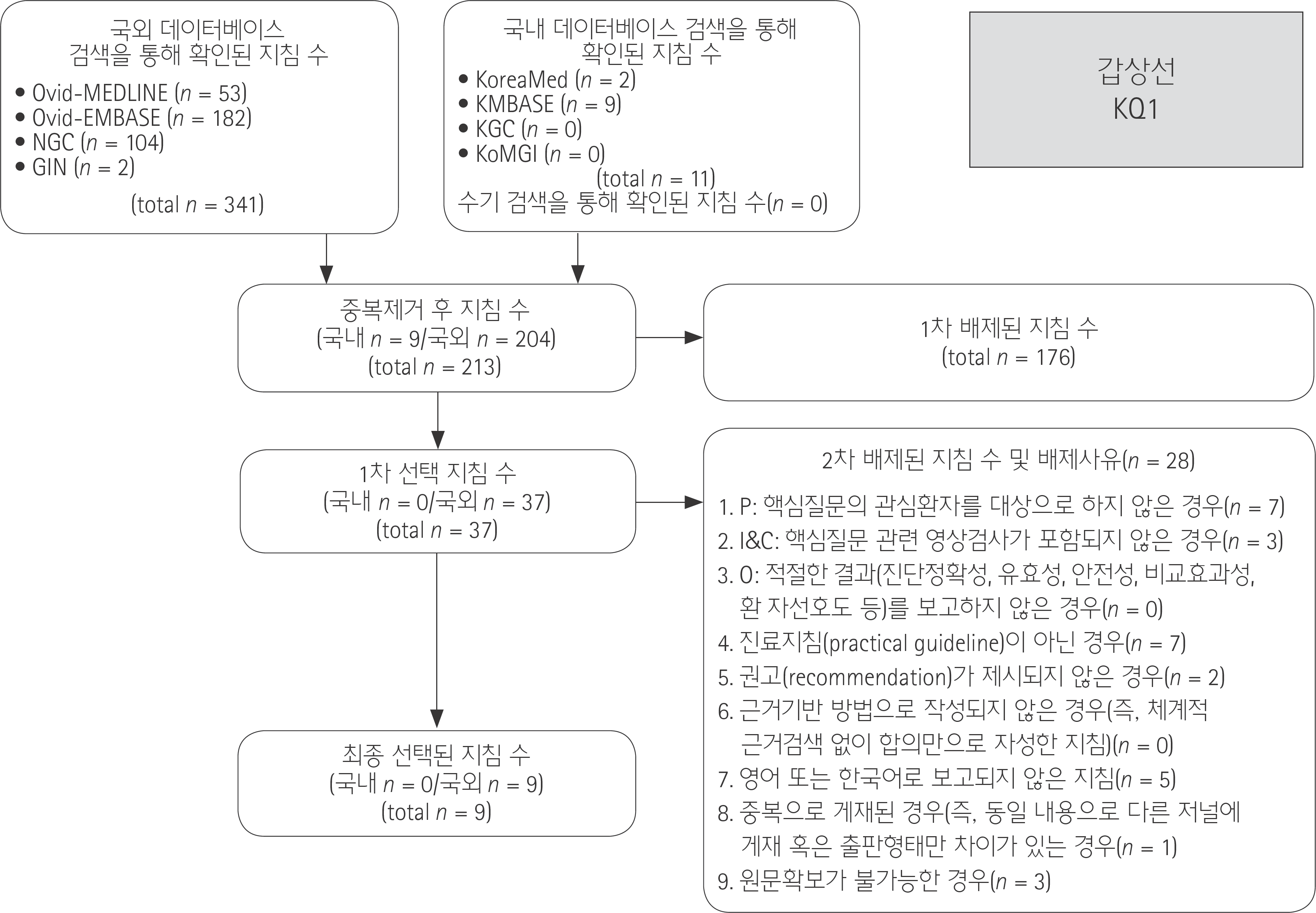
Fig. 1
Flow diagram of guideline selection (Key Question 1).
jksr-79-1f1.tif
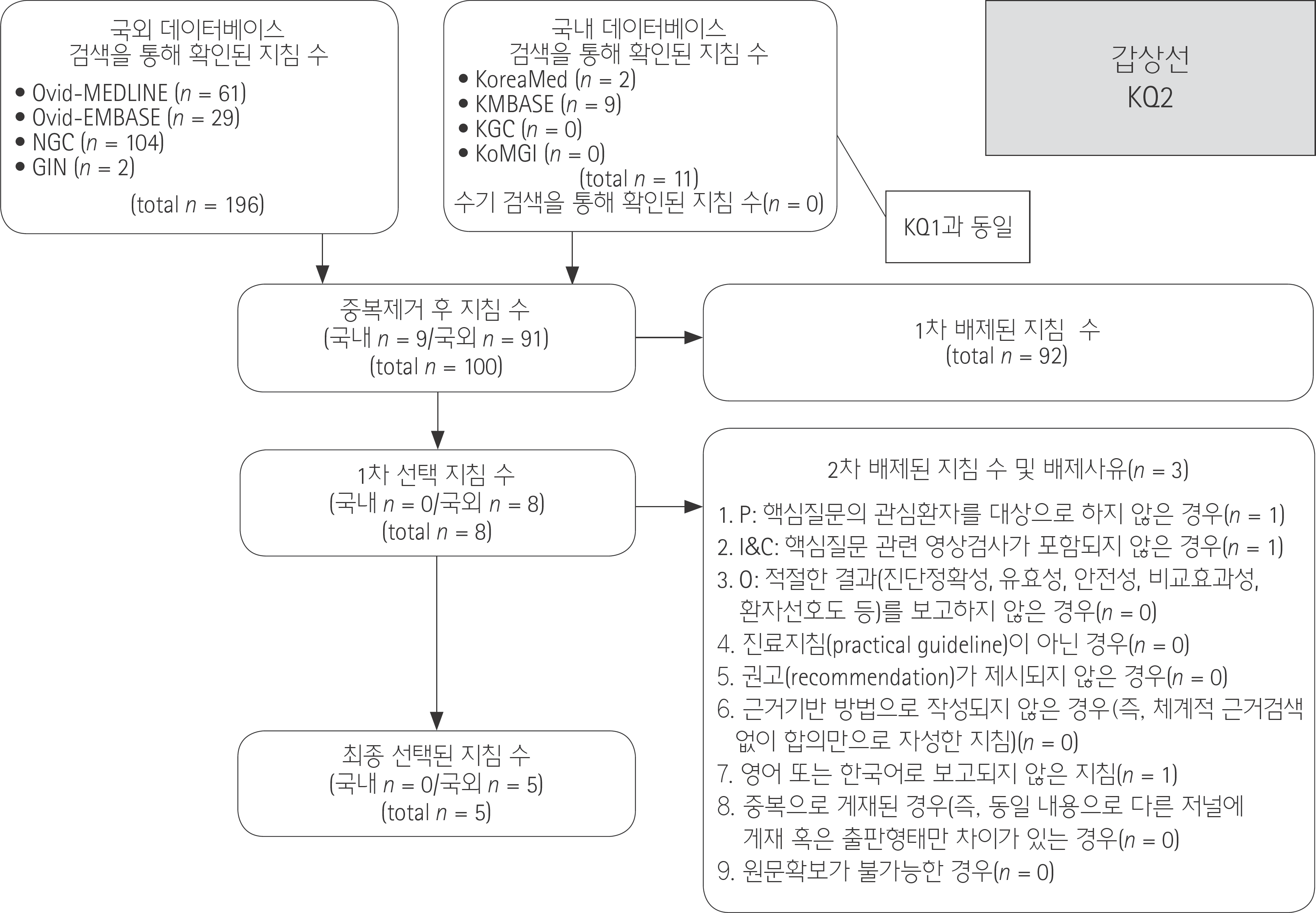
Fig. 2
Flow diagram of guideline selection (Key Question 2).
jksr-79-1f2.tif
Table 1.
Recommendations Matrix of Existing Guidelines (Key Question 1)
Source Guidelines (Publication Year) AACE/AME/ETA Medical Guidelines for Clinical Practice for the Diagnosis and Management of Thyroid Nodules (2010) Ultrasonography and the US-Based Management of Thyroid Nodules: Consensus Statement and Recommendations (2011) British Thyroid Association Guidelines for the Management of Thyroid Cancer (2014) Thyroid Carcinoma, Version 2. 2015 (NCCN Guideline) (2015) 2015 American Thyroid Association Management Guidelines for Adult Patients with Thyroid Nodules and Differentiated Thyroid Cancer (2016)
AGREE II (domain 3. rigour of development) 69 41 84 74 63
Recommendation US evaluation is recommended for
  • Patients at risk for thyroid malignancy

  • Patients with palpable thyroid nodules or MNGs

  • Patients with lymphadenopathy suggestive of a malignant lesion

Among the modern imaging modalities, high-resolution US is the most sensitive diagnostic modality for the detection of the thyroid nodules and it is necessary to perform US for the nodules found after palpation US is an extremely sensitive examination for thyroid nodules. It can be specific for the diagnosis of thyroid carcinoma (particularly papillary carcinoma), and aids decision making about which nodules to perform FNAC
  • 1) All patients being investigated for possible thyroid cancer should undergo an US of the neck in secondary care by an appropriate, competent practitioner

For thyroid nodules known or suspected on clinical or imaging findings, US recommended Thyroid sonography with survey of the cervical lymph nodes should be performed in all patients with known or suspected thyroid nodules
Grading of recommendation Grade B, BEL 3 Not available Good practice point Category 2A Strong recommendation, High-quality evidence

AACE = American Association of Clinical Endocrinologists, AGREE = Appraisal of Guidelines for Research & Evaluation, AME = Associazione Medici Endocrinologi, BEL = best evidence level, ETA = European Thyroid Association, FNAC = fine needle aspiration cytology, MNG = multinodular goiter, NCCN = National Comprehensive Cancer Network, US = ultrasound

Table 2.
Recommendations Matrix of Existing Guidelines (Key Question 2)
Source Guidelines (publication year) AACE/AME/ETA Medical Guidelines for Clinical Practice for the Diagnosis and Management of Thyroid Nodules (2010) Ultrasonography and the US-Based Management of Thyroid Nodules: Consensus Statement and Recommendations (2011) British Thyroid Association Guidelines for the Management of Thyroid Cancer (2014) Thyroid Carcinoma, Version 2. 2015 (NCCN guideline) (2015) 2015 American Thyroid Association Management Guidelines for Adult Patients with Thyroid Nodules and Differentiated Thyroid Cancer (2016)
AGREE II (domain 3. rigour of development) 69 41 84 74 63
Recommendation 3.7.2.1. How to select nodule(s) for FNA biopsy (Grade B, BEL 3):
  • FNA biopsy is recommended for nodule(s):

    • - Of diameter larger than 1.0 cm that is solid and hypoechoic on US

    • - Of any size with US findings suggestive of extracapsular growth or metastatic cervical lymph nodes

    • - Of any size with patient history of neck irradiation in childhood or adolescence; PTC, MTC, or MEN 2 in first-degree relatives; previous thyroid surgery for cancer; increased calcitonin levels in the absence of interfering factors

    • - Of diameter smaller than 10 mm along with US findings associated with malignancy (see section 3.7.1.2.); the coexistence of 2 or more suspicious US criteria greatly increases the risk of thyroid cancer

  • Nodules that are hot on scintigraphy should be excluded from FNA biopsy (see difference in recommendations for children; section 8.4.) 3.7.2.2. FNA Biopsy of Multinodular Glands

  • It is rarely necessary to biopsy more than 2 nodules when they are selected on the basis of previously described criteria (Grade D)

  • If a radioisotope scan is available, do not biopsy hot areas (Grade B, BEL 4)

Figure 8 1) US appearances that are indicative of a benign nodule (U1–2) should be regarded as reassuring not requiring FNAC, unless the patient has a statistically high risk of malignancy (Chapter 3.7) 2) If the US appearances are equivocal, indeterminate or suspicious of malignancy (U3–5), an US guided FNAC should follow THYR-1: Consider FNA based on clinical and sonographic features (a) FNA is the procedure of choice in the evaluation of thyroid nodules, when clinically indicated
Grading of recommendation 3.7.2.1. Grade B, BEL 3 3.7.2.2. Grade D, Grade B, BEL 4; Grade B, BEL 4 Not available 2++, B Category 2A Strong recommendation, High-quality evidence

AACE = American Association of Clinical Endocrinologists, AGREE = Appraisal of Guidelines for Research & Evaluation, AME = Associazione Medici Endocrinologi, BEL = best evidence level, ETA = European Thyroid Association, FNA = fine-needle aspiration, FNAC = fine needle aspiration cytology, MEN = multiple endocrine neoplasia, MTC = medullary thyroid carcinoma, NCCN = National Comprehensive Cancer Network, PTC = papillary thyroid carcinoma, US = ultrasound

TOOLS
Similar articles