

 PDF
PDF ePub
ePub Citation
 Print
Print


INTRODUCTION
Neurinoma or more precisely schwannoma is a benign nerve sheath tumor, which consists of Schwann cells (1). Schwannomas are usually benign tumors and thus rarely show malignant degeneration (2). The tumor is commonly found in the head, neck, posterior mediastinum and extremities. Although three percent of schwannomas develop in the retroperitoneum, they are rarely found in the kidneys and a few cases of these tumors have been reported in the literature. Sometimes renal schwannomas are found in renal parenchyma and mimic renal cell carcinoma (3). Differentiation of renal schwannoma from renal cell carcinoma is difficult at radiologic analysis. Therefore, renal schwannomas are diagnosed during pathologic examination. Here, we report a rare case of renal schwannoma with radiological correlations.
CASE REPORT
A 66-year-old male patient was presented with abdominal pain of 1-month duration. He was referred to our hospital for further examination of solid masses in the liver and upper pole of the right kidney found in an outside abdominal computed tomography (CT) and ultrasonography (US). He had been on medications for ulcerative colitis for 7 years. He had a normal heart rate of 70 beats per minute, with a blood pressure of 130/84 mm Hg. His respiratory rate was 18 breaths per minute and the body temperature was in normal range. The results of laboratory studies, which included a complete blood count, a blood chemistry analysis, urinalysis, and a urine culture, were within normal range except for showing elevated aspartate aminotransferase, alanine aminotransferase (130, 49 IU/L), and protein induced vitamin K antagonist-II (26773 mAU/mL). Serologic examination on admission was negative for hepatitis B surface antigen, hepatitis B and C virus antibodies.
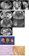
The US, performed at the previous hospital, revealed circumscribed and heterogeneously echogenic mass in the segment eight of the liver without evidence of liver cirrhosis. On CT, a large and heterogeneously enhanced mass with cystic and necrotic portions measuring about 11.2 cm in diameter was detected in the upper pole of the right kidney (Fig. 1A). The renal vessels were deviated due to the mass effect of the large the mass. Presence of vascular invasion was not definite.
After his hospital admission, magnetic resonance imaging (MRI) with contrast material (Primovist, Bayer Healthcare, Berlin, Germany) was performed using 3T system (Skyra, Siemens Healthcare, Erlangen, Germany) to evaluate the mass lesion in the liver and the kidney. MRI revealed a large mass in the right kidney with heterogeneously hypersignal intensity on T2-weighted images and heterogeneously hyposignal intensity on T1-weighted images. Multifocal hypersignal intensities in the mass on T1-weighted images suggested hemorrhagic portions and central necrosis was shown as ill demarcated hypersignal intensity in the mass on T2-weighted images. Contrast-enhanced T1-weighted imaging showed inhomogeneous enhancement in the renal mass on portal phase. Diffusion weighted imaging showed hypersignal intensity with a high b-value (800 s/mm2) but a low apparent diffusion coefficient value at the solid portion of the mass (Fig. 1B). Taken together, the imaging features of a mass in the kidney on CT and MRI suggested a renal cell carcinoma or renal metastasis from the hepatocellular carcinoma. The imaging features of a mass in the liver on CT and MRI suggested a hepatocellular carcinoma. The patient underwent a right anterior sectionectomy and right radical nephrectomy, which was performed without complication.
Gross pathological evaluation of the right nephrectomy specimen revealed an ill-demarcated, yellow multilobulated mass with cystic degeneration, measuring 12.2 cm in maximal dimension (Fig. 1C). On microscopic evaluation, the section from the renal mass is mostly composed of spindle cells (Fig. 1D). Tumor cells were immunoreactive with S-100 protein and non-immunoreactive for smooth muscle actin and Human Melanoma Black-45. Ki-67 labeling index was less than 1%. Based on these findings, a diagnosis of intrarenal schwannoma was determined. In addition, the mass in the liver was pathologically proven to be hepatocellular carcinoma.
DISCUSSION
A schwannoma is a benign tumor that arises from the myelinated nerve. Retroperitoneal schwannomas are rare, accounting for 3% of all schwannomas (4). Its presence in the kidneys is extremely uncommon. In many cases, it originates from renal hilum because parasympathetic nerve fibers of the kidney accompany the renal artery, which enters into the renal hilum (35). Sometimes renal schwannomas are located in renal parenchyma and mimic renal cell carcinoma (3). Renal schwannomas occur most commonly in patients between 40 and 60 years of age, predominantly in females (56). They are slowly growing and asymptomatic, and renal schwannomas are often found incidentally in patients presenting with nonspecific symptoms including abdominal pain, microhematuria, fever, palpable mass (3).
Typically, renal schwannomas appear as solitary, well-circumscribed, rounded masses with occasional lobulation and are associated with hemorrhage, cystic degenerations and calcifications (27). The average size of this tumor has been reported as 10 cm and the masses varied in color from tan to yellow (37).
Histologically, renal schwannoma is similar to that seen in other organs. The tumor consists of compact spindle cellular lesions (Antoni A tissue) and loosely arranged, hypocellular, myxoid lesions with microcystic spaces (Antoni B tissue) (2678). Tumor cells are immunohistochemically reactive with S-100 protein (2678). The current case had typical patterns of a schwannoma and was positive for S-100 on immunostaining.
Image findings of renal schwannoma are documented in previous studies (3689). Ultrasound demonstrates renal schwannomas as hypoechoic and complex cystic lesions (8). Contrast-enhanced CT has been used to demonstrate renal schwannoma as well circumscribed, homogeneous or inhomogeneous, enhancing soft tissue mass (3689). It can also have areas of cystic degeneration, hemorrhage or calcification as can renal cell carcinoma. MRI has revealed that it has iso-intensity on T1-weighted images and hypersignal intensity on T2-weighted images. On Gadolinium-enhanced T1-weighted images, they can show a strong and homogeneous enhancement in the solid part of the tumor (368).
However, these image findings are not disease specific and cannot distinguish a renal schwannoma from other renal neoplasms. Differential diagnostic considerations include renal cell carcinoma, low-grade malignant peripheral nerve sheath tumor, sarcomatoid carcinoma, solitary fibrous tumor, leiomyoma, rhabdomyosarcoma, and angiosarcoma (5). Renal schwannoma in our case showed relatively well-demarcated margin and multinodular appearance. Despite large size of the tumor, invasion of renal vessels and tumor thrombosis, which may be relatively common in renal cell carcinoma, were not identified in our case.
Pathologic examination using fine-needle aspiration or renal biopsy would be the proper preoperative tools to differentiate renal masses. However, renal biopsy can be of limited value in the clinical practice mainly due to the possibility of tumor spread and false negative results (2).
Partial or radical nephrectomy is the treatment of choice for these tumors because these tumors are usually thought to be renal cell carcinoma before surgery (23). Local recurrence and malignant changes are possible with benign schwannomas despite a prior benign diagnosis (10). Therefore, complete resection of the tumor is very crucial and post-surgery follow-ups are necessary (23).
In conclusion, we report a rare case of renal schwannoma with CT and MRI findings. Renal schwannoma presents as a well circumscribed, inhomogeneously enhancing soft tissue mass with cystic degeneration and hemorrhages on CT and MRI. Since these imaging features of renal schwannoma are nonspecific, proper pathologic examinations are important to distinguish a renal schwannoma from other renal neoplasms reliably.
 XML Download