

 PDF
PDF ePub
ePub Citation
 Print
Print


INTRODUCTION
DAVF caused by tear in the inferolateral trunk (ILT) is an uncommon pathology, which mainly involves the cavernous sinus as well as the adjacent draining veins (123). We describe a case of DAVF between the ophthalmic vein and the ILT, which is a rare pathology with symptoms mimicking carotid cavernous fistula, and it was treated successfully by performing transvenous coil embolization via the facial vein.
CASE REPORT
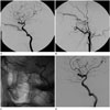
A 31-year-old man was admitted with exophthalmos 7 days after head trauma. He suffered from progressive exophthalmos, bruit and conjunctival chemosis. Cerebral angiography showed a dural arteriovenous fistula (DAVF) fed by the ILT, artery of the foramen rotundum of the internal maxillary artery (IMA) and the small branches of the right middle meningeal artery (MMA), draining mainly into the right superior ophthalmic vein (SOV) and the facial vein (Fig. 1A, B).
After informed consent was obtained, transvenous embolization was chosen in order to avoid the transarterial approach for preventing theprogression of embolic complications. Through the right femoral vein, a 5F guiding catheter (Envoy, Codman Neurovascular, Raynham, MA, USA) was placed in the right internal jugular vein. Initially, transvenous approach via the right cavernous sinus was attempted, but it was unsuccessful due to navigation failure. Next, through the right facial vein, the angular vein, to the SOV, a microcatheter (preshaped J Excelsior, Stryker, Fremont, CA, USA) could be navigated up to the far distal fistulous point under guidance of a 0.014-inch microwire (Synchro, Stryker) and embolization was performed using a single coil (GDC helical 2 × 5 mm, Stryker) (Fig. 1C). Immediately after the procedure, angiography confirmed that the fistula was completely occluded (Fig. 1D).
After the procedure, his ocular symptoms improved without any neurologic complications and he was discharged well. The patient had no recurrent symptoms at the 12-month follow-up.
DISCUSSION
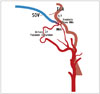
DAVFs associated with the ILT have been rarely reported in the literatures (123). Duncan and Fourie (1) reported a case of traumatic DAVF between the cavernous sinus and the ILT and its management with transarterial embolization. Uchiyama et al. (2) described a spontaneous DAVF fed by the ILT and draining into the superficial sylvian vein with varix formation, which was treated by surgical clipping. Interestingly, the case presented in this report showed a rare type of DAVF fed by the ILT, MMA, and IMA forming a common fistulous tract, draining directly into the confluence of ophthalmic veins (Fig. 2) is be fiwithout cavernous sinus involvement, but with symptoms mimicking carotid cavernous fistula. Horie et al. (3) also reported a similar case, which was treated by transarterial embolization via the ILT.
In some anatomical reports (4), the ILT was identified in 80% of cadaveric studies, and it arose directly from the cavernous part of the ICA in 84% of studies and from the meningohypophyseal trunk in 6% of studies. ILT gives off branches that run to the superior orbital fissure, foramen rotundum, foramen ovale, and foramen spinosum, where they anastomose with branches of the IMA, the MMA and the accessory meningeal artery. Any of these branches can be injured and may be connected to venous structures outside the cavernous sinus after head trauma (25). The anterior ramus of the ILT divides into medial and lateral branches: the former supplies the third, fourth, and fifth cranial nerves and ends as the deep recurrent ophthalmic artery, which interrelates with some branches of the IMA; the latter supplies the dura of the adjacent temporal fossa and the nerve, and it anastomoses with the artery of the foramen rotundum. Unique hemodynamics in this case could be explained by the anatomical proximity of the ILT to the SOV, adjacent narrowing of the transitional zone from the ophthalmic confluence to the cavernous sinus, and topographical complexity of the superior orbital fissure (36).
Endovascular treatment has become the first treatment option for DAVF because it can offer similar results with use of a less invasive approach, while direct vascular surgery or indirect packing may be performed only if the endovascular option fails (25). In this case, we preferred the transvenous approach via the facial vein to transarterial embolization via the ILT, MMA, or IMA due to the possible risk of cranial nerve damage from embolic complications. Some authors (1) reported catheterization failure in the ILT via the transarterial approach and development of neurologic complications following transarterial glue embolization via the MMA or ILT. Kiyosue et al. (7) also described the potential risk of ophthalmic nerve injury following embolization of the distal branches of the IMA.
For transvenous embolization of paracavernous DAVFs, Kim et al. (8) remarked that the facial vein is likely to be a safe and effective route, and our case was also treated successfully through the same route. Caragine et al. (9) described transvenous embolization of DAVFs of the ophthalmic vein fed by the ophthalmic artery and the IMA or the MMA without any neurological deficit.
In conclusion, a DAVF between the ILT and the ophthalmic vein without direct involvement of the cavernous sinus caused by traumatic injury of the ILT is a rare pathology, and it was successfully treated with transvenous coil embolization.
 XML Download