

 PDF
PDF ePub
ePub Citation
 Print
Print


INTRODUCTION
Endometriosis is a common disease, occurring in up to 7% of menstruating females; it is defined as the presence of ectopic endometrial glands and stroma outside the uterine cavity and myometrium (1). Endometriosis most frequently involves the pelvic structures, such as the ovaries, the uterine surface, cul-de-sac, and pelvic peritoneum. However, it may occur in any organ of the body (2). Intestinal endometriosis is the most common extragenital endometriosis, occurring in 3–37% of patients with endometriosis (345). The most commonly involved areas are the sigmoid colon and rectum, representing up to 73% of cases, followed by the ileum, appendix, and cecum (6). The descending colon, however, is a rarely affected site, and only three cases have been reported without computed tomography (CT) imaging (5). In this report, we describe a rare case of intestinal endometriosis involving the descending colon that was misdiagnosed as acute diverticulitis with a small abscess on preoperative multidetector CT. To the best of our knowledge, this is the first reported case with CT findings of descending colon endometriosis beyond the pelvic cavity.
CASE REPORT
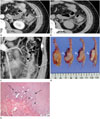
A 47-year-old female patient presented with left lower abdominal pain for 16 months. She had undergone a laparoscopic myomectomy for uterine myomas 2 years earlier. Physical examination revealed focal tenderness in the left lower abdomen. Laboratory findings, including serum white blood cell count and C-reactive protein, were unremarkable. She underwent a colonoscopy for evaluation of the cause of abdominal pain and the colonoscopic findings were also unremarkable. CT of the abdomen and pelvis was performed to evaluate the cause of left lower abdominal pain. CT scans demonstrated a 2.5 × 3.0-cm ovoid exophytic nodule with central low density and peripheral rim enhancement at the antimesenteric border of the descending colon, colonic wall thickening, pericolic fat infiltration, and adjacent fascial thickening (Fig. 1A-C). These CT findings suggested acute colonic diverticulitis with small abscess formation. Due to the longstanding abdominal pain, she underwent a laparoscopic segmental resection of the descending colon. Operative findings showed severe adhesions between the omentum and descending colon and marked retroperitoneal fibrosis. A small pericolic abscess with colon wall thickening was seen, like a diverticular perforation with abscess formation. The surgical specimen was obtained from the mid descending colon, approximately 7.5 cm in length, with intact mucosa. However, the serosal layer of the specimen showed a densely adhered soft tissue mass with focal hemorrhage, measuring about 3.0 × 2.5 × 1.5 cm (Fig. 1D). Microscopic examination demonstrated that the foci of endometrial stroma and endometrial glands were embedded in the hypertrophied muscle layer of the descending colon, suggesting intestinal endometriosis of the descending colon (Fig. 1E). The abdominal pain resolved completely after the surgery.
DISCUSSION
Intestinal endometriosis is characterized by the presence of ectopic endometrial glands and stroma in the intestine (345). The most commonly involved areas are the sigmoid colon and rectum, located in the pelvic cavity (6). However, the descending colon is a rare anatomical site for intestinal endometriosis. A likely reason for this occurrence is that the descending colon is covered by the peritoneum only anteriorly and it is located in the retroperitoneal space.
There have been some theories that explain how ectopic endometrial tissue reaches the intestine, such as retrograde menstruation and implantation, coelomic metaplasia, and lymphatic or vascular dissemination (17). The most widely accepted theory is retrograde menstruation through the fallopian tubes along with viable endometrial tissue and subsequent implantation in the peritoneal cavity (7). Given that this patient had undergone a laparoscopic uterine myomectomy, operative spillage of endometrial tissue and peritoneal implantation may be suggested as an etiological factor.
Although intestinal endometriosis is usually asymptomatic and an incidental finding during pelvic surgery, it may also present with various non-specific gastrointestinal symptoms, such as abdominal pain, nausea, vomiting, alternating diarrhea and constipation, and rectal bleeding (46). Thus, the clinical diagnosis of intestinal endometriosis is challenging, and it is difficult to differentiate it from various gastrointestinal disorders, such as malignancies, inflammatory bowel disease, and ischemic colitis (3). Our patient presented with chronic abdominal pain, confined to the left lower abdomen. Many gastrointestinal disorders, such as diverticulitis, inflammatory bowel disease, colon cancer, ureteric stones, and gynecologic disorders, could be included in the differential diagnosis. CT is the most frequently used diagnostic method in patients with abdominal pain. In this case, CT scans demonstrated focal colon wall thickening and a small exophytic nodule with central low density and peripheral rim enhancement at the antimesenteric border of the descending colon, pericolic fat infiltration and adjacent fascial thickening. These findings suggested acute colonic diverticulitis with abscess formation. We also considered rare subepithelial tumors, such as leiomyoma and benign gastrointestinal stromal tumor, in the differential diagnosis. We did not include intestinal endometriosis because there was neither a visible endometrioma nor a prior history of pelvic endometriosis.
Intestinal endometriosis can show variable CT findings depending on the size and penetrating depth of the endometrial implant, the amount of the bleeding of ectopic endometrial tissue, and the degree of surrounding inflammatory and fibrotic responses (267). Small and superficial endometrial nodules cannot readily be detected on CT scans. Large and deep infiltrating endometriosis can be demonstrated as an extrinsic nodular mass with enhancement, contiguous with or penetrating the thickened colonic wall (2). Intestinal endometriosis usually presents as a subepithelial mass or bowel wall thickening with stricture, because the endometrial tissue implants on the antimesenteric edge of the bowel, and it primarily involves the proper muscle, the subserosa or mesentery. Ectopic endometrial tissue may lead to smooth muscle hypertrophy and fibrosis in the proper muscle and it presents as a mural mass or thickening on CT. In the subserosa or mesentery, endometrial tissue may cause acute and chronic inflammation, fibrosis, and adhesion, representing pericolic inflammatory changes and adhesion on CT (4).
In our case, the densely adhered soft tissue mass with focal hemorrhage in the gross specimen demonstrated concentric hypertrophied muscle with embedded endometrial tissues on microscopic examination. Additionally, the endometrial tissue and surrounding muscle hypertrophy appeared as a hypodense exophytic nodule adjacent to the colon wall on CT, which was mistaken as a diverticular abscess. It may also mimic a subepithelial tumor of the colon such as leiomyoma or gastrointestinal stromal tumor, but pericolic fat infiltration and fascial thickening are rarely seen in these subepithelial tumors.
In conclusion, intestinal endometriosis frequently involves the rectosigmoid colon and ileocecal area, located in the pelvic cavity. However, endometriosis of the descending colon is very rare. There have been several reports describing the CT findings of intestinal endometriosis; however, descending colonic endometriosis and its CT findings have not been reported before. We present the first report of endometriosis of the descending colon that was misdiagnosed as acute diverticulitis with a small abscess on preoperative multidetector CT. Despite its rarity, radiologists should consider intestinal endometriosis in the differential diagnosis when a reproductive-aged woman presents with chronic abdominal pain and CT findings such as bowel wall thickening with an exophytic nodule and an adjacent inflammatory or fibrotic change.
 XML Download