

 PDF
PDF ePub
ePub Citation
 Print
Print


INTRODUCTION
An arteriovenous fistula (AVF) is a direct communication between an artery and a vein without interposition of a capillary bed. AVFs can be found almost anywhere in the body, but those that occur between major abdominal vessels are relatively rare. To our knowledge, there have been only 31 case reports of inferior mesenteric AVFs, including both congenital and iatrogenic forms.
Inferior mesenteric AVF usually presents with abdominal pain, gastrointestinal bleeding, and a palpable mass with thrill or portal hypertension (1). Colonic ischemia is another serious manifestation of inferior mesenteric AVF. The condition may be diagnosed when an abnormal vascular connection is seen on angiography, computed tomography (CT) or magnetic resonance angiography. Colitis can be confirmed via endoscopic biopsy, but it is also easily noted on CT images showing edematous wall thickening and abnormal enhancement of the left hemicolon.
In this report, we present a case of ischemic colitis secondary to idiopathic inferior mesenteric AVF; we also include a review of the literature, focusing on how CT has contributed to the diagnosis of AVF.
CASE REPORT
A 75-year-old male presented to our hospital with non-bloody diarrhea that had occurred on and off over the previous 7 months. The patient complained of abdominal distension, rectal tenesmus, and change in stool caliber. He had hypertension, benign prostatic hyperplasia, and angina, for all of which he was on oral medications. He had no history of previous abdominal surgery or trauma. Physical examination revealed no significant abdominal findings. Laboratory tests including a complete blood count, liver function test, tumor markers, and C-reactive protein levels were within normal ranges.
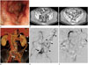
Colonoscopy revealed diffuse edematous wall thickening with hyperemia, located 10 cm to 45 cm above the anal verge (Fig. 1A). Colonoscopic biopsy specimens from the sigmoid colon revealed histopathologically nonspecific findings, which, together with gross morphology, suggested ischemic colitis as the most plausible diagnosis.
A contrast-enhanced CT scan of the abdomen and pelvis revealed marked wall thickening and submucosal edema extending from the splenic flexure to the rectum (Fig. 1B). Abnormally dilated veins, especially at the distal end of the superior rectal branch of the inferior mesenteric vein, raised the possibility of an arteriovenous shunt (Fig. 1C). There was no evidence of portal hypertension, such as varices, hepatosplenomegaly, or collateral vessels, on the CT scan.
An elective angiography was done with the aim of confirming mesenteric AVF, and, if feasible, embolizing the feeder arteries. The angiography agreed with the CT findings: the distal end of the superior rectal artery was markedly dilated, and its receiving counterpart vein showed abnormally early opacification with engorgement (Fig. 1D). The left colic vessels could not be visualized on an inferior mesenteric arteriogram, probably because of rapid drainage of most of the inferior mesenteric flow to the AVF (Fig. 1E). Diversion of blood from the left colic vessels to the AVF was partially compensated for by the marginal artery of Drummond and the meandering arteries in the arc of Riolan. These arcades of collaterals showed prominent engorgement that diverted some superior mesenteric artery blood flow to the left colon on the verge of ischemic insult (Fig. 1F).
The large number of fine fistulas could not be selectively embolized, so the patient underwent low anterior resection instead, in which the entire rectum and sigmoid colon were completely resected. The patient was discharged in good general health.
DISCUSSION
Abdominal AVF is either congenital, resulting from undifferentiated embryonic vessels that fail to regress and interconnect the arterial and venous system, or secondary, arising from accidental or iatrogenic trauma. AVFs in the abdomen commonly involve hepatic, superior mesenteric and splenic arteries (2). On the other hand, AVFs of the inferior mesenteric artery are rare, with only 31 cases thus far reported in the literature.
Mesenteric AVFs manifest with portal hypertension, abdominal pain, bleeding, palpable masses, or bruit; however, sometimes they are asymptomatic (1). Ischemic colitis is another form of mesenteric AVF, and 9 cases of inferior mesenteric AVF with ischemic colitis have been reported in the English literature (1).
Ischemic colitis is a well-known clinical entity with various causes. Of the wide array of etiologies, mesenteric AVF is very rare, making this particular case of ischemic colitis caused by mesenteric AVF especially notable (3). Non-occlusive ischemic colitis caused by mesenteric AVF seems to be secondary to the shunting of arterial flow directly to the veins. The rectum is often spared from ischemic insult, probably due to its rich dual blood supply from the splanchnic and systemic arterial systems. It is logical to think that the mid and distal rectum, which are also supplied by the middle and inferior rectal arteries from the systemic circulation, would be less likely to be influenced by an inferior mesenteric AVF. However, there is a previously reported case of ischemic colitis caused by inferior mesenteric AVF which affected the distal rectum as well as the proximal rectum (1).
Angiography, which directly shows abnormal connections between an artery and a vein, is the most useful tool in diagnosing mesenteric AVF. However, angiography is rarely used in work-up for ischemic colitis, partly due to its relatively low accessibility. Thus, it is clinically important to note the signs of mesenteric AVF on more routinely used CT scans.
Of the 23 studies reported in the English literature that we reviewed, 12 (52%) cases were correctly diagnosed as mesenteric AVFs with the aid of CT (14567). CT findings that indicated mesenteric AVF included abnormal mesenteric vessel dilatation and early opacification of mesenteric veins in the arterial phase, along with diverse findings of ischemic colitis or portal hypertension. In all 12 studies, enlargement of the mesenteric vessels was cited as the key finding that led radiologists to identify AVF. Even though angiography is the optimal modality for confirming AVF, CT findings guide radiologists to suspect AVF, and thus facilitate prompt treatment.
Treatment strategies for AVF involve either surgical intervention or embolization. It is known that fistulas less than 8 mm in diameter can be treated with embolization (8). Surgery is preferred for AVFs with diameters greater than 8 mm because of the increased possibility that embolization material would migrate somewhere else due to high blood flow rate (1). Even though our patient's fistula was not large, embolization was deemed infeasible, because there were many fine feeding vessels that were too difficult to meticulously select. If an arterial approach is difficult, AVF can also be embolized via retrograde injection through an inflated balloon catheter placed at the venous side of the AVF (9) or via direct fluoroscopy-guided puncture of the mesenteric varices (10). The venous approach is only possible in cases where there is a single, straight draining vein. However, in our case, there were multiple tortuous draining veins, so retrograde embolization of the AVF from its venous side was not possible.
To our knowledge, this is the first reported case in Korea of an inferior mesenteric AVF causing ischemic colitis in a patient without any previous surgery or trauma. In the clinical setting of ischemic colitis without any demonstrable etiology, the possibility of a mesenteric AVF should be considered; knowledge of the CT features of inferior mesenteric AVF will aid in prompt diagnosis and treatment in future cases, thus avoiding grave consequences such as portal hypertension and bowel ischemia.
 XML Download