

 PDF
PDF ePub
ePub Citation
 Print
Print


INTRODUCTION
A choledochal cyst is a rare congenital anomaly of the biliary system, manifested as the cystic dilatation of bile ducts, usually occurring in the common bile duct (1234). The Todani classification system is widely used to classify choledochal cysts; however, not all cases fit into this classification system. Here, we present a case of a choledochal cyst that did not fit into this Todani classification system: saccular cystic dilatation of the confluent portion of both intrahepatic ducts (IHDs).
CASE REPORT
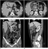
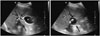
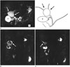
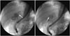
A 45-year-old male patient presented with a sudden onset of severe pain in the upper right abdomen. On physical examination, there was tenderness in the upper right abdominal area, and Murphy's sign was positive. There was no increase in inflammatory markers in the blood tests, except a mild increase of alanine aminotransferase (43 IU/L) and gamma glutamyl transpeptidase (103 IU/L). In the contrast-enhanced abdominal CT performed to rule out biliary colic and diseases such as acute cholecystitis, we detected a 4.5 cm thin-walled unilocular cystic lesion containing multiple calcified stones. The lesion abutted the gallbladder in the porta hepatis without apparent origin (Fig. 1). There was a calcified gallstone without the mural thickening of the gallbladder. Both intrahepatic and common bile ducts were undilated. At this point, the differential diagnosis included the duplication anomalies of the gallbladder with cholelithiasis, an exophytic complicated hepatic cyst originating from segment 4 of the liver, and a choledochal diverticulum with choledocholithiasis. In the abdominal ultrasonography, we detected a 4.5 cm unilocular thin-walled cystic lesion containing multiple echogenic stones, which abutted the gallbladder. The lesion did not show the anatomical layering of its wall, which was detected in the gallbladder (Fig. 2). Magnetic resonance cholangiopancreatography (MRCP) (Fig. 3) and endoscopic retrograde cholangiopancreatography (ERCP) (Fig. 4) showed a saccular dilatation of the confluent portion of both IHDs with multiple internal filling defects of variable sizes. Both IHDs directly arose from the dilated cystic lesion. The length of the common channel of the common bile duct and pancreatic duct was under 1.5 cm, and there was no anomalous pancreaticobiliary ductal union. At laparotomy, a 4 × 3.5 × 3.5 cm thin-walled cyst was found in the confluent portion of both IHDs at the level of the porta hepatis with multiple intracystic stones. Both IHDs directly arose from the cyst. There were no abnormal findings in the common hepatic or bile ducts. The cyst was completely excised and cholecystectomy, choledochojejunostomy, and jejunojejunostomy were performed. Histopathology showed an inflamed cystic structure lined with biliary epithelium, consistent with a choledochal cyst. The patient made an uneventful post-operative recovery.
DISCUSSION
A choledochal cyst is a rare congenital anomaly of the biliary system, involving any part of the bile duct. It is manifested as the cystic dilatation of bile ducts, usually occurring in the common bile duct (1234). It was first reported by Douglas in 1852 (2). The detection of congenital anomalies, including choledochal cysts, may avoid diagnostic errors, help to plan surgery, and prevent unnecessary ductal injury (5).
The symptoms associated with a choledochal cyst are nonspecific, which can lead to a delay in the diagnosis. It usually presents with abdominal pain, jaundice, and a palpable right upper quadrant mass. The symptoms are more prevalent in children than in adults (1). Babbitt et al. (2), described that the symptoms of a choledochal cyst may be similar to those of Mirizzi's syndrome at presentation. The incidence of choledochal cysts is 100 times more common in Asian countries, particularly Japan (one in 1000), than in Western countries (one in 100000-150000). It is more common in women, with a female:male ratio of 4:1 (136). Approximately 2/3 of cases are diagnosed before the age of ten (17), and it is relatively rare for it to occur in an adult, as in the present case.
The Todani classification system is the most widely accepted classification of choledochal cysts based on their anatomical location and cholangiographic morphology. This classification system was expanded from the work of Along-Lej et al., and describes five main types of choledochal cysts, with several sub-types: type I is the most common type (80-90%), involving the dilatation of the entire common hepatic or common bile duct or segments of each. It is subclassified into type IA (cystic dilatation of the common bile duct), type IB (focal segmental dilatation of the distal common bile duct), and type IC (fusiform dilatation of the common hepatic and common bile ducts). Type II is rare (2%), and is a true diverticulum anywhere in the extrahepatic duct. Type III (also called choledochocele) is also rare (1.4-5%), and is confined to the intraduodenal portion of the common bile duct. Type IV is more common (19%) and may be further subclassified into type IVA, which involves both the intra- and extrahepatic bile ducts, and the least common type IVB, where only extrahepatic cysts are observed. Type V, or Caroli's disease, is characterized by the dilatation of single or multiple intrahepatic bile ducts (136).
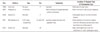
Although most of the choledochal cysts can be classified using the Todani classification system, a few cases have been previously reported that did not fit into the classification system (Table 1). Loke et al. (6), described a case of an atypical choledochal cyst characterized by the cystic dilatation of the cystic duct. They described this as a type VI choledochal cyst. Choledochal cysts involving only the IHD were reported by Prekop et al. (right IHD) (7), Salles et al. (right IHD) (8), and Sadiq et al. (IHD at the level of the porta hepatis) (3). Michaelides et al. (1), described a case of a choledochal cyst (with dilatation of the common bile duct, common hepatic duct, and the proximal portion of the cystic duct as a bicornal configuration); they proposed the classification of this atypical type as a new subtype of Todani I cyst, namely type ID. Similar to these cases, our case of a choledochal cyst also did not fit into the typical Todani classification system because the cyst involved the confluent portion of both IHDs.
Many theories have been proposed regarding the etiology of choledochal cysts, however, none of these theories explain all the different types of choledochal cyst. A few theories have attempted to explain the cause of the common types of choledochal cysts. The first theory, which is the most widely accepted, is that there is an abnormal common channel related to the anomalous union of the pancreaticobiliary duct (AUPBD). This anomalous union has two features relevant to the formation of choledochal cysts. One is that the union of the pancreatic duct and common bile duct is located far from the duodenum, creating a long common channel of a length greater than 1.5 cm. The other is the angle of this junction. According to this theory, the abnormal pancreaticobiliary duct union allows the chronic reflux of pancreatic enzymes into the biliary system. The chemical and enzymatic destruction of the ductal wall results in the weakening and dilatation of the involved bile duct, which subsequently forms a cyst. However, this theory can be mainly applied in Todani classification type I, and does not explain the occurrence of choledochal cysts in the presence of a normal pancreaticobiliary ductal union, as seen in our case. A few previous articles have reported cases of choledochal cysts without accompanied AUPBD. Another study proposed an alternative theory that choledochal cysts represent a spectrum of embryonic malformation of the pancreaticobiliary system, one of which may be an anomalous junction (1245).
The histopathology of choledochal cysts shows a fibrotic wall varying from a few millimeters up to 10 millimeters in thickness. Dense collagenous connective tissue makes up the cystic wall, without a complete epithelial lining. Elastic fibers and smooth muscle bundles can also be occasionally found as constituents of the wall. Choledochoceles (Todani classification type III) are different in that they are lined by either duodenal or bile duct mucosa as an exception. An inflammatory reaction caused by the free flow of pancreatic juice into the common bile duct is often present. The recurrent episodes of inflammation result in thickening of the wall of the involved bile duct (12).
A choledochal cyst can be confused with other cystic lesions. The differential diagnosis includes hepatic cyst, enteric duplication cyst, pancreatic pseudocyst, hepatic artery aneurysm, duplication anomalies of the gallbladder, recurrent pyogenic cholangitis, and spontaneous perforation of the common bile duct (9). Sahoo and Kumar (10), described a case report of a double gallbladder masquerading as a choledochal cyst, contrary to the diagnosis in our case. We had a differential diagnosis of several possible diseases, such as duplication anomalies of the gallbladder with cholelithiasis, an exophytic complicated hepatic cyst originating from segment 4 of the liver, and a choledochal diverticulum with choledocholithiasis. An ultrasonography of the lesion did not depict the mural layering that is detected in the gallbladder, therefore, we were able to rule out the possibility of duplication anomalies of the gallbladder with cholelithiasis. Recurrent pyogenic cholangitis is caused by intrahepatic ductal strictures and intraductal calculi, and is characterized by the recurrent attacks of abdominal pain, fever, and jaundice with mildly elevated bilirubin. In our case, the patient did not have a history of recurrent attacks or laboratory abnormalities.
Various preoperative imaging techniques, such as CT, MRI (including MRCP), ultrasonography, hepatobiliary scan, ERCP, and percutaneous transhepatic cholangiography are available for the evaluation of choledochal cysts (5). Savader et al. (4), described MRCP as an effective non-invasive imaging technique for the diagnosis and preoperative evaluation of a choledochal cyst. Such evaluation is essential for surgical planning. Michaelides et al. (1), described the importance of the surgeon's knowledge regarding the details of the cyst, such as the exact location and length of the common channel.
Common complications of choledochal cysts, including cholecystitis, biliary stricture, recurrent cholangitis, recurrent acute pancreatitis, cholelithiasis, choledocholithiasis, and even malignancy have been reported. Choledocholithiasis is one of the most common complications as seen in our case. Savader et al. (4), reported rare complications such as ectopic pancreas (jejunum, one case), intrahepatic abscesses (one case), and cystolithiasis (one case). Due to the fact that the frequency of biliary malignancy is more common in adults than in children, adult patients with choledochal cysts should be cautiously evaluated. Malignancy may occur in the remaining stump after resection, although the excision of the cyst greatly decreases the risk of malignancy (15).
In conclusion, we described an unusual type of choledochal cyst involving the confluent portion of both IHDs with choledocholithiasis. Our report may help to increase the awareness of the unusual types of choledochal cysts that do not fit into the Todani classification system.
 XML Download