

 PDF
PDF ePub
ePub Citation
 Print
Print


INTRODUCTION
Acute ischemic stroke with the occlusion of internal carotid artery terminus (ICAT) is associated with a high rate of mortality and poor functional outcome in survivors (12). Early, complete revascularization of the occluded internal carotid artery (ICA) is essential to improve outcome in this condition (3). However, pharmacological treatment using intravenous (IV) or intra-arterial (IA) thrombolysis is associated with lower recanalization rates in ICAT occlusion, mainly because of large clot burden (1456). Additional to the IA thrombolysis, several methods of mechanical clot disruption using microcatheter, microguidewire balloon or stent are attempted to improve recanalization rates for large vessel occlusions. These methods facilitate pharmacologic thrombolysis by fragmenting the clot and increasing the surface area exposed to lytic agents. Some previous studies showed higher recanalization rates of these multimodal approaches than IA thrombolysis alone, but the clinical benefit is unclear (78910).
Recent advances in IA techniques and thrombectomy devices such as Penumbra System (PS) (Penumbra Inc., Alameda, CA, USA) and stent retrievers (Solitaire FR, ev3, Irvine, CA, USA or Trevo retriever, Concentric Medical Inc., Mountain View, CA, USA) lead to high rate of recanalization and have changed the concept of intra-arterial therapy (IAT) from pharmacological thrombolysis to mechanical thrombectomy (111213).
In our institution, the PS and Solitaire stent have been available since December, 2008 and July, 2010, respectively. These advances provide promising measures toward improving results of endovascular revascularization therapy (ERT). We aimed to compare the results of multimodal ERT approaches based on thrombolytic therapy versus mechanical thrombectomy, in patients with cardioembolic ICAT occlusion.
MATERIALS AND METHODS
Patients
This study was conducted with the approval of our Institutional Review Board, and informed consent from the patients or their legal representatives was waived.
We screened consecutive patients with acute ischemic stroke who underwent ERT at the Seoul National University Bundang Hospital from September 2003 to January 2015. Clinical and radiologic data were reviewed from a total of 614 patients who were treated by ERT for acute ischemic stroke in our medical center. Clinical data included the following: demographic information; time from stroke onset or last normal time to treatment; baseline National Institutes of Health Stroke Scale (NIHSS) and modified Rankin Scale (mRS) score at 3 months. Computed tomography (CT) and/or magnetic resonance image (MRI) obtained before ERT were also reviewed. All the patients underwent stroke MR imaging to select eligible patients for ERT within 8 hours, with the exception of those patients who had contraindication of MRI. Multimodal CT scans (non-contrast CT, CT angiography, and perfusion CT) were performed in patients 1) who presented < 3 hours after symptom onset, or 2) with contraindication to MR imaging. Angiograms were reviewed for the location of the occlusion, clot burden, and reperfusion status after ERT.
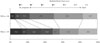
Patients were excluded from the analysis if they 1) had not angiographically demonstrated complete occlusion of the ICAT (n = 459) that was defined as the ICA occlusion involving the ICA bifurcation area, 2) had non-cardioembolic stroke (n = 76), 3) had symptom onset to hospital arrival of > 8 hours (n = 16), 4) had an NIHSS score < 4 and no prior neurologic event (n = 2) or ≥ 2 premorbid mRS (n = 6) (Fig. 1).
ERT Procedure and the Technical Strategy for ICAT Occlusion
All procedures were performed using femoral artery approach and local anesthesia. A 6-Fr coaxial guiding system (Shuttle SL Flexor; Cook Medical, Bloomington, IN, USA and Envoy 6-Fr, Cordis, Miami Lakes, FL, USA) or a 9-Fr balloon guide catheter (Optimo; Tokai Medical Products, Inc., Japan or Cello; Medtronic, Irvine, CA, USA) was introduced through a femoral sheath into the carotid artery of concern. A heparinized saline solution was continuously perfused through the catheter during the procedure.
Multimodal combination technique generally used for treating patients with ICAT occlusion consist of 1) pharmacological thrombolysis, 2) mechanical disruption using a microguidewire or a balloon catheter, 3) mechanical thrombectomy using a guiding catheter, PS or Solitaire stent, and 4) permanent stenting with a balloon-expandable or self-expandable stents (Neuroform, Boston Scientific, Fremont, CA, USA; Enterprise, Codman Neurovascular, Miami Lakes, FL, USA; Solitaire FR, ev3, Irvine, CA, USA).
The technical strategy of the multi-modal approach of ERT has been changed in our institution due to the introduction of th-rombectomy devices such as PS or Solitaire FR. Before the th-rombectomy device was available, the ICAT occlusion was initially treated by suction thrombectomy using a guiding catheter to decrease the thrombus burden. Residual thrombus was then treated by pharmacological thrombolysis and mechanical disruption. If recanalization was not achieved, permanent stenting was considered for recanalization. After PS and Solitaire stent became available, mechanical thrombectomy using a guiding catheter, PS or Solitaire stent was initially attempted for recanalization on a case-by-case basis. The technique of forced-suction thrombectomy using a reperfusion catheter of PS without a separator was used and described by Kang et al. (14). If mechanical thrombectomy failed, pharmacological thrombolysis, mechanical disruption or permanent stenting were considered. Therefore, we divided patients into the thrombolytic-based IAT group (TLG) and the thrombectomy-based IAT group (TEG) according to the primary method for ERT.
Urokinase was used as the thrombolytic agent with a maximum dose of 800000 U, and Glycoprotein IIb/IIIa receptor antagonist was also used (Reopro 10 mg or tirofiban 10 µg/kg as the maximum dose). Urokinase was manually infused in 20000 U aliquots at 1 or 2-minute intervals between doses and directly into or near the clots.
Imaging Analysis
Two experienced neuroradiologists (B. S. C. and J. H. K.) who had 7 and 22 years, respectively, of clinical experience and who were unaware of the patients' histories or treatments, scored the Albert Stroke Program Early CT Score (ASPECTS) using the diffusion weighted image (DWI) or cerebral blood volume (CBV) map of the perfusion CT (1516).
The ICAT occlusions were categorized as an L or T type lesion, based on functional nature of collateral flow patterns on angiography (17). If the ipsilateral anterior cerebral artery (ACA) territory was filled by collateral flow from contralateral ICA, it was defined as a functional L type occlusion; and if the ipsilateral ACA flow was also compromised, it was defined as a functional T type occlusion.
The angiographic clot burden score (ACBS) was defined according to the clot extent and location determined by cerebral angiography including super-selective angiography with a microcatheter performed during ERT. The ACBS was modified from the previous report in order to determine the clot burden score using CT angiography (18) as follows: 1) ICA was divided as intradural and extradural segments by ophthalmic artery and then given 2 points to its segment. More specifically, 1 point was given, if less than half length of intradural segment was involved or only 1 segment was involved between cavernous, petrosal and cervical segment ICA. 2) M1 was divided as proximal and distal half segment that was given 1 point. 3) M2, A1, and A2 were also given 1 point, respectively, if thrombus was located at each of the arteries regardless of thrombus length and number of involv-ed arteries.
Outcome Evaluations
Clinical outcome measurement at 3 months after stroke onset included mortality and disability, measured according to mRS score. We dichotomized outcome according to the functional de-pendency. Patients with a mRS score ≤ 2 were in the favorable outcome (functionally independent) group and those with a mRS score > 2 were in the unfavorable outcome (functionally dependent) group. The complete reperfusion was defined as modified treatment in cerebral infarction (mTICI) grade 2b or 3 and futile recanalization was defined by the occurrence of unfavorable outcome despite complete reperfusion (19). The reperfusion time was defined as the time interval from the puncture to the final reperfusion with TICI ≥ 2a. Procedure-related complications, such as vascular perforation or arterial dissection, were also assessed. Procedural distal emboli (PDE) into the ipsilateral or contralateral ACA were also evaluated and defined as a new site occlusion distal to A1 segment of ACA.
Symptomatic intracranial hemorrhage was defined as any he-morrhagic transformation seen on the 24-hour CT scan and as-sociated with a decline of ≥ 4 points in the NIHSS score within 24 hours or leading to a patient's death (20). All other hemorrhages were defined as asymptomatic.
Statistical Tests
The baseline characteristics for the TLG versus the TEG were compared using the χ2 test for categorical variables and the Student t-test or the Mann-Whitney U test for continuous variables. Multivariate analyses were performed to identify predictors for unfavorable outcome at 3 months. Receiver operating characteristic curves were constructed to obtain the cutoff DWI or CBV ASPECTS for discriminating between patients with and without functional dependency at 3 months. Significance was set at the 2-tailed p < 0.05 level. We presented the values as frequencies (percentages), means (standard deviations), or medians [interquartile ranges (IQRs)], as appropriate. All statistical analyses were performed using SPSS 17.0.1 (SPSS Inc., Chicago, IL, USA).
RESULTS
Among the screened 614 patients, 55 patients with ICA occlusion met the inclusion and exclusion criteria for this study. Eighteen patients were included in the TLG and the remaining 37 patients were included in the TEG, respectively. The patients' baseline characteristics were summarized in Table 1. There were no statistical differences in baseline characteristics between the groups. Cardioembolic sources of stroke were atrial fibrillation in 50 patients (90.9%), large patent foramen ovale in 2 patients, cardiac arrhythmia except atrial fibrillation that required pacemaker in 1 patient, ischemic heart disease accompanied by inferior wall akinesia in 1 patient, and prosthetic mitral valve in 1 patient.
In the TLG, pharmacological thrombolysis and mechanical disruption were attempted in all cases. Suction thrombectomy with a guiding catheter was performed in 3 cases in order to de-crease the thrombus burden in the ICA; and permanent stenting of the intracranial artery was performed in 3 cases as the final option. In the TEG, reperfusion could be achieved in 12 of 37 cases only by mechanical thrombectomy using PS and/or the Solitaire stent without thrombolytics. Pharmacological thrombolysis and mechanical disruption were also conducted in 9 cases. One case in the thrombectomy group had permanent stenting that was used as a rescue method for procedure-related arterial dissection in the cervical segment of the ICA.
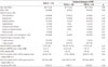
The procedural and clinical outcomes were summarized in Table 2. The median dose of urokinase was significantly lower in TEG than in TLG (median [IQR], 20 [0-100] vs. 300 [87.5-500], × 103 U; p < 0.01). The reperfusion time was also significantly shorter in TEG than in TLG (median [IQR], 38 [18-78] vs. 142 [102-169], min; p < 0.01). The rate of complete reperfusion with mTICI of 2b or 3 was significantly higher in TEG than in TLG (78.4% vs. 44.4%; p = 0.01). The rate of futile recanalization was relatively lower in TEG than in TLG, but without statistical significance (58.6% vs. 87.5%, p = 0.13). The statistical trend showed that TEG had more chance to achieve the favorable clinical outcome with mRS ≤ 2 at 3 months than TLG (Fig. 2, Table 2). Symptomatic intracranial hemorrhage and all-cause mortality at 3 months showed no statistical difference between 2 groups. Although PDE into the ACA did not show statistical difference between the 2 groups, all patients with PDE into the ACA had unfavorable outcome at 3 months. Mild procedural complications occurred in 4 cases of the TLG and in 6 of the TEG.
Forty of 55 patients (72.7%) with mRS score > 2 at 3 months were categorized as the "unfavorable outcome" group in dichotomized analysis. In the multivariate logistic regression analysis taking functional outcome at 3 months as a dependent variable, age [adjusted odds ratio (OR), 1.11 per 1 year increase; 95% confidence interval (CI), 1.02 to 1.21; p = 0.02], baseline ASPECT score (adjusted OR, 0.41 per 1 point increase; 95% CI, 0.22 to 0.77; p < 0.01), and time from groin puncture to reperfusion or completion of procedure (adjusted OR, 1.03 per 1 minute increase; 95% CI, 1.01 to 1.06; p < 0.01) were significantly associated with unfavorable outcome at 3 months (Table 3). This model included only the statistically significant variables from the univariate logistic regression model.
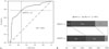
The optimal cutoff DWI or CBV ASPECTS to predict patients with favorable outcome at 3 months was ≥ 5, with a sensitivity of 79%, specificity of 74%, and an area under the curve (AUC) of 0.835 (Fig. 3). On excluding 6 patients without DWI, the optimal cutoff was also ≥ 5, with a sensitivity of 80%, specificity of 72.4%, and an AUC of 0.809. Overall, 19 (67.9%) of 28 patients with baseline ASPECT ≥ 5 and 5 (17.2%) of 29 patients with baseline ASPECT < 5 had mRS scores ≤ 2 at 3 months (p < 0.01) (Fig. 3). There was no significant difference in the rate of symptomatic intracranial hemorrhage between the groups [6 of 29 (20.7%) in patients with ASPECTS < 5 vs. 3 of 26 (11.5%) of patients with ASPECTS ≥ 5, p = 0.36].
Ten of 55 patients (18.2%) were categorized as a functional T type occlusion and all had an unfavorable outcome at 3 months. Of these, 5 patients had mRS 5 at 3 months, and the remaining 5 patients died before 3 months. The DWI or CBV ASPECTS were significantly lower in patients with functional T type occlusion than in patients with L type occlusion, although there was no difference in NIHSS at admission between the groups. In addition, the rate of complete reperfusion was significantly lower in patients with functional T type occlusion than those with the L type occlusion.
DISCUSSION
Our study showed that thrombectomy based multimodal ERT (TE-MERT) for cardioembolic ICAT occlusion has potential advantages over thrombolytic therapy based multimodal ERT (TL-MERT) to improve the clinical outcome. First, TE-MERT can reduce the time from groin puncture to reperfusion as well as the total dose of urokinase. Second, the reperfusion rate with mTICI ≥ 2b in TE-MERT (78.4%) was higher than that in TL-MERT (44.4%).
Recently, new devices such as PS and stent-retrievers showed their superior efficacy on reperfusion rate than Merci retrievers or other IA thrombolytics, especially in anterior circulation (71112131421). The systematic review of IAT for acute ischemic stroke due to ICA occlusion showed higher recanalization rate (69% vs. 48%, p = 0.001) and favorable outcome (34% vs. 12%, p < 0.001) in mechanical thrombectomy group than for IA thrombolysis (21). Our result supported the conclusion of the systematic review, although we included only isolated ICAT occlusions from a suspected cardioembolic source.
The results indicated that baseline ASPECTS, age and time from groin puncture to reperfusion were independent predictors for unfavorable outcome at 3 months after stroke. Recently, it is emphasized that procedure time is a very important factor for reducing the number of futile recanalization, as well as for successful recanalization (22). Rai et al. (23) showed that procedure predictors affecting the favorable outcome included successful recanalization (p < 0.01), collateral support (p = 0.0008), distal occlusion (p = 0.001), and a shorter procedure duration (p = 0.01) (23). Hassan et al. (22) showed that the rates of favorable outcome in cases with a procedure time of ≥ 60 minutes were lower than the rates observed in the placebo group in the Prourokinase for Acute Ischemic Stroke Trial. Moreover, a recently published clinical trial showed the beneficial effect of embolectomy using stent retriever on clinical outcome in patients with proximal artery occlusion in the anterior circulation; this also emphasized that rapid endovascular treatment and early reperfusion improved functional outcomes and reduced mortality (24). In our series, the time from the groin puncture to reperfusion time was significantly reduced in the TEG.
We used the DWI-ASPECTS to assess the lesion volume, due to the MR-based protocol of patient selection for ERT, except for patients with contraindication of MR study. In our series, 29 of 55 patients (52.7%) had baseline DWI or CBV ASPECTS ≤ 4, despite the relative short duration of onset to hospital arrival time (median [IQR], 80 [49-170], minutes). The DWI-ASPECTS tends to be higher than ASPECTS measured by CT, because of the high sensitivity of the DWI to detect early ischemic changes in acute ischemic stroke (15). Furthermore, it could also be facilitated by poor collateral circulation in ICAT occlusion. However, we attempted to treat these patients with low ASPECTS if they had no or only subtle signal changes on fluid-attenuated inversion recovery image because of the high rate of mortality and poor functional outcome in survivors of ICAT occlusion (25). As a result, the baseline ASPECTS ≥ 5 of DWI was predictive for favorable outcomes at 3 months with a sensitivity of 80%, and specificity of 72.4%, respectively. In studies of IV thrombolysis, the cutoff value of DWI ASPECTS was < 5 for a poor clinical outcome and ≥ 7 for good clinical outcome (2627), while the cutoff values of ASPECTS for good and poor clinical outcomes have not been defined in the ERT of cardioembolic ICAT occlusion.
It is notable that the rate of complete reperfusion and favorable outcome at 3 months were significantly lower in patients with functional T type occlusion. Patients with functional T type occlusion had lower baseline ASPECT score than those in patients with L type occlusion, despite the lack of differences in st-roke severity at admission. It showed that the collateral flow patterns at initial presentation could distinguish the nature and im-pact of ICA occlusion on reperfusion and subsequent clinical outcomes in AIS, as demonstrated in the previous study (17).
The clinical significance of PDE has been occasionally overlooked in many studies for ERT (19). Table 4 summarized the case series that describes the incidence of PDE according to the methods and devices used in ERT (282930). Mechanical disruption has the highest incidence of PDE suggesting that the incidence of PDE varies depending on the techniques and devices used. Todo et al. (30) suggested that PDE requires additional procedure time for complete recanalization and is associated with the clinical outcome as well as patient's mortality. In our series, PDE into ACA was more frequent in the TLG, and was associated with mortality and poor functional outcome; this was in agreement with another report analyzing the effect of new embolic ACA occlusion (31). Therefore, any effort to reduce PDE into the ACA would improve clinical outcome, regardless of the techniques or devices used for ERT.
Our study had some limitations due to its retrospective design. First, the number of cases in both groups was too small to achieve statistical significance regarding the clinical outcome. Second, as the devices used to remove the thrombus were heterogeneous, we could not assess the efficacy of a single device. Therefore, further studies are essential to demonstrate which thrombectomy device is more effective and safe for use in recanalization of cardioembolic ICAT occlusion, e.g., the stent retrievers or the PS. Third, we included large proportion of patients with low baseline DWI or CBV ASPECTS, who were not included in the clinical trial. Despite the possible reasons for the high rate of futile recanalization, our study reflected real practice and utilized unbiased sample from target population with acute ICAT occlusion. Fourth, as the TLG was an historical control group, selection bias was inevitable and the devices such as the guiding catheter, microcatheter, and microwire, except for the thrombectomy devices, could not be controlled.
In conclusion, the TE-MERT of cardioembolic ICAT occlusion may have technical advantages over TL-MERT in that the former 1) reduces the time from puncture to reperfusion, and 2) achieves more complete reperfusion. As a result, these technical advantages of the TE-MERT could be expected to achieve a favorable outcome.


 XML Download