

 PDF
PDF ePub
ePub Citation
 Print
Print


INTRODUCTION
Parathyroid carcinoma is an uncommon disease, accounting for only 1-3% of all patients with primary hyperparathyroidism (1). Intrathyroidal parathyroid carcinoma arising from ectopic intrathyroidal parathyroid tissue is even rarer, and to the best of our knowledge, only 8 cases have been reported (2). Radiologically, preoperative diagnosis of parathyroid carcinoma remains a challenge because its imaging features are nonspecific and they overlap with those of more frequently occurring disease such as parathyroid adenoma. We report a rare case of parathyroid carcinoma arising from intrathyroidal parathyroid tissue and review the radiologic findings of parathyroid carcinoma.
CASE REPORT
A 33-year-old woman was referred to our hospital for evaluation of hypercalcemia. Four years ago, she had undergone right nephrectomy for recurrent renal stones. Laboratory tests revealed an elevated calcium level of 13.9 mg/dL (reference range, 8.4-10.2 mg/dL), a low phosphorous level of 1.2 mg/dL (reference range, 2.6-4.6 mg/dL), and a markedly elevated intact parathyroid hormone level of 965.8 pg/mL, but a relatively normal level of 1, 25 dihydroxyvitamin D of 32.9 pg/mL, suggesting primary hyperparathyroidism.
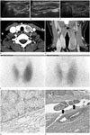
Ultrasonogram revealed a 1-cm sized, well-defined, hypoechoic nodule located in the posterior aspect of the mid to upper right thyroid gland, which was considered to be a right parathyroid adenoma. Another 5-cm sized, elongated, mixed echoic nodule was noted in the left thyroid gland (Fig. 1A, B). Color Doppler examination demonstrated increased vascularity in the lesion, and it was considered to be a thyroid adenoma (Fig. 1C).
Non-enhanced computed tomography (CT) revealed a 1-cm sized nodule considered to be a parathyroid adenoma in the right parathyroid gland as well as a 5-cm sized, hypoattenuated intrathyroidal mass in the left thyroid gland. After contrast enhancement, heterogeneous moderate enhancement was observed in both lesions, with tiny hypodense foci, presumably due to cystic degeneration or necrosis, in the left thyroid gland mass (Fig. 1D). Coronal reformatted images showed an intrathyroid nodule with a positive beak sign on the surface in contact with the thyroid gland (Fig. 1E).
The patient underwent a sequential technetium 99m (Tc-99m) pertechnetate and Tc-99m methoxyisobutylisonitrile (MIBI) scan. Tc-99m pertechnetate scan revealed a large cold nodule in the left thyroid gland, which was correlated with the intrathyroidal mass observed in the previous ultrasonography and CT scans. This finding suggests that the left thyroid gland mass was hormonally non-functional. On the Tc-99m MIBI scan, diffuse even uptake was noted in the left thyroid gland mass similar to that in the adjacent normal thyroid parenchyma, suggesting that the mass was solid with vascularity. On the 2-hour delayed image, diffuse washout was noted in both lobes of the thyroid gland and the left thyroid gland mass without focal abnormal hot uptake (Fig. 1F).
The patient underwent total thyroidectomy with right upper parathyroidectomy. During surgery, relatively well-marginated, encapsulated masses were found in the left thyroid gland and the right upper parathyroid gland, measuring approximately 6 × 3.2 × 2.3 cm and 0.8 × 0.5 cm, respectively. Immunohistochemical analysis revealed strong immunoreactivity for parathyroid hormone and no reactivity for thyroid transcription factor 1, suggesting that the left thyroid mass originated in the parathyroid gland (Fig. 1G). The results of immunohistochemical staining for CD10, synaptophysin, vimentin, and galectin 3 were negative. On histopathologic examination, parathyroid cell proliferation was observed, suggesting the possibility of parathyroid neoplasm or hyperplasia; however, both nodules were considered to be malignant due to the presence of vascular invasion in both nodules (Fig. 1H). Based on these findings, the tumor was diagnosed as parathyroid carcinoma.
DISCUSSION
Parathyroid carcinoma is a rare cause of primary hyperparathyroidism, accounting for approximately 1-3% of all cases. The imaging features of parathyroid carcinoma are nonspecific, and therefore, it is often diagnosed after surgery via histopathologic examination.
Although the preoperative diagnosis of parathyroid carcinoma is quite difficult, parathyroid carcinomas are generally larger than adenomas. A previous review of 286 parathyroid carcinoma cases found that the average tumor size was 3.3 cm (3), and another review of 17 cases reported that tumors were between 2 and 7 cm in size (4). Therefore, a parathyroid mass, greater than 3 cm in size should be considered to be parathyroid carcinoma rather than parathyroid adenoma because the size of parathyroid carcinoma is generally larger than that of parathyroid adenoma. Clinically, parathyroid carcinoma is often associated with markedly increased levels of intact parathyroid hormone and serum calcium (5). However, another study that included 8 parathyroid carcinoma patients found that the serum calcium and intact parathyroid hormone levels were slightly lower than those in patients with benign lesions, and 5 of the 8 patients had a tumor smaller than 3 cm (6). Therefore, size of the mass and level of intact parathyroid hormone alone cannot help to make a definite diagnosis of parathyroid carcinoma.
The imaging features of parathyroid carcinoma are nonspecific. The lesions often have a lobulated contour, heterogeneous internal architecture, calcifications, and an internal cystic component, but the only reliable diagnostic finding preoperatively is the presence of local invasion or nodal metastasis (7).
In our case, the patient presented with a mass within the left lobe of the thyroid gland and another small nodule in her right parathyroid gland. Based on the overall imaging findings of the lesions and clinical manifestations of the patient, we considered it probable that the left thyroid gland lesion was a non-functional thyroid adenoma and the right upper parathyroid gland lesion was a parathyroid gland neoplasm associated with hypercalcemia. The final pathologic diagnosis was intrathyroidal parathyroid carcinoma with possible extension into the right upper parathyroid gland. Intrathyroidal parathyroid carcinoma is a rare variant of parathyroid carcinoma and is extremely uncommon, and to the best of our knowledge, only 8 cases have been reported previously (2). Intraparathyroidal carcinoma is thought to originate from ectopic intrathyroidal parathyroid tissue, which is present in 0.2% of the population. The parathyroid glands arise from the third and fourth pharyngeal pouches and complete their migration by the seventh week of gestation. It has been suggested that variations in this migration result in ectopic parathyroid gland location (8).
Metastasis of parathyroid carcinoma at initial presentation is rare, and very few patients with parathyroid carcinoma have metastases either to regional lymph nodes (< 5%) or distant sites (< 2%). Although the imaging findings are nonspecific, heterogeneous mixed echogenicity, marked increased vascularity on color Doppler examination, and a relatively large tumor size may indicate parathyroid carcinoma.
Tc-99m MIBI imaging is generally considered to be a more sensitive diagnostic tool for the evaluation of parathyroid adenoma compared with high-resolution ultrasonography. The reported sensitivity and specificity of Tc-99m MIBI scans was 56-100% and 83-99%, respectively. Owing to the rarity of this malignancy, the diagnostic accuracy of Tc-99m MIBI imaging for parathyroid carcinoma has been evaluated in only a few studies. Kitapçi et al. (9) reported that a patient with parathyroid carcinoma showed increased Tc-99m MIBI accumulation on preoperative scanning. However, Ruan et al. (10) reported a case of a patient with parathyroid carcinoma with normal findings on Tc-99m MIBI scan. In our case, a Tc-99m MIBI scan did not show any abnormal hot uptake on a 2-hour delayed image. Unlike parathyroid adenoma that generally shows markedly increased uptake on a Tc-99m MIBI scan, parathyroid carcinoma seems to have a variable uptake, and its uptake rate is assumed to be lower than that of parathyroid adenoma. Therefore, a negative Tc-99m MIBI scan in a patient with a parathyroid neoplasm and with clinical signs of hyperparathyroidism may suggest parathyroid carcinoma rather than parathyroid adenoma. Further well-designed studies are required to evaluate the diagnostic potential of Tc-99m MIBI uptake in patients with parathyroid carcinoma.
Although the clinical and imaging features of parathyroid carcinoma are nonspecific, parathyroid carcinoma should be included in the differential diagnosis of a case with a relatively large tumor size (> 3 cm), heterogeneous internal tumor architecture, a lobulated contour mass with marked increased vascularity, and a negative MIBI scan in patients with clinical signs of hyperparathyroidism.
 XML Download