

 PDF
PDF ePub
ePub Citation
 Print
Print


INTRODUCTION
Transarterial chemoembolization (TACE) is widely considered as the first-line therapy for patients with non-resectable hepatocellular carcinoma (HCC) and those who are surgically contraindicated for other reasons (1, 2). Known complications of TACE include postembolization syndrome (3), transient or irreversible liver failure (4), liver infarction and abscess formation (5), ischemic bile duct injury (6), and pulmonary oil embolism due to systemic shunting of excessive amounts of ethiodized oil during the procedure (7). Herein, we report a rare complication of TACE manifesting as an empyema secondary to the migration of Lipiodol content from the liver into the ipsilateral pleural cavity.
CASE REPORT
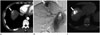
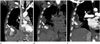
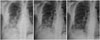
A 60-year-old male patient was referred to our hospital from an outside hospital due to refractory pleural effusion and hemoptysis that had begun six weeks before his referral. The patient was a hepatitis B carrier who had undergone neoadjuvant TACE combined with left lobectomy at our hospital eight years ago due to a huge HCC in the left lobe of the liver. Regular surveillance for possible recurrence had been performed several times. The patient remained free from recurrence for the next seven years. However, six months before he was re-admitted on this occasion, CT revealed a recurrent mass in the right liver dome measuring at 2.9 cm in diameter, demonstrating typical radiological features of HCC (Fig. 1A). At the time, TACE was repeated using an emulsion of doxorubicin and Lipiodol (ethiodized oil; Guerbet, Aulnay-Sous-Bois, France) followed by gelatin sponge particles suspended in contrast media. They were administered via a 2.0-Fr microcatheter (Progreat, Terumo, Tokyo, Japan) after superselection of the feeding arteries of the mass (Fig. 1B). Chemoembolization was not performed in any extrahepatic arteries because no extrahepatic collaterals was revealed on angiography. Cone-beam CT scan acquired immediately after TACE demonstrated compact uptake of Lipiodol within the tumor surrounded by oily portograms (Fig. 1C). Other than transient fever that spontaneously subsided after a few days, the remainder of the in-hospital period was uneventful for the patient. He was discharged without further problems. Three months after TACE, the patient visited a local clinic complaining of fever, cough, and blood-tinged sputum. Therefore, the patient underwent conservative management. Plain radiography of his chest revealed loculated pleural effusion above the right diaphragm. Chest CT was performed to disclose an area of infarcted liver around the Lipiodol-laden mass in the liver dome and focal herniation of Lipiodol through the right diaphragm (Fig. 2A). The patient's blood cell counts and routine blood chemistry were unremarkable. The only serologic abnormality was that serum C-reactive protein level was elevated to 4.61 mg/dL. In general, the patient was clinically stable. Underlying co-morbidities associated with liver cirrhosis restricted the patient from undergoing surgical treatment. As a result, the patient underwent intensive antibiotic therapy. Follow-up chest CT was performed sequentially four and five weeks later which showed progressive migration of the lump of Lipiodol until it had completely evacuated into the pleural cavity to become enclosed within an empyema (Fig. 2B, C). However, there was no residual Lipiodol in the liver. Only a region of low attenuation indicating where the Lipiodol accumulation once had been. No viable tumor was demonstrated either within the liver or in the pleural cavity on arterial phase of contrast-enhanced CT. A drainage catheter was inserted into the empyema via percutaneous approach through which turbid and yellowish material was drained. After further medical treatment and serial follow-up of chest radiographs, the patient's symptoms regressed and the empyema was found to progressively resolved (Fig. 3). The drainage catheter was finally removed ten days after its insertion. Cytology of the pleural fluid was negative for malignancy. Bronchoscopy was performed to exclude other possible causes of hemoptysis. It only revealed old blood clots within the airway. Bronchial wash cytology did not reveal any evidence of malignancy. Positron emission tomography (PET)-CT disclosed no evidence of viable tumor in the chest or the liver. However, it did reveal distant metastasis to the right supraclavicular and diaphragmatic lymph nodes. Following full symptomatic recovery, the patient was transferred back to the local clinic for palliative care. He has not returned since being discharged four months prior to the preparation of this report.
DISCUSSION
Various complications associated with TACE have been reported in the literature (8, 9). A frequent complication of TACE is postembolization syndrome (3), which encompasses symptoms such as abdominal pain, nausea, and vomiting. Such symptoms are usually self-limiting. They will subside with conservative management. Irreversible liver failure is a more severe complication of TACE (4) that may occur in the presence of portal vein thrombosis or after extensive embolization of a large volume of the liver. Other severe complications include ischemic bile duct injury (6), liver infarction, and intrahepatic abscess (5). As an extrahepatic complication of TACE, pulmonary oil embolism (7) has frequently been reported to occur secondary to shunting of the ethiodized oil in the hepatic arteries into the systemic circulation, particularly when a large dose of ethiodized oil is administered. The current case of pleural empyema is, in a sense, a pulmonary complication of chemoembolization not related to pulmonary embolism. The plausible explanation for migration of intrahepatic Lipiodol into the pleural cavity in our case could be infarction around the HCC in the liver dome after chemoembolization that resulted in the rupture of the liver capsule. Even though chemoembolization was not performed via the right inferior phrenic artery, we postulate that infarction of the diaphragm abutting the HCC may have occurred due to the shunting of the chemoembolic agent through the peripheral anastomoses between the right hepatic artery and the right inferior phrenic artery. As a result, the Lipiodol lump is thought to have herniated through the diaphragmatic defect into the ipsilateral pleural cavity. The resulting presence of chemoembolic material in the pleural space most probably triggered an inflammatory reaction that created an empyema.
With regards to treatment, surgical debridement and repair of the ruptured diaphragm was initially considered for our patient. This would have revealed the nature of the Lipiodol lump in the pleural cavity and histological information, especially whether seeding of HCC had occurred. Unfortunately, histological confirmation was not available in this case because a decision was reached to treat the patient medically and drain the empyema by percutaneous approach. Fortunately, such treatment sufficed in treating the empyema. No further complication developed in either the chest or the abdomen. There was no evidence to suggest viable tumor in the chest or liver on cytology of the pleural fluid and PET-CT.
Transdiaphragmatic herniation and consequent evacuation of ethiodized oil into the pleural cavity is an extremely rare complication of TACE. To our knowledge, only one similar case has been reported previously in the literature (10). Although rare, the possible development of liver abscess with or without diaphragm rupture should be considered when a patient develops pleural effusion or empyema after undergoing chemoembolization.
 XML Download