

 PDF
PDF ePub
ePub Citation
 Print
Print


Abstract
Pericoccygeal epidermoid cyst is a rare benign congenital lesion lined with keratinized squamous epithelium. We report the magnetic resonance imaging findings of an epidermoid cyst at the precoccygeal tip as a cause of coccygodynia in a 32-year-old woman and a retrococcygeal epidermoid cyst in a 27-year-old man. We also describe the pericoccygeal lesions and coccygodynia.
Figures and Tables
Fig. 1
A 32-year-old woman with a precoccygeal epidermoid cyst.
A. Coccyx plain lateral view shows bony erosion (arrows) at the coccygeal tip.
B, C. Enhanced CT scan (B) reveals a well defined nonenhanced mass with thin wall, anterior to the coccyx (arrows). Bone setting CT scan (C) demonstrates bony erosion at the coccyx (arrows).
D. Sagittal T2 weighted magnetic resonance image shows a well defined, heterogeneous hypointense signal mass (arrow) with a low signal rim between rectum and coccygeal tip.
E. On postcontrast fat suppressed T1-weighted sagittal image, this mass shows no remarkable enhancement (arrow).
F. Photomicrograph shows some keratinous materials lined by stratified squamous epithelium with a well-formed granular layer (H&E, × 100).
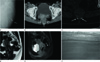
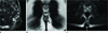
Fig. 2
Magnetic resonance imaging in a 27-year-old man with a retrococcygeal epidermoid cyst.
A. Sagittal T1-weighted image (WI) shows a well defined soft tissue mass (arrow) with a slightly increased signal intensity to surrounding muscle in the retrococcygeal region.
B. This mass (arrow) is located in the subcutaneous region of the intergluteal fold and shows a heterogeneous intermediate to high signal intensity with a high signal rim on coronal T2WI.
C. Postcontrast fat suppressed T1-weighted axial image demonstrates no remarkable enhancement (arrow) of this mass, suggesting cystic lesion.
References
1. Chin SC, Chen CY, Zimmerman RA. Pericoccygeal hidrocystoma. AJNR Am J Neuroradiol. 1998; 19:587–588.
2. Chen ML, Su JM, Cheng YM, Chou CY, Kuo PL. Presacral epidermoid cyst with right hydronephrosis. Taiwan J Obstet Gynecol. 2006; 45:155–158.
3. Yang DM, Yoon MH, Kim HS, Kim HS, Chung HS, Chung JW, et al. CT and MR findings of presacral epidermoid cyst: a case report. J Korean Radiol Soc. 1999; 41:545–547.
4. Kocaoglu M, Frush DP. Pediatric presacral masses. Radiographics. 2006; 26:833–857.
5. Harrist TJ, Gang DL, Kleinman GM, Mihm MC Jr, Hendren WH. Unusual sacrococcygeal embryologic malformations with cutaneous manifestations. Arch Dermatol. 1982; 118:643–648.
6. Yang DM, Yoon MH, Kim HS, Oh YH, Ha SY, Oh JH, et al. Presacral epidermoid cyst: imaging findings with histopathologic correlation. Abdom Imaging. 2001; 26:79–82.
7. Jaiswal A, Shetty AP, Rajasekaran S. Precoccygeal epidermal inclusion cyst presenting as coccygodynia. Singapore Med J. 2008; 49:e212–e214.
8. Nathan ST, Fisher BE, Roberts CS. Coccydynia: a review of pathoanatomy, aetiology, treatment and outcome. J Bone Joint Surg Br. 2010; 92:1622–1627.
9. Patijn J, Janssen M, Hayek S, Mekhail N, Van Zundert J, van Kleef M. 14. Coccygodynia. Pain Pract. 2010; 10:554–559.
10. Ueda K, Tsunoda A, Nakamura A, Kobayashi H, Shimizu Y, Kusano M, et al. Presacral epidermoid cyst: report of a case. Surg Today. 1998; 28:665–668.
 XML Download