

 PDF
PDF ePub
ePub Citation
 Print
Print


Amyloidoma or amyloid tumor is a localized form of amyloid deposit limited to a single organ or tissue without involvement of any other site in the body presenting as a tumorous mass (1). It can involve any anatomic sites including head and neck, orbit, breast, mediastinum, heart, liver and nervous system (1). However, localized involvement of retroperitoneal lymph nodes is a rare manifestation of this disease. Only a few cases of retroperitoneal amyloidoma involving lymph nodes have been reported (234).
We experienced a case of retroperitoneal amyloidoma that represented extensive masses in the retroperitoneum mimicking lymphoma, other lymphoproliferative disease or metastatic disease. For this report, we aimed to describe radiologic and pathologic findings of this case.
Case Report
A 56-year-old man visited our institution for evaluation of abdominal masses that had incidentally been found one month previously. He had underlying hypertension, but no other health problems including chronic inflammatory conditions. Family history was negative. On physical examination, the masses were palpable as hard and smooth objects; mild tenderness was elicited. Laboratory studies revealed the following values: white blood cell count, 4.8×10³/mm3; hemoglobin, 14.8 g/dL; hematocrit, 43.5%; platelet count, 265,000/mm; differential leukocyte count, lymphocyte-28.7%, monocyte-7.5%, eosinophil-1.7%; total protein, 6.5 g/dL; albumin, 3.9 g/dl. Tumor markers including CEA, CA19-9 and PSA were within normal limits.
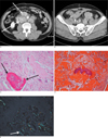
We performed abdominal computed tomography (CT) with contrast enhancement. There were multiple con glomerated nodular masses in the retroperitoneum from the level of the kidneys to the pelvic cavity with surrounding infiltration (Figs. 1A, B). The masses encased the aorta and inferior vena cava. The attenuation of the masses was similar or higher than that of muscle. Some of the masses had punctuate and amorphous calcifications.
A surgeon did an explorative laparotomy to determine the histologic diagnosis of the retroperitoneal masses. Pathology returned a diagnosis of 'amyloid tumor' in the retroperitoneum.
Gross pathologic examination showed a well demarcated and encapsulated lobular mass with areas of calcification on the cut surface. Microscopic examination revealed extensive amyloid deposits within the masses and walls of the blood vessels (Fig. 1C). On Masson's trichrome stain and Congo red stain (Fig. 1D), the amyloid nature of the deposits was identified by the applegreen birefringence under polarizing microscopy (Fig. 1E). There was no sensitivity to potassium permanganate, suggesting an AL type of amyloidosis.
The patient underwent a follow up CT scan three years after the initial diagnosis. The retroperitoneal masses had increased in size and number as compared with the previous study. He had not received any treatment in the interim; subsequently, he was lost to follow-up.
Discussion
Amyloidosis is a heterogeneous group of diseases characterized by the deposition of a pathologic proteinaceous substance, 'amyloid' (56). The mechanism and biological behavior of this amyloid deposit has not been clear until recently.
Classically, amyloidosis is categorized as either a secondary process that is associated with chronic infections or inflammatory disorders, or a primary process in the absence of such associations (5). Histologically, this entity is classified as type 'AA'(serum protein A) or type 'AL' (immunoglobulin light chains) based on the chemical type that forms the fibrils (5). Amyloidosis can also be subdivided clinically into a systemic or localized form (5). The above classifications can be summarized as follows: a) primary amyloidosis with no pre-existing disease except multiple myeloma or other plasma cell dyscrasia; b) secondary amyloidosis with co-existent underlying infective or neoplastic pathology; c) focal single organ involvement without any systemic involvement; d) familial; e) senile (7).
Amyloidoma or amyloid tumor is a localized or isolated form of amyloidosis that is frequently associated with immunocystic dyscrasia, long-term hemodialysis, and chronic infectious or inflammatory diseases (17). In some cases, clinical evidence of preexisting disease may be absent (1). Our patient had no preexisting conditions either. Therefore, our case would be amyloidosis of the primary form. Clinically, amyloidoma can cause localized signs and symptoms (5). Its diagnosis should be confirmed through biopsy. Grossly, amyloid tumors are represented by lobulated nodules or masses with whiteyellow or pink-yellow waxy cut surfaces (1). On immunohistochemical examination, they demonstrate characteristic apple-green birefringence with polarized light and positive Congo red staining. The majority of cases are composed of type AL (immunoglobulin light chain) amyloid (5).
The retroperitoneum is a rare site of amyloid involvement. To our knowledge, only a few cases have been reported (234). Retroperitoneal involvement of amyloidosis can be subdivided into infiltrative or nodal mass lesions (23). An infiltrative lesion implies that there is retroperitoneal fibrosis by amyloid deposition in the retroperitoneal fat tissue. On the other hand, a nodal mass lesion represents involvement of a retroperitoneal lymph node (310). Previous cases in the literature regarding retroperitoneal lymph node involvement usually reported calcified nodules or masses with inhomogeneous enhancement (24). Amyloidomas in any site may have calcification with an indolent growth pattern of the masses (458). However, their clinical significance or molecular etiology is unknown (8). The CT appearance of calcifications within amyloidomas has been characterized as showing various patterns, including speckled, punctate or eggshell (24).
The differential diagnosis includes lymphoma, Castleman's disease, and metastatic lymphadenopathy. In patients with lymphoma, the masses usually show homogeneous attenuation and rarely are accompanied by calcifications (9). In Castleman's disease, vivid enhancement of nodal masses is a characteristic feature (10). Although features of metastatic lymph nodes can be varied, soft tissue attenuation and homogeneous enhancement are usually noted.
Although pathognomonic radiologic findings of this rare disease have not been definitively established, an inhomogeneous enhancement pattern with amorphous calcifications may be a clue that differentiates it from other mimicking diseases.
 XML Download