Journal List > J Korean Med Sci > v.34(Suppl 1) > 1126518

Park, Shin, Kim, Song, Jung, Lee, You, Lim, Seo, Kim, Zo, Kim, and on behalf of Korean Cardiac Arrest Research Consortium (KoCARC) Investigators: Clinical Factors Associated with Obstructive Coronary Artery Disease in Patients with Out-of-Hospital Cardiac Arrest: Data from the Korean Cardiac Arrest Research Consortium (KoCARC) Registry

Abstract

Background

Although coronary artery disease (CAD) is a major cause of out-of-hospital cardiac arrest (OHCA), there has been no convinced data on the necessity of routine invasive coronary angiography (ICA) in OHCA. We investigated clinical factors associated with obstructive CAD in OHCA.

Methods

Data from 516 OHCA patients (mean age 58 years, 83% men) who underwent ICA after resuscitation was obtained from a nation-wide OHCA registry. Obstructive CAD was defined as the lesions with diameter stenosis ≥ 50% on ICA. Independent clinical predictors for obstructive CAD were evaluated using multiple logistic regression analysis, and their prediction performance was compared using area under the receiver operating characteristic curve with 10,000 repeated random permutations.

Results

Among study patients, 254 (49%) had obstructive CAD. Those with obstructive CAD were older (61 vs. 55 years, P < 0.001) and had higher prevalence of hypertension (54% vs. 36%, P < 0.001), diabetes mellitus (29% vs. 21%, P = 0.032), positive cardiac enzyme (84% vs. 74%, P = 0.010) and initial shockable rhythm (70% vs. 61%, P = 0.033). In multiple logistic regression analysis, old age (≥ 60 years) (odds ratio [OR], 2.01; 95% confidence interval [CI], 1.36–3.00; P = 0.001), hypertension (OR, 1.74; 95% CI, 1.18–2.57; P = 0.005), positive cardiac enzyme (OR, 1.72; 95% CI, 1.09–2.70; P = 0.019), and initial shockable rhythm (OR, 1.71; 95% CI, 1.16–2.54; P = 0.007) were associated with obstructive CAD. Prediction ability for obstructive CAD increased proportionally when these 4 factors were sequentially combined (P < 0.001).

Conclusion

In patients with OHCA, those with old age, hypertension, positive cardiac enzyme and initial shockable rhythm were associated with obstructive CAD. Early ICA should be considered in these patients.

Graphical Abstract

jkms-34-e159-abf001

INTRODUCTION

The out-of-hospital cardiac arrest (OHCA) is a global health problem with prevalence ranging from 50 to 90 cases per 100,000 person-years worldwide.1 Although the major advance has been achieved in the emergency medical service system and cardiac arrest resuscitation techniques, the overall survival rate in patients with OHCA is still low, especially in Asians.123 Among various etiologies of OHCA, coronary artery disease (CAD) has been reported as the most common cause of OHCA, and over 50% of OHCA patients without definite evidence for non-cardiac causes had significant CAD.456789 Numerous studies have attempted to assess the clinical benefit of invasive coronary angiography (ICA) with coronary revascularization after resuscitation; however, their results were inconsistent.2567101112 Although the current guideline recommended ICA for those with ST-segment elevation on the post-resuscitation electrocardiogram (ECG),13 the decision on ICA based on ST-segment changes in post-resuscitation ECG has limitations because of a low diagnostic yield of ECG.141516 In addition, clinical presentation is similar between patients with arrest due to hemorrhagic stroke and those with cardiac arrest. In such cases, physicians sometimes performed unnecessary ICA and antithrombotic treatment, which could delay accurate diagnosis and lead to deleterious outcome.1718 Moreover, due to its invasiveness, patients would be exposed to additional complication risk following ICA such as vascular injury, hemorrhage, myocardial ischemia, infection or event death.19 On the contrary, delaying ICA can be fatal in OHCA patients who have a significant stenosis or occlusion of the coronary artery by prohibiting maximal benefit from life-saving coronary revascularization. Therefore, it is very important to identify clinical factors guiding physicians to perform appropriate ICA on OHCA patients. The aim of the current study was to investigate clinical factors related to obstructive CAD among OHCA patients at initial presentation.

METHODS

Study subjects

Study subjects and clinical data were obtained from the database of The Korean Cardiac Arrest Research Consortium (KoCARC) registry. The KoCARC is a collaborative research network organized in 2014 which consists of seven research committees including Epidemiology and Preventive Research Committee (EPR), Community Resuscitation Research Committee (CRR), EMS Resuscitation Research Committee (ERR), Hospital Resuscitation Research Committee (HRR), Hypothermia and Post-resuscitation Care Research Committee (HPR), Cardiac Care Resuscitation Committee (CCR) and Pediatric Resuscitation Research Committee (PRR). The KoCARC has administrated multicenter registry database in the field of OHCA resuscitation (NCT03222999). The following conditions were excluded from the registry enrollment: 1) those with terminal illness, 2) those under hospice care, 3) those in pregnancy, 4) those with pre-documented ‘Do Not Resuscitate (DNR)’ card, 5) those who disagreed for registration and 6) those with definite non-cardiac cause comprising trauma, drowning, poisoning, burn, asphyxia or hanging. From October 2015 to June 2017, a total of 4,757 OHCA patients who were transported to 32 participating emergency departments with resuscitation efforts were registered. For the current study, 1,523 (32.0%) patients achieved a return of spontaneous circulation; of these patients, 516 (33.9%) underwent ICA during hospitalization.

Data collection

Clinical characteristics, including age and gender as well as the history of cardiovascular risk factors such as hypertension, diabetes mellitus, dyslipidemia, current smoking and family history of CAD were acquired. Hypertension was defined as previous history of hypertension or use of antihypertensive medication. Diabetes mellitus was defined as prior history of diabetes mellitus or use of antidiabetic medication. Dyslipidemia was also defined as previous history of dyslipidemia or anti-dyslipidemic medication. Current smoking was defined as regular smoking habit in the past 12 months. Patients with family members who were diagnosed for CAD with or without coronary revascularization were defined as positive family history of CAD. Data on resuscitation comprising bystander cardiopulmonary resuscitation (CPR), bystander application of automated external defibrillator (AED) and time duration of CPR was also obtained. Initial ECG was reviewed for the presence of shockable rhythm (ventricular fibrillation or pulseless ventricular tachycardia) or non-shockable rhythm (pulseless electrical activity or asystole). A positive cardiac enzyme was defined based on initial laboratory data on cardiac enzymes such as creatine kinase-MB (CK-MB) (> 5 ng/mL) or cardiac troponin I (> 0.028 ng/mL). Data on the treatment after ICA including percutaneous coronary angiography (PCI), coronary artery bypass graft surgery (CABG), intra-aortic balloon pump (IABP), implantable cardioverter defibrillator (ICD), and extracorporeal membrane oxygenation (ECMO) was also obtained.

ICA

The severity of coronary artery stenosis and the extent of CAD such as multi-vessel disease or left main CAD were evaluated at the time of ICA. Further treatment strategies such as revascularization and medical treatment were determined by attending physicians' preference based on the ICA results. Obstructive CAD was defined as the lesion with % diameter stenosis ≥ 50% in 1 or more major epicardial coronary arteries.

Statistical analysis

Baseline characteristics, including demographic information, cardiovascular risk factors, information on cardiac resuscitation, laboratory results and post-ICA treatment, were compared between patients according to ICA and the presence of obstructive CAD. Continuous variables are presented as mean with standard deviation or median with interquartile range, and were compared using Student's t-test for parametric variables or Mann–Whitney U test for non-parametric variables. Categorical variables are presented as numbers with percentage and were compared by the χ2 test. To investigate independent factors associated with obstructive CAD, multivariable logistic regression analysis was performed. The following potential confounders were controlled in the multivariable analysis: age, gender, hypertension, diabetes mellitus, dyslipidemia, current smoking, family history of CAD, positive result of initial cardiac enzyme and presence of shockable rhythm at initial ECG. To assess the discrimination ability of clinical information on obstructive CAD, 4 prediction models were constructed. Baseline prediction model (model 1) was derived from age and gender. Next, model 2 was designed by combining cardiovascular risk factors (model 1 + hypertension + diabetes mellitus + dyslipidemia + current smoking + family history of CAD). Then, model 3 was derived by combining positive results of the cardiac enzyme (model 2 + positive cardiac enzyme). The final model (model 4) included the results of initial ECG (model 3 + initial shockable rhythm). The discriminatory ability of each prediction model was assessed using the area under the receiver operator characteristics curve (AUC) with 10,000 permutation resampling method. The distributions of estimated AUC for incremental models from models 1 to 4 were compared. To evaluate the association of ICA with long-term prognostic benefit among OHCA patients, 1-year cumulative risk of death was assessed for patients who underwent ICA (n = 516) and those who did not (n = 1,007) after resuscitation by Kaplan-Meier survival analysis and compared using Cox proportional hazard model adjusting baseline characteristics. All analyses were performed 2-sided, and a P value of < 0.05 was considered statistically significant. All data was analyzed using SPSS version 21.0 for Windows (IBM, Armonk, NY, USA) and R, version 3.5.3 (R Development Core Team, Vienna, Austria).

Ethics statement

The study protocol complied with the Declaration of Helsinki, and was reviewed and approved by the Institutional Review Board (IRB) of the Seoul Metropolitan Government-Seoul National University Boramae Medical Center (IRB No. 16-2014-32). The study was waived for informed consent by the IRB.

RESULTS

Clinical characteristics of the study patients

Of 4,757 OHCA patients, 516 underwent ICA, and 254 (49%) of them had obstructive CAD. Table 1 shows clinical characteristics of patients according to ICA and those with and without obstructive CAD. Patients who underwent ICA were younger (58 vs. 66 years, P < 0.001), had a higher proportion of men, and had higher prevalence of hypertension (45% vs. 39%, P < 0.001), dyslipidemia (7% vs. 4%, P = 0.008), current smoking (32% vs. 10%, P < 0.001), and family history of CAD (4% vs. 1%, P < 0.001). Regarding resuscitation, ICA group had a higher proportion of bystander CPR (56% vs. 43%, P < 0.001), bystander AED (2.1% vs. 0.4%, P < 0.001) and shorter duration of CPR. At initial presentation, those who underwent ICA showed higher rates of positive cardiac enzymes (79% vs. 46%, P < 0.001) and shockable rhythm on ECG (66% vs. 12%, P < 0.001). Comparisons of clinical characteristics between patients with and without obstructive CAD is also demonstrated in Table 1. Patients with obstructive CAD were older (61 vs. 55 years, P < 0.001), and had higher prevalence of hypertension (54% vs. 36%, P < 0.001) and diabetes mellitus (29% vs. 21%, P = 0.032) than those without. There was no difference in the proportion of bystander CPR, bystander AED and duration of CPR between the 2 groups. Compared to patients without obstructive CAD, those with obstructive CAD showed higher rates of having positive cardiac enzymes (84% vs. 74%, P = 0.010) and shockable rhythm at initial ECG (70% vs. 61%, P = 0.033). Among the patients with obstructive CAD, 58.3% (n = 148), 5.5% (n = 14), 4.7% (n = 12), 1.6% (n = 4), and 7.9% (n = 20) received treatment with PCI, CABG, IABP, ICD, and ECMO, respectively. Those with obstructive CAD showed a significantly higher rate of PCI (58% vs. 10%, P < 0.001) and IABP (5% vs. 2%, P = 0.018), whereas a lower rate of ICD insertion (2% vs. 11%, P < 0.001) compared to those without.
Table 1

Clinical characteristics of study patients

jkms-34-e159-i001
Characteristics Overall (n = 4,757) With ICA (n = 516) Without ICA (n = 4,241) P value Obstructive CAD (n = 254) No obstructive CAD (n = 262) P value
Demographics
Age, yr 65.4 ± 18.9 57.9 ± 14.2 66.3 ± 19.1 < 0.001 60.8 ± 12.6 55.1 ± 15.2 < 0.001
Age ≥ 60 yr 3,183 (66.9) 243 (47.1) 2,940 (69.3) < 0.001 142 (55.9) 101 (38.5) < 0.001
Men 3,088 (66.9) 430 (83.3) 2,658 (62.7) < 0.001 217 (85.4) 213 (81.3) 0.238
Cardiovascular risk factors
Hypertension 1,879 (39.5) 230 (44.6) 1,649 (38.9) 0.013 136 (53.5) 94 (35.9) < 0.001
Diabetes mellitus 1,159 (24.4) 128 (24.8) 1,031 (24.3) 0.828 74 (29.1) 54 (20.6) 0.032
Dyslipidemia 223 (4.7) 37 (7.2) 186 (4.4) 0.008 24 (9.4) 13 (5.0) 0.060
Current smoking 608 (12.8) 165 (32.0) 443 (10.4) < 0.001 86 (33.9) 79 (30.2) 0.396
Family history of CAD 83 (1.7) 22 (4.3) 61 (1.4) < 0.001 13 (5.1) 9 (3.4) 0.389
CPR event
Bystander CPR 2,094 (44.0) 289 (56.0) 1,805 (42.6) < 0.001 144 (56.7) 145 (55.3) 0.790
Bystander AED 28 (0.6) 11 (2.1) 17 (0.4) < 0.001 5 (2.4) 5 (1.9) 0.769
CPR over 20 min 1,972 (41.5) 89 (17.2) 1,883 (44.4) < 0.001 47 (18.5) 42 (16.0) 0.486
CAD extent
Left main disease 4 (1.5) 4 (1.5) - - 4 (1.5) - -
One-vessel disease 101 (39.8) 101 (39.8) - - 101 (39.8) - -
Two-vessel disease 65 (25.6) 65 (25.6) - - 65 (25.6) - -
Three-vessel disease 84 (33.1) 84 (33.1) - - 84 (33.1) - -
Initial cardiac enzyme
CK-MB, ng/mL 4.40 (2.20–10.09) 3.8 (2.00–7.90) 4.52 (2.20–10.76) 0.003 3.86 (2.02–7.78) 3.72 (2.00–8.11) 0.789
Cardiac troponin I, ng/mL 1.03 (0.19–5.55) 0.81 (0.15–6.00) 1.06 (0.19–5.30) 0.576 0.23 (0.06–1.04) 0.19 (0.04–2.28) 0.504
Positive cardiac enzyme 2,336 (49.1) 406 (78.7) 1,930 (45.5) < 0.001 212 (83.5) 194 (74.0) 0.010
Initial ECG rhythm < 0.001 0.033
Shockable 845 (17.8) 338 (66.0) 507 (12.1) 178 (70.1) 160 (61.1)
Non-shockable 3,841 (80.7) 174 (34.0) 3,667 (87.9) 76 (29.9) 102 (38.9)
Post CAG care
PCI 173 (3.64) 173 (33.5) - - 148 (58.3) 25 (9.5) < 0.001
CABG 21 (0.4) 20 (3.9) 1 (0.0) < 0.001 14 (5.5) 6 (2.3) 0.069
IABP 16 (0.3) 15 (2.9) 1 (0.0) < 0.001 12 (4.7) 3 (1.1) 0.018
ICD insertion 40 (0.8) 33 (6.4) 7 (0.2) < 0.001 4 (1.6) 29 (11.1) < 0.001
ECMO 48 (1.0) 37 (7.2) 11 (0.3) < 0.001 20 (7.9) 17 (6.5) 0.610
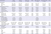
Values are given as mean ± standard deviation, median (interquartile range, 25th and 75th percentile) or number (%) unless otherwise indicated.
ICA = invasive coronary angiography, CAD = coronary artery disease, CPR = cardiopulmonary resuscitation, AED = automated external defibrillator, CK-MB = creatine kinase-MB, ECG = electrocardiogram, PCI = percutaneous coronary intervention, CABG = coronary artery bypass graft surgery, IABP = intra-aortic balloon pump, ICD = implantable cardioverter defibrillator, ECMO = extracorporeal membrane oxygenation.

Independent risk factors associated with obstructive CAD

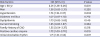
Independent risk factors associated with obstructive CAD are shown in Table 2. In binary multiple logistic regression analysis, older age (≥ 60 years) (odds ratio [OR], 2.01; 95% confidence interval [CI], 1.36–3.00; P = 0.001), hypertension (OR, 1.74; 95% CI, 1.18–2.57; P = 0.005), positive cardiac enzyme (OR, 1.72; 95% CI, 1.09–2.70; P = 0.019) and initial shockable rhythm (OR, 1.71; 95% CI, 1.16–2.54; P = 0.007) were associated with obstructive CAD even after controlling for potential confounders.
Table 2

Independent risk factors associated with obstructive CAD

jkms-34-e159-i002
Risk factors OR (95% CI) P value
Age ≥ 60 yr 2.01 (1.36–3.00) 0.001
Men 1.38 (0.83–2.31) 0.220
Hypertension 1.74 (1.18–2.57) 0.005
Diabetes mellitus 1.07 (0.67–1.70) 0.772
Dyslipidemia 1.73 (0.82–3.63) 0.149
Current Smoking 1.25 (0.83–1.87) 0.284
Family history of CAD 1.35 (0.54–3.37) 0.516
Positive cardiac enzyme 1.72 (1.09–2.70) 0.019
Initial shockable rhythm 1.71 (1.16–2.54) 0.007
CAD = coronary artery disease, OR = odds ratio, CI = confidence interval.

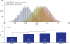
Four different prediction models for obstructive CAD

Table 3 and Fig. 1 present the discrimination ability of prediction models from the various combination of clinical information. Model 1 showed modest performance in prediction for obstructive CAD (AUC, 0.601; 95% CI, 0.600–0.602). The discriminant ability was improved when cardiovascular risk factors were added to model 1 (“model 2”) (AUC, 0.601 to 0.658; P < 0.001). Model 2 with cardiac enzyme (“model 3”) also showed significantly improved discriminant ability compared to model 2 (AUC, 0.658 to 0.673; P < 0.001). When information on initial shockable rhythm was combined to model 3 (“model 4”), the highest performance of discrimination was found (AUC, 0.673 to 0.687; P < 0.001).
Table 3

Prediction models for obstructive CAD

jkms-34-e159-i003
Model AUC 95% CI
Model 1: Age + gender 0.601 0.600–0.602
Model 2: Model 1 + cardiovascular risk factors 0.658 0.657–0.659
Model 3: Model 2 + cardiac enzyme 0.673 0.672–0.674
Model 4: Model 3 + shockable rhythm 0.687 0.686–0.689
CAD = coronary artery disease, AUC = area under the curve, CI = confidence interval.
Fig. 1

Comparison of the AUC of prediction model for obstructive coronary artery disease.

AUC = area under the curve, CI = confidence interval.
jkms-34-e159-g001

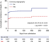
Prognostic impact of ICA among OHCA patients

OHCA patients who underwent ICA was associated with a significantly lower rate of 1-year mortality compared to those who did not (log-rank P < 0.001, Fig. 2). After adjusting baseline characteristics, ICA group consistently showed a lower risk of mortality (adjusted hazard ratio, 0.72; 95% CI, 0.57–0.91).
Fig. 2

Comparison of 1-year mortality according to ICA among patients with OHCA.

HR = hazard ratio, ICA = invasive coronary angiography, OHCA = out-of-hospital cardiac arrest.
jkms-34-e159-g002

DISCUSSION

The present study showed that old age, hypertension, positive cardiac enzyme and shockable rhythm at initial ECG were independently related to higher risk of obstructive CAD in patients with ROSC after OHCA. Our study also demonstrated incremental prediction abilities for obstructive CAD when these 4 factors were sequentially added.

The role of ICA in patients with OHCA

As CAD is the most common cause of OHCA, previous studies have assessed the clinical benefit of early ICA on survival or good neurologic outcome among patients after OHCA.25671112 Although some studies have demonstrated the prognostic benefit of early ICA in patients after OHCA,256712 others showed no benefit of early invasive approaches.11 As previous studies were not randomized but retrospectively designed, discordance among studies could be attributed to different baseline characteristics. Indeed, as the prevalence of obstructive CAD was higher in studies showing a positive prognostic impact of ICA,56 compared to those presenting no benefit of ICA in OHCA patients.11 This implies that immediate ICA for patients who are highly suspected for obstructive CAD will maximize the prognostic benefit of ICA. However, imprudent application of early ICA after OHCA may increase not only false positive test results but also delay proper differential diagnosis.20 Moreover, ICA is sometimes associated with an increased risk of fatal bleeding following unnecessary antithrombotic treatment,172122 and its invasiveness increases the mortality rate.18 Therefore, appropriate discrimination of patients who have obstructive CAD is also essential to minimize the potential risk of early ICA among OCHA patients. In our study results, OHCA patients who underwent ICA showed a significantly lower rate of 1-year mortality. Also, we found 4 clinical factors associated with obstructive CAD in patients with OHCA, which can be obtained at early phases on initial presentation. Identification of these 4 factors may have prognostic values in augmenting clinical benefit of ICA among patients with OHCA.

Traditional risk factors and ECG changes

Traditional cardiovascular risk factors and ECG changes can be clues to obstructive CAD in patients with OHCA. It has been suggested that patients with acute coronary syndrome (ACS) or obstructive CAD tend to have higher rate of cardiovascular risk factors compared to those without.23 ST segment changes in post-resuscitation ECG have been widely adopted as a guideline for deciding coronary revascularization,13 although several studies have raised questions for its value in prediction.141516 Abe et al.24 reported a prediction model for identifying patients who have angiographically normal coronary arteries after OHCA by retrospectively analyzing 47 consecutive adult survivors who underwent immediate ICA after resuscitation. In that study, patients without diabetes mellitus, previous history of ACS and ST-segment abnormalities were associated with normal coronary arteries. Higny et al.9 demonstrated clinical factors related to significant CAD in patients without ST-segment elevation after OHCA, which were the previous history of CAD, and the presence of cardiovascular risk factors including diabetes mellitus and dyslipidemia. In line with those studies, our results also showed that, among cardiovascular risk factors, old age and the presence of hypertension were independently associated with obstructive CAD in OHCA patients. Previously, patients at younger age were reported to be related to better neurologic and functional outcome after OHCA, which may have been attributed to the relatively lower burden of cardiovascular risk factors.2526 We suggest a possibility that poor prognoses in elderly patients with OHCA could be partially due to a lower rate of ICA,27 despite higher risk of obstructive CAD as in our study results. Recently, Legriel et al.28 indicated the benefit of early ICA in elderly patients after OHCA, suggesting that elderly patients should be more optimally managed with appropriate treatment.
Hypertension is one of the traditional cardiovascular risk factors. Compared to those without hypertension, patients with hypertension were related to increased prevalence and severity of CAD including extent and calcification, and were associated with higher risk of adverse cardiovascular events.2930 Also, previous studies have reported hypertension as an independent risk factor for OHCA among patients with CAD as well as poor in-hospital and subsequent long-term outcome after resuscitation.3132 Although the pathophysiologic association between hypertension and OHCA in patients with CAD is not completely understood, several explanations have been proposed.32 Hypertension leads to myocardial hypertrophy which augments myocardial ischemia caused by obstructive CAD as coronary arteries fail to grow with a sufficient rate to supply hypertrophic myocardium.33 Additionally, hypertrophic myocardium is arrhythmogenic compared to normal myocardium, which is more vulnerable to ectopic trigger leading to lethal arrythmias.34 Increased activity of sympathetic nerves and the renin-aldosterone system in hypertensive status could also involve the arrhythmogenic condition of the myocardium.35

Initial shockable rhythm

Patients with initial shockable rhythm have considered to be associated with higher risk of myocardial infarction (MI) as an origin for OHCA and are likely to be referred for ICA.2736 Youngquist et al.37 investigated 151 autopsy cases of OHCA patients with failed resuscitation and found that 43% of the OHCA cases were related to coronary occlusion. They also stated that old age and initial shockable rhythm were associated with CAD, which is consistent with ours. Also, previous studies of patients who had intracranial hemorrhage as a cause of OHCA found that the prevalence of shockable rhythm was significantly lower compared to those who did not.1738 Although regarding the role of shockable rhythm as a positive prognostic factor is not fully understood in MI patients, a short time to administration of medical assistance and a relatively lower burden of myocardial ischemia may contribute to better outcome in those with shockable rhythm.39 This may be supported by a previous experimental study showing that cardiac arrest secondary to severe hypoxemia or respiratory arrest causes asystole rather than shockable rhythm and severe global ventricular dysfunction.40

Clinical implications

Identifying patients with OHCA related to obstructive CAD is important for not only maximizing the clinical benefit of early coronary revascularization but also avoiding unnecessary invasive procedures. In the current study, we found 4 clinical factors associated with obstructive CAD in patients with OHCA, which included old age, hypertension, positive cardiac enzyme and initial shockable rhythm on ECG. As information on all these clinical factors can be obtained easily in a short time period at initial presentation, identification of these 4 factors may be clinically valuable in selecting patients and maximizing benefit of ICA in the setting of OHCA.

Study limitations

There are several limitations in this study. First, although we screened patients from the multicenter OHCA registry, the number of patients included for analysis is relatively small. Secondly, as this study was retrospectively designed, some concerns could be raised regarding the selection of patients who survived to undergo ICA. Therefore, it could be limited to generalize our study results for overall OHCA patients. Thirdly, the association of traditional cardiovascular risk factors with obstructive CAD may differ in other populations with a different prevalence rate of the risk factors. Finally, clinical outcome data is not available in our cross-sectional analysis.
In summary, among the patients with OHCA and ROSC, old age, hypertension, positive cardiac enzyme test and initial shockable rhythm may be associated with obstructive CAD. Early ICA should be considered in this group of patients with these risk factors.

ACKNOWLEDGMENTS

We would like to acknowledge and thank to investigators from all participating hospitals of KoCARC: Do Kyun Kim (Seoul National University Hospital), Sang Kuk Han (Kangbuk Samsung Medical Center), Phil Cho Choi (Kangbuk Samsung Medical Center), Sang O Park (Konkuk University Medical Center), Jong Won Kim (Konkuk University Medical Center), Han Sung Choi (Kyung Hee University Hospital), Jong Seok Lee (Kyung Hee University Hospital), Sung Hyuk Choi (Korea University Guro Hospital), Young Hoon Yoon (Korea University Guro Hospital), Soo Jin Kim (Korea University Anam Hospital), Min Seob Sim (Samsung Medical Center), Gun Tak Lee (Samsung Medical Center), Shin Ahn (Asan Medical Center), Sang Hyun Park (Seoul Medical Center), Keun Hong Park (Seoul Medical Center), In Cheol Park (Yonsei University Severance Hospital), Yoo Seok Park (Yonsei University Severance Hospital), Tae Young Kong (Yonsei University Gangnam Severance Hospital), Kyoung Won Lee (Inje University Seoul Paik Hospital), Chu Hyun Kim (Inje University Seoul Paik Hospital), Youngsuk Cho (Hallym University Kangdong Sacred Heart Hospital), Gu Hyun Kang (Hallym University Kangnam Sacred Heart Hospital), Yong Soo Jang (Hallym University Kangnam Sacred Heart Hospital), Tai Ho Im (Hanyang University Seoul Hospital), Jae Hoon Oh (Hanyang University Seoul Hospital), Seok Ran Yeom (Pusan National University Hospital), Sang Kyoon Han (Pusan National University Hospital), Jae Hoon Lee (Dong-A University Hospital), Jeong Bae Park (Kyungpook National University Hospital), Hyun Wook Ryoo (Kyungpook National University Hospital), Kyung Woo Lee (Daegu Catholic University Medical Center), Tae Chang Jang (Daegu Catholic University Medical Center), Jae-hyug Woo (Gachon University Gil Medical Center), Woon Jeong Lee (The Catholic University of Korea Incheon St. Mary's Hospital), Seon Hee Woo (The Catholic University of Korea Incheon St. Mary's Hospital), Sung Hyun Yun (Catholic Kwandong University International St. Mary's Hospital), Tae Jin Cho (Catholic Kwandong University International St. Mary's Hospital), Sun Pyo Kim (Chosun University Hospital), Yong Jin Park (Chosun University Hospital), Jin Woong Lee (Chungnam National University Hospital), Wonjoon Jeong (Chungnam National University Hospital), Sung Soo Park (Konyang University Hospital), Jae Kwang Lee (Konyang University Hospital), Ryeok Ahn (Ulsan University Hospital), Wook Jin Choi (Ulsan University Hospital), Young Gi Min (Ajou University Hospital), Eun Jung Park (Ajou University Hospital), You Hwan Jo (Seoul National University Bundang Hospital), Joong Hee Kim (Seoul National University Bundang Hospital), In Byung Kim (Myongji Hospital), Ki Ok Ahn (Myongji Hospital), Han Jin Cho (Korea University Ansan Hospital), Seung Cheol Lee (Dongguk University Ilsan Hospital), Sang Hun Lee (Dongguk University Ilsan Hospital), Young Sik Kim (Bundang Jesaeng Hospital), Young Rock Ha (Bundang Jesaeng Hospital), Jin Sik Park (Sejong Hospital), Myoung Woo Lee (Sejong Hospital), Dai Han Wi (Wonkwang University Sanbon Hospital), Ok Jun Kim (Cha University Bundang Medical Center), Tae Nyoung Chung (Cha University Bundang Medical Center), Soon Joo Wang (Hallym University Dongtan Sacred Heart Hospital), Hang A Park (Hallym University Dongtan Sacred Heart Hospital), Jun Hwi Cho (Kangwon National University Hospital), Chan Woo Park (Kangwon National University Hospital), An Mu Eob (Hallym University Chuncheon Sacred Heart Hospital), Tae Hun Lee (Hallym University Chuncheon Sacred Heart Hospital), Sang Chul Kim (Chungbuk National University Hospital), Hoon Kim (Chungbuk National University Hospital), Han Joo Choi (Dankook University Hospital), Chan Young Koh (Dankook University Hospital), Jung Won Lee (Soonchunhyang University Cheonan Hospital), Dong Wook Lee (Soonchunhyang University Cheonan Hospital), Tae Oh Jung (Chonbuk National University Hospital, Jae Chol Yoon (Chonbuk National University Hospital), Dai Hai Choi (Dongguk University Gyeongju Hospital), Jung Tae Choi (Dongguk University Gyeongju Hospital), Jin Hee Jeong (Gyeongsang National University Hospital), Soo Hoon Lee (Gyeongsang National University Hospital), Ji Ho Ryu (Pusan National University Yangsan Hospital), Maeng Real Park (Pusan National University Yangsan Hospital), Won Kim (Cheju Halla General Hospital), Sung Wook Song (Jeju National University Hospital), Woo Jung Kim (Jeju National University Hospital), Joon-myoung Kwon (Mediplex Sejong Hospital), Eui Hyuk Kang (Mediplex Sejong Hospital), Sang Chan Jin (Keimyung University Dongsan Medical Center), Tae-kwon Kim (Keimyung University Dongsan Medical Center), Hyuk Joong Choi (Hanyang University Guri Hospital), Seong Chun Kim (Gyeongsang National Univerisy Changwon Hospital).
And to steering committee, comprised of following individuals: Sung Oh Hwang (Chair, Wonju Severance Christian Hospital), Sang Do Shin (Chair of Steering Committee, Seoul National University Hospital), Hyuk Jun Yang (Advisory Committee, Gachon University Gil Hospital), Sung Phil Chung (Data Safety and Management Board, Gangnam Severance Hospital), Sung Woo Lee (Security and Monitoring Board, Korea University Anam Hospital), Seung Sik Hwang (Epidemiology and Prevention Research Committee, Seoul National University), Gyu Chong Cho (Community Resuscitation Research Committee, Hallym University Kangdong Sacred Heart Hospital), Sung Woo Moon (Emergency Medical Service Resuscitation Research Committee, Korea University Ansan Hospital), Kyoung Chul Cha (Hospital Resuscitation Research Committee, Wonju Severance Christian Hospital), Won Young Kim (Hypothermia and Post-Resuscitation Care Research Committee, Asan Medical Center), Sang Hoon Na (Cardiac Care Resuscitation Research Committee, Seoul National University Hospital), Young Ho Kwack (Pediatric Resuscitation Research Committee, Seoul National University Hospital).
And lastly to member of secretariat: Joo Yeong Kim (Korea University Ansan Hospital), Jeong Ho Park (Seoul National University Hospital), Sun Young Lee (Seoul National University Hospital), and Jung Eun Kim (Seoul National University Hospital).

Notes

Disclosure The authors have no potential conflict of interest to disclose.

Author Contributions

  • Conceptualization: Park J, Shin J, Kim HL.

  • Data curation: Shin J, Kim HL.

  • Formal analysis: Park J, Kim HL.

  • Investigation: Shin J, Kim HL.

  • Methodology: Park J, Shin J, Kim HL.

  • Software: Park J, Kim HL.

  • Validation: Park J, Kim HL.

  • Writing - original draft: Park J, Shin J.

  • Writing - review & editing: Park J, Shin J, Kim HL, Song KJ, Jung JH, Lee HJ, You KM, Lim WH, Seo JB, Kim SH, Zo JH, Kim MA.

References

1. Berdowski J, Berg RA, Tijssen JG, Koster RW. Global incidences of out-of-hospital cardiac arrest and survival rates: systematic review of 67 prospective studies. Resuscitation. 2010; 81(11):1479–1487.
crossref
2. Jaeger D, Dumas F, Escutnaire J, Sadoune S, Lauvray A, Elkhoury C, et al. Benefit of immediate coronary angiography after out-of-hospital cardiac arrest in France: a nationwide propensity score analysis from the RéAC Registry. Resuscitation. 2018; 126:90–97.
crossref
3. Ong ME, Shin SD, De Souza NN, Tanaka H, Nishiuchi T, Song KJ, et al. Outcomes for out-of-hospital cardiac arrests across 7 countries in Asia: The Pan Asian Resuscitation Outcomes Study (PAROS). Resuscitation. 2015; 96:100–108.
crossref
4. Larsen JM, Ravkilde J. Acute coronary angiography in patients resuscitated from out-of-hospital cardiac arrest--a systematic review and meta-analysis. Resuscitation. 2012; 83(12):1427–1433.
crossref
5. Davies MJ, Thomas A. Thrombosis and acute coronary-artery lesions in sudden cardiac ischemic death. N Engl J Med. 1984; 310(18):1137–1140.
crossref
6. Spaulding CM, Joly LM, Rosenberg A, Monchi M, Weber SN, Dhainaut JF, et al. Immediate coronary angiography in survivors of out-of-hospital cardiac arrest. N Engl J Med. 1997; 336(23):1629–1633.
crossref
7. Vyas A, Chan PS, Cram P, Nallamothu BK, McNally B, Girotra S. Early coronary angiography and survival after out-of-hospital cardiac arrest. Circ Cardiovasc Interv. 2015; 8(10):e002321.
crossref
8. Martínez-Losas P, Salinas P, Ferrera C, Nogales-Romo MT, Noriega F, Del Trigo M, et al. Coronary angiography findings in cardiac arrest patients with non-diagnostic post-resuscitation electrocardiogram: a comparison of shockable and non-shockable initial rhythms. World J Cardiol. 2017; 9(8):702–709.
crossref
9. Higny J, Guédès A, Jamart J, Hanet C, Gabriel L, Dangoisse V, et al. Early prognosis and predictor analysis for positive coronary angiography after out-of-hospital cardiac arrest (OHCA). Acta Cardiol. 2018; 1–8.
crossref
10. Moutacalli Z, Georges JL, Ajlani B, Cherif G, El Beainy E, Gibault-Genty G, et al. Immediate coronary angiography in survivors of out-of-hospital cardiac arrest without obvious extracardiac cause: Who benefits? Ann Cardiol Angeiol (Paris). 2017; 66(5):260–268.
crossref
11. Staudacher II, den Uil C, Jewbali L, van Zandvoort L, Zijlstra F, Van Mieghem N, et al. Timing of coronary angiography in survivors of out-of-hospital cardiac arrest without obvious extracardiac causes. Resuscitation. 2018; 123:98–104.
crossref
12. Welsford M, Bossard M, Shortt C, Pritchard J, Natarajan MK, Belley-Côté EP. Does early coronary angiography improve survival after out-of-hospital cardiac arrest? a systematic review with meta-analysis. Can J Cardiol. 2018; 34(2):180–194.
crossref
13. O’Gara PT, Kushner FG, Ascheim DD, Casey DE Jr, Chung MK, de Lemos JA, et al. 2013 ACCF/AHA guideline for the management of ST-elevation myocardial infarction: a report of the American College of Cardiology Foundation/American Heart Association Task Force on Practice Guidelines. Circulation. 2013; 127(4):e362–e425.
crossref
14. Sideris G, Voicu S, Dillinger JG, Stratiev V, Logeart D, Broche C, et al. Value of post-resuscitation electrocardiogram in the diagnosis of acute myocardial infarction in out-of-hospital cardiac arrest patients. Resuscitation. 2011; 82(9):1148–1153.
crossref
15. Stær-Jensen H, Nakstad ER, Fossum E, Mangschau A, Eritsland J, Drægni T, et al. Post-resuscitation ECG for selection of patients for immediate coronary angiography in out-of-hospital cardiac arrest. Circ Cardiovasc Interv. 2015; 8(10):e002784.
crossref
16. Zanuttini D, Armellini I, Nucifora G, Grillo MT, Morocutti G, Carchietti E, et al. Predictive value of electrocardiogram in diagnosing acute coronary artery lesions among patients with out-of-hospital-cardiac-arrest. Resuscitation. 2013; 84(9):1250–1254.
crossref
17. Arnaout M, Mongardon N, Deye N, Legriel S, Dumas F, Sauneuf B, et al. Out-of-hospital cardiac arrest from brain cause: epidemiology, clinical features, and outcome in a multicenter cohort. Crit Care Med. 2015; 43(2):453–460.
18. Fanari Z, Abraham N, Kolm P, Doorey J, Herman A, Hoban A, et al. Aggressive measures to decrease “door to balloon” time and incidence of unnecessary cardiac catheterization: potential risks and role of quality improvement. Mayo Clin Proc. 2015; 90(12):1614–1622.
crossref
19. Tavakol M, Ashraf S, Brener SJ. Risks and complications of coronary angiography: a comprehensive review. Glob J Health Sci. 2012; 4(1):65–93.
crossref
20. Doorey A, Garratt KN, Weintraub WW. Coronary angiography after out-of-hospital cardiac arrest. JACC Cardiovasc Interv. 2017; 10(9):963.
crossref
21. Baber U, Mehran R, Giustino G, Cohen DJ, Henry TD, Sartori S, et al. Coronary thrombosis and major bleeding after PCI with drug-eluting stents: risk scores from PARIS. J Am Coll Cardiol. 2016; 67(19):2224–2234.
22. Böttiger BW, Arntz HR, Chamberlain DA, Bluhmki E, Belmans A, Danays T, et al. Thrombolysis during resuscitation for out-of-hospital cardiac arrest. N Engl J Med. 2008; 359(25):2651–2662.
crossref
23. Pizzi C, Xhyheri B, Costa GM, Faustino M, Flacco ME, Gualano MR, et al. Nonobstructive versus obstructive coronary artery disease in acute coronary syndrome: a meta-analysis. J Am Heart Assoc. 2016; 5(12):e004185.
crossref
24. Abe T, Watanabe S, Mizuno A, Toyama M, Totten VY, Tokuda Y. A model for predicting angiographically normal coronary arteries in survivors of out-of-hospital cardiac arrest. J Intensive Care. 2015; 3(1):32.
crossref
25. Oh SJ, Kim JJ, Jang JH, Hwang IC, Woo JH, Lim YS, et al. Age is related to neurological outcome in patients with out-of-hospital cardiac arrest (OHCA) receiving therapeutic hypothermia (TH). Am J Emerg Med. 2018; 36(2):243–247.
crossref
26. Deasy C, Bray J, Smith K, Harriss L, Bernard S, Cameron P. VACAR Steering Committee. Functional outcomes and quality of life of young adults who survive out-of-hospital cardiac arrest. Emerg Med J. 2013; 30(7):532–537.
crossref
27. Lam DH, Glassmoyer LM, Strom JB, Davis RB, McCabe JM, Cutlip DE, et al. Factors associated with performing urgent coronary angiography in out-of-hospital cardiac arrest patients. Catheter Cardiovasc Interv. 2018; 91(5):832–839.
crossref
28. Legriel S, Bougouin W, Chocron R, Beganton F, Lamhaut L, Aissaoui N, et al. Early in-hospital management of cardiac arrest from neurological cause: Diagnostic pitfalls and treatment issues. Resuscitation. 2018; 132:147–155.
crossref
29. Kronmal RA, McClelland RL, Detrano R, Shea S, Lima JA, Cushman M, et al. Risk factors for the progression of coronary artery calcification in asymptomatic subjects: results from the Multi-Ethnic Study of Atherosclerosis (MESA). Circulation. 2007; 115(21):2722–2730.
30. Nakanishi R, Baskaran L, Gransar H, Budoff MJ, Achenbach S, Al-Mallah M, et al. Relationship of hypertension to coronary atherosclerosis and cardiac events in patients with coronary computed tomographic angiography. Hypertension. 2017; 70(2):293–299.
crossref
31. de Vreede-Swagemakers JJ, Gorgels AP, Weijenberg MP, Dubois-Arbouw WI, Golombeck B, van Ree JW, et al. Risk indicators for out-of-hospital cardiac arrest in patients with coronary artery disease. J Clin Epidemiol. 1999; 52(7):601–607.
32. Bunch TJ, West CP, Packer DL, Panutich MS, White RD. Admission predictors of in-hospital mortality and subsequent long-term outcome in survivors of ventricular fibrillation out-of-hospital cardiac arrest: a population-based study. Cardiology. 2004; 102(1):41–47.
crossref
33. Houghton JL, Carr AA, Prisant LM, Rogers WB, von Dohlen TW, Flowers NC, et al. Morphologic, hemodynamic and coronary perfusion characteristics in severe left ventricular hypertrophy secondary to systemic hypertension and evidence for nonatherosclerotic myocardial ischemia. Am J Cardiol. 1992; 69(3):219–224.
crossref
34. Levy D, Anderson KM, Plehn J, Savage DD, Christiansen JC, Castelli WP. Echocardiographically determined left ventricular structural and functional correlates of complex or frequent ventricular arrhythmias on one-hour ambulatory electrocardiographic monitoring. Am J Cardiol. 1987; 59(8):836–840.
crossref
35. Toyoshima H, Park YD, Ishikawa Y, Nagata S, Hirata Y, Sakakibara H, et al. Effect of ventricular hypertrophy on conduction velocity of activation front in the ventricular myocardium. Am J Cardiol. 1982; 49(8):1938–1945.
crossref
36. Zeyons F, Jesel L, Morel O, Kremer H, Messas N, Hess S, et al. Out-of-hospital cardiac arrest survivors sent for emergency angiography: a clinical score for predicting acute myocardial infarction. Eur Heart J Acute Cardiovasc Care. 2017; 6(2):103–111.
crossref
37. Youngquist ST, Hartsell S, McLaren D, Hartsell S. The use of prehospital variables to predict acute coronary artery disease in failed resuscitation attempts for out-of-hospital cardiac arrest. Resuscitation. 2015; 92:82–87.
crossref
38. Yamashina Y, Yagi T, Ishida A, Mibiki Y, Sato H, Nakagawa T, et al. Differentiating between comatose patients resuscitated from acute coronary syndrome-associated and subarachnoid hemorrhage-associated out-of-hospital cardiac arrest. J Cardiol. 2015; 65(6):508–513.
crossref
39. Taglieri N, Saia F, Bacchi Reggiani ML, Ghetti G, Bruno AG, Rosetti C, et al. Prognostic significance of shockable and non-shockable cardiac arrest in ST-segment elevation myocardial infarction patients undergoing primary angioplasty. Resuscitation. 2018; 123:8–14.
crossref
40. McCaul CL, McNamara P, Engelberts D, Slorach C, Hornberger LK, Kavanagh BP. The effect of global hypoxia on myocardial function after successful cardiopulmonary resuscitation in a laboratory model. Resuscitation. 2006; 68(2):267–275.
crossref
TOOLS
ORCID iDs

Jiesuck Park
https://orcid.org/0000-0001-6924-7106

Jonghwan Shin
https://orcid.org/0000-0002-7121-2137

Hack-Lyoung Kim
https://orcid.org/0000-0002-6703-1472

Kyoung Jun Song
https://orcid.org/0000-0002-0027-6352

Jin Hee Jung
https://orcid.org/0000-0002-2005-8088

Hui Jai Lee
https://orcid.org/0000-0002-9064-4680

Kyoung Min You
https://orcid.org/0000-0002-4687-8818

Woo-Hyun Lim
https://orcid.org/0000-0001-9298-8500

Jae-Bin Seo
https://orcid.org/0000-0001-5347-8313

Sang-Hyun Kim
https://orcid.org/0000-0001-8026-1582

Joo-Hee Zo
https://orcid.org/0000-0003-1417-1049

Myung-A Kim
https://orcid.org/0000-0002-3064-7118

Similar articles