

 PDF
PDF Citation
 Print
Print


INTRODUCTION
Widespread use of computed tomographies (CTs) and magnetic resonance images (MRIs) has spurred development of sectional anatomy.12 The sectional planes of cadavers are the key reference for interpretation of the clinical images. Therefore, the learning tools to show the sectional planes have been developed as books and software packages. One developed by the authors is browsing software of the horizontal sectioned images of a cadaver.3
From the anatomical viewpoint, the sectional planes show the morphological features of the neighboring structures that are missed by the conventional cadaver dissection.24 Classical planes including horizontal, coronal, and sagittal planes are not curved. If the human body is sectioned in curves, it will disclose new features on the anatomy. For instance, unlike the conventional planes, curved sectional planes can show the sawtooth-like appearance of the conjoining area of the external oblique muscle and the serratus anterior. Among the many curved sectional planes, those parallel to the skin were selected in the present study because the planes provide information on how deep the structures are located from the skin concerning surface anatomy.
Based on the sectioned images of cadavers (Visible Korean Project), the authors had experience in making volume models of the chosen regions or organs. Serial peeling of the volume models from the surface to produce the curved sectional planes has been described. 5678 Moreover, the corresponding segmented images were processed with the same methods to add anatomical information.910 In this research, the region was expanded from a part of body to a half of body.
The purpose of this study was to verify whether the curved sectional planes contribute to the understanding of surface anatomy of the human body. For that, the volume model reconstructed from a male cadaver's sectioned images was successively peeled; the resultant curved sectional planes were put into the convenient browsing software and observed.
METHODS
Sectioned images and color-filled images
In the Visible Korean Project, 8,590 horizontal sectioned images (intervals 0.2 mm; pixel size 0.2 mm; resolution 3,040 × 2,008; color depth 24 bit) were acquired from a male cadaver (33 years old) who was not obese (statue 1.64 m; weight 55 kg). Pathological findings were pneumonia and splenomegaly due to leukemia. The cadaver was not embalmed, so that there was no artificial thickening of subcutaneous tissue and other soft tissue.11
Every five sectioned images were selected at 1 mm intervals for 1,702 images, where the outlines of the skin, muscles, bones, and other organs (937 different structures) had been traced, mostly by hand.12 The outlines were filled with different colors according to the individual components; the color-filled outlines were superimposed on the sectioned images (Fig. 1).
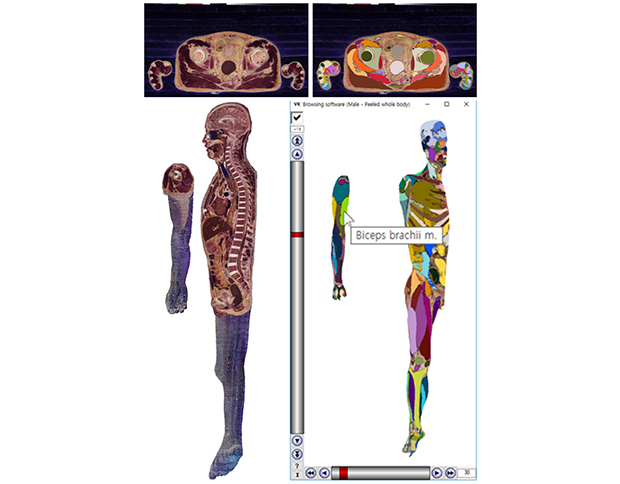
Fig. 1
Two-dimensional images of anatomical structures. (A) A horizontal sectioned image of male cadaver. (B) The sectioned image overlapped with the color-filled image made from segmentation.

The sectioned images and color-filled sectioned images were processed as follows. Pixel size was increased to 1 mm (as large as their intervals). The right half of the body was chosen for this study because the authors wanted to demonstrate the sagittal planes of the subject also. The upper limb was separated from the rest of the body (head, neck, trunk, and lower limb) because the authors intended to prevent the upper limb and trunk from hiding each other when the model was rotated (Fig. 2).
Peeled volume models
After stacking the sectioned images, a volume model of the right upper limb and another volume model of the rest of the body were produced (voxel size 1 mm). When representing each volume model, the parallel view was used rather than the perspective view. For the privacy of the subject, the eyes were hidden (Figs. 2, 3, 4).
Fig. 3
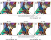
Operation of the browsing software of the peeled volume model. (A) Anterior view. (B) Rotated model. (C) More peeled model with the name of a pointed structure. (D) Color-filled model.
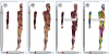
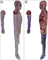
Fig. 4
Collection of models made from an identical cadaver. (A) Unpeeled volume model with the underlying structures labeled manually. (B) Peeled color-filled volume model. (C) Surface models.
On the basis of an established method,9 peeling of the volume model (thickness 1 mm) was continuously carried out to a maximum of 49 mm; as a result, 50 peeled models were obtained. Each volume model was rotated at every 30 degree angle. For the 50 peeled models and 12 viewing angles, 600 images could be screen captured (Fig. 3).
With the color-filled sectioned images, the same procedure of volume reconstruction, peeling, and rotation was repeated to acquire the corresponding 600 captured views (Fig. 3).
Browsing software of peeled volume models
For the small file size and real time operation of the browsing software, the captured views were cropped and resized (resolution 989 × 2,050 pixels), and were saved as portable network graphics (PNG) format.
New browsing software equipped with convenient interfaces has already been programmed.13 On the browsing software, the peeled volume models (intervals 1 mm) and rotated volume models (intervals 30 degrees) were arranged to be selected. The color-filled volume models were also arranged to appear instead of the original volume models (Fig. 3).
RESULTS
The software could be downloaded from the Visible Korean homepage (anatomy.co.kr) without charge or registration14 by clicking the menu entitled “Browsing software (Male - Peeled whole body for surface anatomy) (ver. 2) (391 MB).” After extracting the ZIP compressed file, the software operated without installation. The software ran off-line with no time delay. The operation method of the software of this study was similar to those of previous studies.1013
On the software, the volume model was peeled at 1 mm thicknesses with the vertical scroll bar, while the volume model was rotated at 30 degrees with the horizontal scroll bar. In the box just above the vertical scroll bar, the peeling thickness (mm) was displayed (e.g., −4, −13) (Fig. 3).
It was beneficial that the tissue colors in the peeled volume model were consistent with those in the original sectioned images. As an artifact, the dark blue color of embedding agent outside the skin was visible together (Figs. 1 and 2).11
The original volume model was switched to the corresponding color-filled volume model by use of the checkbox over the vertical scroll bar. If the mouse pointer was located on an outlined structure (e.g., biceps brachii m.) in a color-filled volume model, the structure name was shown as the tool tip text beside the mouse pointer. The structure name was also shown in the original volume model because the two kinds of volume models corresponded to each other (Fig. 3C and D).
While the model contains all the body regions, the following regions were explored as representatives, to find their surface anatomy features of clinical significance.
In the neck region, the sternocleidomastoid muscle was located at a depth of 2 to 21 mm, with the internal jugular vein located at a depth of 16 to 26 mm. When performing trigger point injection at the muscle, the performer should beware of the overlapping 16 to 21 mm depth, so as to avoid the vein (Fig. 5).
Fig. 5
Lateral views of volume model (right side of neck). (A) 15 mm-peeled model. (F) 20 mm-peeled model.
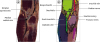
On the volume model, the pronator teres and the brachioradialis, which are the borders of the cubital fossa, could be observed. The brachialis and the supinator, which constitute the floor of the cubital fossa, could be observed as well. The bicipital aponeurosis, which is the roof of the cubital fossa, was located around 4 mm depth. Underneath the aponeurosis, the brachial artery and the median nerve were present. On the other hand, superficial to the aponeurosis, the median cubital vein, which is frequently used for venous blood sampling, was present (Fig. 6).
Fig. 6
Anterior view of volume models of the right cubital fossa. (A) 4 mm-peeled model made from sectioned images. (B) 5 mm-peeled volume made from color-filled images.
Regarding the femoral triangle, two muscular borders, which are the adductor longus and the sartorius, were color-coded, while the inguinal ligament was not color-coded but recognizable on the volume model. The femoral artery is frequently used for vascular access in cardiac catheterization and intervention, whereas the femoral vein is used for intravenous injection. The vessels were visible from −13 mm depth in the triangle, while the femoral nerve was visible from −18 mm depth. The nerve, artery, and vein were arranged from the lateral to medial side. By knowing the arrangement of the nerve and vessels, practitioners can avoid damaging them when performing biopsy or resection of the deep inguinal nodes (Fig. 7A).
Fig. 7
Volume models presenting the pelvis region. (A) Anterior view of 21 mm-peeled color-filled volume model of right femoral triangle. (B) Posterior view of 45 mm-peeled color-filled volume model of the right gluteal region that is overlapped by the unpeeled volume model.
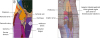
For the intramuscular injection at the gluteus maximus, superior lateral quadrant of the gluteal region is used, so as not to damage the sciatic nerve.15 Its anatomical background was presented by the overlapped volume models. Moreover, for performing trigger point injection at the piriformis, the depth and position of the muscle should be understood. The piriformis could be observed from the depth of 21 mm and was located superior to the sciatic nerve (Fig. 7B).
DISCUSSION
The present study elaborated and released the software to browse the curved sectional planes of the entire body. The software has strong points that are not achievable with other learning tools.
First, the volume model in the software gives rise to different viewpoints of surface anatomy. Surface anatomy is the study to learn locations of the underlying structures without dissection. Depths of the structures that are influenced by thickness of the subcutaneous tissue and others are important.1617 On the software, locations and depths of the various body structures can be recognized systematically (Figs. 3, 5, 6, 7).
Second, the volume model in the software has been made from the sectioned images of a non-embalmed cadaver; therefore, it shows the structures with real color and high resolution (Fig. 1). Furthermore, the software presents automatic labeling of the structures on the basis of the color-filled outlines. The automatic labeling is superior to the classical labeling because the extent of each structure can be grasped (Fig. 3).
Third, the volume model in the software can be viewed together with the surface models in portable document format (PDF) file. The PDF file of the whole body's surface models can be downloaded on the same website. Their comparison is valuable since the volume and surface models have been reconstructed from the same male cadaver (Fig. 4).18
While the volume model reveals the real body color, the surface models show only the allocated colors. While the volume model can be peeled from the skin, the surface models can be concealed organ by organ (Fig. 4). While the volume model can be rotated at 30 degrees along one axis (Fig. 3), the surface models can be rotated freely.18
The complimentary browsing software is expected to have many applications. One application is education for medical students learning surface anatomy. When the students learn various medical procedures, they need to know the locations and depths of the related components (Figs. 5, 6, 7).19 When the students do physical examination of a patient, they should imagine the underneath muscles and bones.20 When the students dissect the cadaver, they need to grasp the holistic distribution of subcutaneous tissue.21 Automatic labeling of assorted structures (mainly, muscles) below the subcutaneous tissue is a powerful function for the students to review surface anatomy by themselves (Fig. 3).
Another application might be the education for patients suffering from several diseases. An example is obesity. The software enables anyone to grasp the thickness of subcutaneous tissue in various regions (e.g., abdomen and gluteal region) (Fig. 3).22 Using the function, clinicians can easily and efficiently explain obesity to patients. The patients may review the explanation using the same software that is installed on their own computers.
In order to increase the education effect of the peeled volume model and surface models of this study, recent technologies including 3-dimensional (3D) printing or virtual reality can be applied in the future. Once the skin is 3D printed with materials which are semitransparent or can be grounded, medical procedures will be more realistically practiced. On the other hand, the authors have experience to assist virtual reality simulator for the training of lumbar punctures by providing the Visible Korean Project data set.23
It is hoped to apply the computer technique of this study to CTs or MRIs of individual patients. The peeled volume models of CTs or MRIs can visualize the personalized surface anatomy,24 which may contribute to new interpretation of a lesion. Since current CTs or MRIs do not contain real color and high resolution, their curved sectional planes need to be observed along with those from the cadaver images provided by this research.
In a study planned for the near future, the peeled volume models of a male will be compared with those of a female. The sectioned images of the pelvis of a female cadaver25 and the whole body of another female cadaver26 are available, and outlining of the hundreds of structures has been finished. A mobile version of the software to be used regardless of time and place will be another challenge.
Anatomy includes two subfields: sectional anatomy24 and surface anatomy.1617 The two fields have been devised and continuously developed according to the clinical needs. This study proposes the combination of the sectional anatomy and surface anatomy by use of the curved sectional planes. This combination might be accepted as a new paradigm of the morphologic study of human body in the era of disciplinary convergence.
 XML Download