Journal List > J Korean Med Sci > v.34(Suppl 1) > 1121879

Lee, Lee, Ahn, Ryoo, Park, Kim, Shin, Hwang, and on behalf of the Korean Cardiac Arrest Research Consortium (KoCARC): New Termination-of-Resuscitation Models and Prognostication in Out-of-Hospital Cardiac Arrest Using Electrocardiogram Rhythms Documented in the Field and the Emergency Department

Abstract

Background

Electrocardiogram (ECG) rhythms, particularly shockable rhythms, are crucial for planning cardiac arrest treatment. There are varying opinions regarding treatment guidelines depending on ECG rhythm types and documentation times within pre-hospital settings or after hospital arrivals. We aimed to determine survival and neurologic outcomes based on ECG rhythm types and documentation times.

Methods

This prospective observational study of 64 emergency medical centers was performed using non-traumatic out-of-hospital cardiac arrest registry data between October 2015 and June 2017. From among 4,608 adult participants, 4,219 patients with pre-hospital and hospital ECG rhythm data were enrolled. Patients were divided into 3 groups: those with initial-shockable, converted-shockable, and never-shockable rhythms. Patient characteristics and survival outcomes were compared between groups. Further, termination of resuscitation (TOR) validation was performed for 6 combinations of TOR criteria confirmed in previous studies, including 2 rules developed in the present study.

Results

Total survival to discharge after cardiac arrest was 11.7%, and discharge with good neurologic outcomes was 7.9%. Survival to discharge rates and favorable neurologic outcome rates for the initial-shockable group were the highest at 35.3% and 30.2%, respectively. There were no differences in survival to discharge rates and favorable neurologic outcome rates between the converted-shockable (4.2% and 2.0%, respectively) and never-shockable groups (5.7% and 1.9%, respectively). Irrespective of rhythm changes before and after hospital arrival, TOR criteria inclusive of unwitnessed events, no pre-hospital return of spontaneous circulation, and asystole in the emergency department best predicted poor neurologic outcomes (area under the receiver operating characteristic curve of 0.911) with no patients classified as Cerebral Performance Category 1 or 2 (specificity = 1.000).

Conclusion

Survival outcomes and TOR predictions varied depending on ECG rhythm types and documentation times within pre-hospital filed or emergency department and should, in the future, be considered in treatment algorithms and prognostications of patients with out-of-hospital cardiac arrest.

Trial Registration

ClinicalTrials.gov Identifier: NCT03222999

Graphical Abstract

jkms-34-e134-abf001

INTRODUCTION

Survival in patients after out-of-hospital cardiac arrest (OHCA) is dependent on factors including no flow time, presence of bystander, bystander cardiopulmonary resuscitation (CPR), early defibrillation, shockable electrocardiogram (ECG) rhythms, and return of spontaneous circulation (ROSC) prior to hospital arrival.12 ECG rhythms play an important role in out-of-hospital and in-hospital cardiac arrest treatment and survival. Cardiac arrest ECG rhythms are classified into 1 of 4 rhythms on monitored ECG: shockable rhythms, such as ventricular fibrillation (VF) and pulseless ventricular tachycardia (pVT), and non-shockable rhythms, such as asystole and pulseless electrical activity (PEA). Whichever category the cardiac arrest rhythm falls into, the duration and variations in rhythms should also be considered when planning cardiac arrest treatment.34
Shockable rhythms require different treatment algorithms according to advanced life support (ALS) guidelines and are known to be strongly associated with improved survival rate compared to those of non-shockable rhythms.23 Regardless of arrest duration or first recorded rhythms, there are no algorithm changes directed at VF/pVT in the American Herat Association's 2015 CPR guidelines update. When a non-shockable rhythm converts to a shockable rhythm during resuscitation, immediate shock is recommended instead of administering CPR.3 However, results from some recent studies have suggested that in these cases there is no benefit from early defibrillations and that new treatment strategies may be necessary.456 Controversially, other studies have demonstrated that patients with subsequent shockable rhythm conversion achieved better neurologic outcomes than patients with remaining in non-shockable rhythm, despite more frequent pauses in CPR due to defibrillation attempts.78910 Termination of resuscitation (TOR) guidelines also recommend to continue resuscitative efforts for patients with initial or subsequent shockable rhythm conversion.101112 However, results from the SOS-KANTO 2012 study suggest that the initial asystole rhythms documented in the field or emergency department (ED) affected TOR rules after OHCA.13
In Korea, the Korean Cardiac Arrest Research Consortium (KoCARC), a collaborative research network developed to coordinate various studies conducted on OHCA resuscitation was organized in 2015 after recruiting hospitals willing to participate voluntarily in the consortium. The KoCARC registry is a data collection system comprising OHCA risks and prognostic variables that was developed to provide a platform for research conducted in the KoCARC and to strengthen the cooperative effort in conducting these studies.
In this study, our primary objective was to identify relationships between survival and neurologic outcomes with the documented location and timing of the initial ECG using the KoCARC registry stratified by ECG rhythms documented in the field and the ED setting. Our secondary objective was to perform an external validation of existing TOR criteria against cases in the KoCARC and present new TOR criteria, inclusive of ECG rhythms.

METHODS

Study design and registry implementation

We conducted a multicenter prospective observational study of patients from 64 participating hospitals using the non-traumatic nationwide OHCA registry data.14 The present study evaluated the epidemiology and outcomes of patients that suffered OHCA with a presumed medical etiology in Korea and is registered with ClinicalTrials.gov, number NCT03222999. To enhance the effectiveness in research, several research committees have been organized. In addition, the Data Safety and Monitoring Board Committee was organized to provide data quality control.14

Study population and data collection

Data were obtained from the KoCARC-pro registry database in Korea for cardiac arrest events that occurred between October 1, 2015 and June 30, 2017. Patients older than 18 years with OHCA who were transported to participating hospitals via emergency medical service (EMS) with resuscitation efforts initiated and whose arrest was identified as being of a presumed medical etiology by emergency physicians in the ED were included. Patients with obvious signs of death (e.g., rigor mortis or dependent lividity), terminal illnesses documented in their medical records, those under hospice care, pregnant women, and those with pre-documented ‘Do Not Resuscitate’ cards were excluded. Additionally, those with non-cardiac etiology arrest, those with missing hospital outcomes, patients with a missing first recorded rhythm and cases of trauma, poisoning, electrocution, primary respiratory arrest, drowning, asphyxia, hanging, and other injuries were also excluded.14
The following information was extracted from the KoCARC database: demographics, including gender, age, home address, and location of event; CPR-related characteristics such as presence of a witness, CPR by a bystander, initial ECG rhythms obtained pre-hospital arrival or in the ED, and provision of CPR by EMS or in the ED (epinephrine, advanced airway, antiarrhythmic, extracorporeal membrane oxygenation [ECMO]); CPR-related time variables such as basic life support (BLS) interval (time from collapse to initiation of chest compression attempt at the scene), defibrillation interval (time from collapse to initiation of electrical shock), and pre-hospital interval (time from collapse to arrival in an ED); and clinical outcomes, including presence of ROSC, ED outcomes (admitted, died, or transferred), hospital outcomes (discharged alive, died, or transferred), neurological outcomes at the time of discharge, and whether post cardiac arrest treatment (i.e., target temperature management [TTM] or percutaneous coronary intervention [PCI]) was provided.

Primary variable of the subgroup setting

Patients with a first documented ECG rhythm in the field or in the ED were divided accordingly into 3 rhythm categories: initial shockable (IS), converted to shockable (CS), and never shockable (NS).4 We extracted patients with OHCA meeting the criteria for each of the TOR rules and calculated the sensitivities, specificities, positive predictive values (PPV), and negative predictive values (NPV) and their respective 95% confidence intervals (CI) to identify OHCA patients with a risk of mortality and poor neurologic outcomes at hospital discharge.1115
The 6 combinations of TOR criteria were as follows (Table 1):
Table 1

Defined combination criteria of previous researchers' and KoCARC TOR rules

jkms-34-e134-i001
TOR rules Witness status Initial pre-hospital rhythm Pre-hospital shock Pre-hospital ROSC Others
International BLS rule Unwitnessed by EMT - No pre-hospital shock (AED) No prehospital ROSC -
International ACLS rule Unwitnessed by bystander/EMT - No pre-hospital shock (AED) No prehospital ROSC No bystander CPR
Goto's TOR rule Unwitnessed by bystander Initial non-shockable rhythm - No prehospital ROSC -
SOS-KANTO's TOR rule Unwitnessed by bystander Asystole in the field - - Asystole in the hospital
New TOR model 1 Unwitnessed by bystander Asystole in the field - No prehospital ROSC -
New TOR model 2 Unwitnessed by bystander - - No prehospital ROSC Asystole in the hospital
KoCARC = Korean Cardiac Arrest Research Consortium, TOR = termination of resuscitation, ROSC = return of spontaneous circulation, BLS = basic life support, EMT = emergency medical technicians, AED = automated external defibrillator, ACLS = advanced cardiac life support, CPR = cardiopulmonary resuscitation.
  • 1) International BLS TOR rule (TOR-BLS): the combinations were unwitnessed by EMS personnel, no shock delivered, and no prehospital ROSC.11

  • 2) International ALS TOR rule (TOR-ALS): the combinations were unwitnessed by bystanders or EMS personnel, no bystander CPR, no shock delivered, and no prehospital ROSC.11

  • 3) Goto's TOR rule: the combinations were unwitnessed by a bystander, non-shockable rhythm in the field, and no pre-hospital ROSC.12

  • 4) SOS-KANTO's TOR rule: the combinations were unwitnessed by a bystander, asystole in the field and the hospital.13

  • 5) New TOR model 1: the combinations were unwitnessed by a bystander, asystole in the field, and no pre-hospital ROSC.

  • 6) New TOR model 2: the combinations were unwitnessed by a bystander, asystole in the hospital, and no pre-hospital ROSC.

Main outcome measurement

Survival to discharge was defined as final discharge to home or transfer to another facility after admission to the hospital. Neurological status was quantified using Cerebral Performance Category (CPC) scores, which are based on a 5-point scale in which scores of 1 (good recovery) and 2 (moderate disability) are defined as favorable neurologic outcomes.
The primary outcome measures were based on the group proportions of survival to hospital discharge and of survival with a favorable neurologic outcome in the 3 ECG rhythm categories. The secondary outcome measure was validation of previously researched TOR rules.

Statistical analysis

Data were analyzed using IBM SPSS Statistics version 23.0 (IBM corp., Armonk, NY, USA) and MedCalc 17.4.4 version (MedCalc Software, Mariakerke, Belgium). Descriptive statistics are presented as medians with interquartile ranges (interquartile range [IQR]: 25th and 75th percentiles) and categorical variables are presented as counts and percentages.
The significance of differences between the 3 groups (IS, NS, and CS) was tested using the Kruskal-Wallis test or analysis of variance with a post-hoc analysis (correcting for multiple comparisons with the Bonferroni method) for continuous variables, and the χ2 test or Fisher's exact test for categorical variables.
Before conducting the multivariable analysis, we conducted a co-linearity test of variables associated with OHCA events and time-related parameters. In this study, the tolerance of each variable was larger than 0.1 (range, 0.72–0.98), while the variance inflation factor was less than 10 (range, 1.02–1.40). This demonstrates that the variables introduced into the model generally did not exhibit multi-collinearity. We used logistic regression for the multivariable analyses, and the outcome of interest was survival to hospital discharge. To determine the logistic model calibration, we calculated the Hosmer-Lemeshow goodness of fit. Odds ratios (OR) greater than 1 were indicative of a beneficial effect on survival. ORs and 95% CIs were derived for all covariates. The characteristics of the adjusted OR are described using Forest plots. P < 0.05 was considered statistically significant.

Ethics statement

The study was approved by the Institutional Review Board of Kyungpook National University Hospital (KNUH 2015-11-013), which waived the requirement of informed consent. Approval for this study was obtained from the institutional review boards of each of the 64 participating hospitals.

RESULTS

Characteristics of the entire study subjects

Of 4,757 patients registered in the KoCARC-pro registry database during the 21 months of registration, 4,608 were analyzed after excluding 6 patients with in-hospital cardiac arrest and 143 patients younger than 18 years. Among the 4,608 patients, 65.1% were men, and 59.5% had a witnessed OHCA (Fig. 1 and Supplementary Table 1). Furthermore, 46.7% received CPR from a bystander. A total of 13.3% of the patients had ROSC prior to hospital arrival. The percentages of patients who underwent PCI, TTM, or ECMO were 11.7%, 9.3%, and 2.9%, respectively.
Fig. 1

Study population and analysis of outcomes after out-of-hospital cardiac arrest.

KoCARC = Korean Cardiac Arrest Research Consortium, OHCA = out-of-hospital cardiac arrest, ECG = electrocardiogram, ROSC = return of spontaneous circulation.
jkms-34-e134-g001

Characteristics of patients with initial ECG at field and in the ED
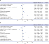
Of the 4,608 OHCA patients, 389 cases who had unmatched medical records or whose pre-hospital time variables had not been recorded were excluded. Overall 4,219 patients were enrolled into the survival analysis. According to the patients' confirmed ECG rhythms and any subsequent rhythm changes, participants were divided into the IS (n = 821), CS (n = 498), and NS rhythm groups (n = 2,900) (Fig. 1 and Table 2). The IS group was relatively younger with a higher proportion of men, public events, witnessed cardiac arrests, bystander CPR, and pre-hospital defibrillation rates (Table 2). The CS and NS groups had similar rates of pre-hospital witnesses, bystander CPR, and degrees of acidosis in hospital. The CS group had the lowest pre-hospital ROSC prior to ED arrival and survival to discharge (5% and 4.2%, respectively) among the 3 groups, and, similar to the IS group, favorable neurologic outcomes to discharge was low.
Table 2

Patient characteristics by type of documented ECG rhythm in the field and in-hospital (n = 4,219)
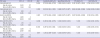
jkms-34-e134-i002
Covariates Initial shockable rhythm (n = 821) Converted shockable rhythm (n = 498) Never shockable rhythm (n = 2,900) P value
Age, yr 60.0 (50–70)a 68.0 (55–78)b 73.0 (60–81)c < 0.001
Gender, men 670 (81.6) 327 (65.7) 1,778 (61.3) < 0.001
Location of OHCAd < 0.001
Home/residency 384 (46.8) 318 (63.9) 1,991 (68.7)
Public 390 (47.5) 149 (29.9) 725 (25.0)
Other 35 (4.3) 22 (4.4) 164 (5.7)
Co-morbidities
Hypertension 317 (38.6) 201 (40.4) 1,229 (42.4) 0.135
Diabetes mellitus 165 (20.1) 131 (26.3) 786 (27.1) < 0.001
Hyperlipidemia 47 (5.7) 22 (4.4) 139 (4.8) 0.472
Personal history of current smoking 201 (24.5) 55 (11.0) 321 (11.1) < 0.001
Personal history of alcohol intake 292 (35.6) 115 (23.1) 630 (21.7) < 0.001
Prehospital parameters
Witnessed event 633 (78.3) 291 (59.3) 1,567 (54.9) < 0.001
Bystander CPR 513 (62.5) 232 (46.6) 1,299 (44.8) < 0.001
Prehospital defibrillation 781 (95.1) 97 (19.5) 202 (7.0) < 0.001
Prehospital advanced airway 453 (55.2) 262 (52.6) 1,585 (54.7) 0.639
Prehospital drug administration 159 (19.4) 62 (12.4) 427 (14.7) < 0.001
Time variables, min
Response time 7 (5–10) 7 (5–10) 7 (5–10) 0.155
Shock delivery 2 (1–3)a 6 (2–12)b - < 0.001
Down time 49 (22–69)a,c 60 (45–71)b 51 (39–62)a,c 0.021
ROSC before ED arrival 331 (40.3) 25 (5.0) 175 (6.0) < 0.001
Resuscitation in ED
pH (NR, 7.35–7.45) 7.11 (6.90–7.28)a 6.92 (6.81–7.03)b,c 6.92 (6.80–7.03)b,c < 0.001
Lactate, mmol/L (NR, 0.7–2.1) 10.2 (7.1–13.9) 13.0 (10.2–16.0) 13.1 (9.9–17.0) 0.093
Troponin, ng/mL (NR, < 0.1) 0.11 (0.03–0.59) 0.15 (0.04–0.43) 0.13 (0.040–0.63) 0.191
PCI 327 (39.8) 33 (6.6) 117 (4.0) < 0.001
ECMO 64 (7.8) 20 (4.0) 39 (1.3) < 0.001
TTM 186 (22.7) 28 (5.6) 191 (6.6) < 0.001
Survival outcomes after OHCA
Survival-to-admission 454 (55.3) 103 (20.7) 562 (19.4) < 0.001
Survival-to-discharge 290 (35.3) 21 (4.2) 166 (5.7) < 0.001
Good cerebral performance 248 (30.2) 10 (2.0) 55 (1.9) < 0.001
Data are presented as median (interquartile range) or number (%).
ECG = electrocardiogram, OHCA = out-of-hospital cardiac arrest, CPR = cardiopulmonary resuscitation, ROSC = return of spontaneous circulation, ED = emergency department, NR = normal range, PCI = percutaneous coronary intervention, ECMO = extracorporeal membrane oxygenation, TTM = target temperature management.
a,b,cThe same letters indicate non-significant difference between groups based on post-hoc multiple comparison test; dUnknown or not determined data (n = 41).
The total survival to discharge rate was 11.7%, and survival with good neurologic outcomes was 7.9%. In the IS group, these rates were 35.3% and 30.2%, respectively, showing that those in the IS group had better neurologic outcomes than those in the NS group which had a total survival to discharge rate of 5.7% and survival with good neurologic outcome rate of 1.9% (crude OR, 9.0 [95% CI, 7.28–11.12] and 22.39 [95% CI, 16.49–30.39], respectively, Table 3). In the CS group, rates of total survival to discharge and survival with good neurologic outcomes (4.2% and 2.0%, respectively) were not significantly different than those of the NS group (crude OR, 0.73 [95% CI, 0.47–1.15] and 1.06 [0.54–2.09]).
Table 3

Results of univariate and multivariate logistic regression analyses of factors associated with favorable survival outcomes
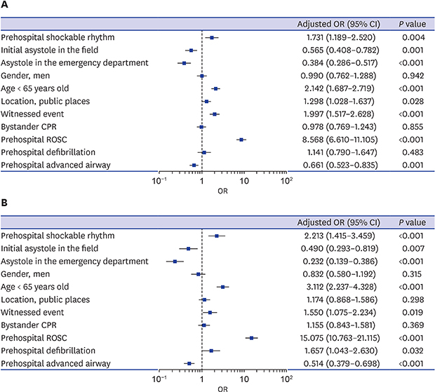
jkms-34-e134-i003
Variables Survival to discharge Good neurologic outcome
Crude OR Adjusted ORa Crude OR Adjusted ORa
Cardiac arrest rhythm
Remain in non-shockable rhythm Reference Reference Reference Reference
Conversion to shockable rhythm 0.73 (0.47–1.15) 0.55 (0.33–0.90) 1.06 (0.54–2.09) 0.90 (0.42–1.91)
Initial shockable rhythm 9.00 (7.28–11.12) 1.88 (1.15–3.06) 22.39 (16.49–30.39) 3.86 (2.01–7.41)
Initial asystole in the field 0.12 (0.09–0.15) 0.56 (0.40–0.80) 0.05 (0.04–0.08) 0.59 (0.33–1.04)
Asystole in the EDs 0.09 (0.07–0.11) 0.35 (0.26–0.48) 0.03 (0.02–0.05) 0.24 (0.14–0.40)
Gender, men 1.83 (1.48–2.25) 0.99 (0.75–1.31) 2.12 (1.64–2.75) 0.78 (0.53–1.16)
Age < 65 yr 4.06 (3.34–4.93) 2.03 (1.58–2.62) 6.47 (4.99–8.40) 2.93 (2.05–4.18)
Location, public places 2.38 (1.98–2.86) 1.32 (1.03–1.69) 2.58 (2.08–3.21) 1.16 (0.84–1.61)
Witnessed event 3.65 (2.90–4.60) 1.99 (1.48–2.67) 3.73 (2.81–4.96) 1.67 (1.12–2.51)
Bystander CPR 1.59 (1.33–1.91) 0.89 (0.69–1.14) 2.20 (1.76–2.75) 1.02 (0.72–1.44)
ROSC before ED arrival 24.30 (19.62–30.11) 8.07 (6.12–10.66) 54.66 (40.94–72.96) 14.51 (10.09–20.87)
Prehospital defibrillation 6.47 (5.35–7.83) 0.92 (0.58–1.49) 13.26 (10.25–17.14) 1.11 (0.60–2.04)
Prehospital advanced airway 0.71 (0.59–0.85) 0.65 (0.51–0.83) 0.65 (0.53–0.81) 0.52 (0.38–0.72)
Data are presented as number (median).
OR = odds ratio, ED = emergency department, CPR = cardiopulmonary resuscitation, ROSC = return of spontaneous circulation, VIF = variance inflation factor.
aAll variables of collinearity statistics: VIF < 10.
Multivariate analysis identified ECG rhythms, age (< 65 years), place of cardiac arrest, presence of a witness, and ROSC before ED arrival as factors related to survival to discharge (Fig. 2A). Factors related to discharge with good neurologic outcomes were ECG rhythm, age, presence of a witness, and ROSC before ED arrival (Fig. 2B). Although gender and bystander CPR were significant in the univariate analysis, they were not significantly associated with survival outcomes in the multivariate analysis (Table 3).
Fig. 2

Adjusted odds ratios for survival outcomes after out-of-hospital cardiac arrest. (A) Forest plot showing survival to discharge. (B) Forest plot showing favorable neurologic outcomes at discharge.

OR = odds ratio, CI = confidence interval, CPR = cardiopulmonary resuscitation, ROSC = return of spontaneous circulation, VIF = variance inflation factor.
All variables of collinearity statistics: VIF < 10.
jkms-34-e134-g002

Validation of various TOR rules for predicting outcomes at discharge

We validated the TOR criteria against the 4,608 adult cardiac arrest cases in this study. When the traditional TOR-BLS and TOR-ALS rules, which do not include ECG rhythms, were applied to our patient pool, the numbers of patients who met TOR criteria but survived to discharge were 118 (2.6%) and 17 (0.4%), respectively (Table 4). Additionally, the number of patients who were discharged with a CPC score of 1 or 2 were 27 (0.6%) and 4 (0.1%), respectively (Table 5). When the Goto's TOR rule, which considers TOR for non-shockable rhythms in the field with unwitnessed events and no pre-hospital ROSC, was applied, the number of patients who met TOR criteria but survived to discharge was 27 (0.6%), and the number of patients who were discharged with CPC scores of 1 or 2 was 4 (0.1%). However, all patients who met the SOS-KANTO's TOR rule (asystole in the field and in the ED), new TOR model 1 (asystole in the field), or new TOR model 2 (asystole in the ED) criteria, had poor neurologic outcomes of CPC 3 or higher, with none of the patients showing favorable neurologic state with CPC 1 or 2 (specificity = 1.000). Regarding their effectiveness at detecting poor outcomes based on the area under the curve (AUC), the international TOR-BLS, Goto's TOR rule, new TOR model 1, and new TOR model 2 criteria (which include the absence of pre-hospital ROSC) had higher AUCs than the SOS-KANTO criteria, which does not include the absent pre-hospital ROSC condition (Tables 4 and 5). Overall, the new TOR model 2 criteria were the most effective at predicting in-hospital mortality and unfavorable neurologic outcomes, with AUCs of 0.853 and 0.911, respectively. Once TOR was established based on these criteria, none of the patients met criteria for CPC 1 or 2 (all specificity and PPV = 1.000).
Table 4

Performance of the new TOR rules for predicting death prior to discharge (n = 4,608)
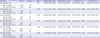
jkms-34-e134-i004
TOR rules Death Survival AUC Sensitivity (95% CI) Specificity (95% CI) PPV (95% CI) NPV (95% CI)
International BLS 0.811 0.718 (0.704–0.732) 0.781 (0.743–0.815) 0.961 (0.955–0.967) 0.268 (0.255–0.281)
Met all criteria 2,921 118
Did not fulfill criteria 1,149 420
International ACLS 0.795 0.213 (0.201–0.226) 0.968 (0.950–0.982) 0.961 (0.970–0.988) 0.140 (0.137–0.143)
Met all criteria 868 17
Did not fulfill criteria 3,202 521
Goto's rule (2013) 0.831 0.388 (0.373–0.403) 0.950 (0.928–0.967) 0.983 (0.976–0.988) 0.170 (0.166–0.175)
Met all criteria 1,579 27
Did not fulfill criteria 2,491 511
SOS-KANTO's (2017) 0.812 0.271 (0.257–0.285) 0.983 (0.969–0.992) 0.992 (0.985–0.996) 0.151 (0.149–0.154)
Met all criteria 1,102 9
Did not fulfill criteria 2,968 529
New TOR model 1 0.843 0.306 (0.292–0.320) 0.974 (0.957–0.986) 0.989 (0.874–0.892) 0.157 (0.153–0.160)
Met all criteria 1,245 14
Did not fulfill criteria 2,825 524
New TOR model 2 0.853 0.323 (0.309–0.338) 0.980 (0.964–0.990) 0.992 (0.985–0.995) 0.161 (0.157–0.164)
Met all criteria 1,316 11
Did not fulfill criteria 2,754 527
Data are presented as median or number.
TOR = termination of resuscitation, AUC = area under the receiver operating characteristic curve, CI = confidence interval, PPV = positive predictive value, NPV = negative predictive value, BLS: basic life support, ACLS = advanced cardiac life support, SOS-KANTO = survey of survivors after cardiac arrest conducted in the Kanto area in 2012 (2017).
Table 5

Neurologic outcomes of patients after out-of-hospital cardiac arrest matching each of 6 rules (n = 4,608)

jkms-34-e134-i005
TOR rules CPC ≥ 3 CPC 1/2 AUC Sensitivity (95% CI) Specificity (95% CI) PPV (95% CI) NPV (95% CI)
International BLS 0.904 0.709 (0.696–0.723) 0.925 (0.893–0.950) 0.991 (0.987–0.994) 0.216 (0.204–0.223)
Met all criteria 3,012 27
Did not fulfill criteria 1,234 335
International ACLS 0.876 0.208 (0.195–0.220) 0.989 (0.972–0.997) 0.996 (0.988–0.998) 0.096 (0.095–0.098)
Met all criteria 881 4
Did not fulfill criteria 3,365 358
Goto's rule (2013) 0.901 0.377 (0.363–0.392) 0.989 (0.972–0.997) 0.997 (0.993–0.999) 0.119 (0.116–0.122)
Met all criteria 1,602 4
Did not fulfill criteria 2,644 358
SOS-KANTO's (2017) 0.848 0.262 (0.248–0.275) 1.000 (0.990–1.000) 1.000 0.104 (0.102–0.105)
Met all criteria 1,111 0
Did not fulfill criteria 3,135 362
New TOR Model 1 0.900 0.297 (0.159–0.182) 1.000 (0.990–1.000) 1.000 0.108 (0.106–0.110)
Met all criteria 1,259 0
Did not fulfill criteria 2,987 362
New TOR Model 2 0.911 0.313 (0.299–0.327) 1.000 (0.990–1.000) 1.000 0.110 (0.108–0.112)
Met all criteria 1,327 0
Did not fulfill criteria 2,919 362
Data are presented as median or number.
TOR = termination of resuscitation, CPC = cerebral performance category, AUC = area under the ROC curve, CI = confidence interval, PPV = positive predictive value, NPV = negative predictive value, BLS = basic life support, ACLS = advanced cardiac life support, SOS-KANTO = survey of survivors after cardiac arrest conducted in the Kanto area in 2012 (2017).

DISCUSSION

In this study, we analyzed the effect of pre-hospital and in-hospital initial ECG rhythms on survival outcomes in a multicenter observational study and presented new predictive power by identifying new TOR rules including ECG rhythms using the KoCARC database. Regarding survival outcomes, we found that patients with pre-hospital non-shockable rhythms showed poor outcomes similar to those of the NS group, even when ECGs converted to shockable rhythms in the hospital. Survival outcomes and TOR predictions also varied among pre-hospital cardiac arrest patients depending on the type of ECG rhythms and time of ECG documentation (in the field or the ED). The criteria suggesting TOR for patients with asystole in the ED, no ROSC before ED arrival, and unwitnessed events showed a specificity of 1.00 for predicting poor neurologic outcomes, which was higher in sensitivity than other criteria.
These results are similar to those of Mader et al.,4 Hallstrom et al.,5 and Thomas et al.6 wherein the CS group showed no superiority in the survival to discharge outcome than the NS group. It is possible that the reason our results differed from those of previous studies, which observed good outcomes in cases with conversion to a shockable rhythm, may be attributable to differences in baseline characteristics. For example, previous studies that reported good outcomes for the converted group reported that, overall, 5%–20% of non-shockable rhythms subsequently converted into shockable rhythms in the field78910; which was different that the rate we observed in the present study (11.8%). Further, in a study by Zheng et al.9 the median response time in the initial non-shockable group was 5.1 minutes, which was shorter than that for the same group in our study (7 minutes). In addition, compared to our study, Wah et al.10 also reported a lower percentage of non-witnessed cardiac arrests, and the percentage of events with a response time of less than 8 minutes also differed from that in our study. Another baseline difference between our study and these other studies was that the pre-hospital ROSC rate in the conversion to shockable group (5.0%) was lower than the rate in the NS group (6.0%). In contrast, previous studies reporting good outcomes among patients whose non-shockable rhythms subsequently converted reported that those patients had pre-hospital ROSC rates (range, 7.6%–26.7%) that were higher than those in the NS group.8910
Further evidence that baseline characteristics may contribute to outcomes of those with subsequently converted rhythms can be seen by comparing our study to those with similar results. Specifically, our study showed similar results to those of Mader et al.4 in terms of arrest location and survival at discharge. Prehospital resources may also vary across study populations or countries, and this may influence the pre-hospital ROSC and survival outcomes.
In this study, similar to previous studies, multivariate analysis showed that initial asystole in the field or asystole in the ED was associated with survival outcomes. However, we only had a small number of cases with which to perform a subgroup analysis based on initial PEA or asystole. Previous studies have had discordant results. Kitamura et al.16 reported that initial PEA rhythms are associated with better neurologic outcomes than those with asystole. On the other hand, Zheng et al.,9 who reported that patients with subsequently shockable rhythm that initially showed asystole had good survival outcomes, whereas initial PEA rhythms were not associated with survival outcomes. However, in the aforementioned study, the pre-hospital defibrillation rate was only 19.5% while the in-hospital defibrillation rate was much higher, suggesting that shock delivery was relatively delayed. Furthermore, the conversion to shockable rhythm group had longer downtimes than those of other groups, which may have negatively affected survival outcomes. Additional studies are needed to investigate the effects of ECG rhythm changes on survival outcomes and differences in survival among the various initial ECG subgroups.
In this study, bystander CPR was not associated with survival to discharge or discharge with good neurologic outcomes. These results are similar to those reported by some previous studies,610 however the literature reports incongruous results. For example, other studies have reported that bystander CPR increased the survival rate and decreased risks of brain damage or nursing home admission compared to when bystander CPR was not performed.1718 Other studies reported that if the initial shock after bystander CPR is delivered within 20 minutes, subsequent shock delivery is associated with improved post-arrest survival outcomes.1019 In this study, we were unable to confirm the time of bystander CPR initiation, nor the depth and rate of compressions because they are not recorded in KoCARC. Thus, we cannot fully analyze the potential effects of bystander-administered CPR, which may have affected the survival outcomes of this study.
In Japan, ED-based TOR criteria are not applied until patients arrive at the hospital because paramedics are not authorized to declare death before hospital arrival, even among patients with a low possibility of recovery.1019 Similarly, EMS personnel in Korea cannot legally terminate their resuscitation efforts in the field except for when death is obvious. However, EMS personnel in Korea use a manual defibrillator and can analyze ECGs prior to hospital arrival. Therefore, we were able to apply various survival outcome changes and TOR models based on ECG rhythms documented pre-hospital compared to those in the ED. In this study, we determined TOR based on the KoCARC model that included pre-hospital and in-hospital ECG rhythms, prehospital ROSC, and the presence of a witness, which were found to be associated with outcomes in the multivariate analysis.
Current guidelines recommend that diagnostic tests designed to guide decisions to cease life-saving efforts must be accurate and reliable with a false positive rate close to 0% with a 95% CI below 0.9.2021 We validated TOR criteria against our patient pool and found that BLS-TOR and Goto's TOR rule did not fit these criteria, as the lower limits of the 95% CIs were below 95%. The SOS-KANTO's TOR rule (combination of unwitnessed, asystole in the field and the ED) and new TOR model 2 (unwitnessed, asystole in the ED, and no prehospital ROSC) met the 95% lower limits for specificity and had PPVs above 0.95 for predicting poor neurologic outcomes and in-hospital mortality. Particularly, the criteria had good predictive power for poor CPC, with a specificity of 1.000 (100%), and showed the best ability to predict poor neurologic outcomes and in-hospital mortality of all the TOR criteria compared in this study. However, the number of patients that met the SOS-KANTO's TOR rule was the lowest and its discriminative power in the AUC was poorer compared to that of new TOR model 2.

Limitations

This study had a few limitations. First, as with other multicenter observational studies, the integrity, effectiveness, and ascertainment bias of the data are potential limitations. We strove to reduce the potential biases in this multicenter observational study by implementation of the quality control committee in KoCARC. Second, facility or regional differences in EMS resources, CPR quality, and post-cardiac arrest care may affect survival outcomes; however this study did not analyze differences in inter-hospital variabilities. These variables may have potentially introduced confounding effects. Third, the specificity of the criteria for in-hospital mortality was not 1.0. If ECMO-CPR is performed or a new CPR-device is applied according to technological developments, the TOR rules could be changed.22 Withdrawal or termination guidelines for ECMO-CPR have not been studied previously, and the volume of ECMO cases in this study was as low as 2.9%, and thus could not be analyzed separately. TOR rules in patients with ECMO criteria and socio-ethical issues should be discussed and a consensus process will be needed. Therefore, externally validating the new TOR criteria obtained in this study using KoCARC data collected in a particular period in the future through a prospective study would be necessary to apply the criteria to patients with a low possibility of inpatient or other clinical situation recoveries.
This study found that patients with non-shockable rhythms on the pre-hospital initial ECGs that were converted to shockable rhythms still had poor survival outcomes, comparable to those of the NS group. Furthermore, survival outcomes and TOR predictions varied widely depending on the type of ECG rhythms and time of ECG documentation. In the future, types of ECG rhythms as well as time of ECG documentation should be considered in treatment algorithms and prognostication among OHCA patients.

Notes

Disclosure The authors have no potential conflicts of interests to disclose.

Author Contributions

  • Conceptualization: Lee DE, Lee MJ, Kim WY, Shin SD, Hwang SO.

  • Formal analysis: Lee DE, Lee MJ, Ahn JY, Ryoo HW, Park J.

  • Methodology: Kim WY, Shin SO, Hwang SO.

  • Writing - original draft: Lee DE, Lee MJ.

  • Writing - review & editing: Lee DE, Lee JM, Kim WY.

Availability of data and materials The data are only available to the participating organizations for approval by the KoCARC committee.

References

1. Yang HJ, Kim GW, Kim H, Cho JS, Rho TH, Yoon HD, et al. Epidemiology and outcomes in out-of-hospital cardiac arrest: a report from the NEDIS-based cardiac arrest registry in Korea. J Korean Med Sci. 2015; 30(1):95–103.
crossref pmid
2. Wissenberg M, Lippert FK, Folke F, Weeke P, Hansen CM, Christensen EF, et al. Association of national initiatives to improve cardiac arrest management with rates of bystander intervention and patient survival after out-of-hospital cardiac arrest. JAMA. 2013; 310(13):1377–1384.
crossref pmid
3. Hasegawa M, Abe T, Nagata T, Onozuka D, Hagihara A. The number of prehospital defibrillation shocks and 1-month survival in patients with out-of-hospital cardiac arrest. Scand J Trauma Resusc Emerg Med. 2015; 23(1):34.
crossref pmid pmc
4. Mader TJ, Nathanson BH, Millay S, Coute RA, Clapp M, McNally B, et al. Out-of-hospital cardiac arrest outcomes stratified by rhythm analysis. Resuscitation. 2012; 83(11):1358–1362.
crossref pmid
5. Hallstrom A, Rea TD, Mosesso VN Jr, Cobb LA, Anton AR, Van Ottingham L, et al. The relationship between shocks and survival in out-of-hospital cardiac arrest patients initially found in PEA or asystole. Resuscitation. 2007; 74(3):418–426.
crossref pmid
6. Thomas AJ, Newgard CD, Fu R, Zive DM, Daya MR. Survival in out-of-hospital cardiac arrests with initial asystole or pulseless electrical activity and subsequent shockable rhythms. Resuscitation. 2013; 84(9):1261–1266.
crossref pmid pmc
7. Kajino K, Iwami T, Daya M, Nishiuchi T, Hayashi Y, Ikeuchi H, et al. Subsequent ventricular fibrillation and survival in out-of-hospital cardiac arrests presenting with PEA or asystole. Resuscitation. 2008; 79(1):34–40.
crossref pmid
8. Goto Y, Maeda T, Nakatsu-Goto Y. Prognostic implications of conversion from nonshockable to shockable rhythms in out-of-hospital cardiac arrest. Crit Care. 2014; 18(5):528.
crossref pmid pmc
9. Zheng R, Luo S, Liao J, Liu Z, Xu J, Zhan H, et al. Conversion to shockable rhythms is associated with better outcomes in out-of-hospital cardiac arrest patients with initial asystole but not in those with pulseless electrical activity. Resuscitation. 2016; 107:88–93.
crossref pmid
10. Wah W, Wai KL, Pek PP, Ho AF, Alsakaf O, Chia MY, et al. Conversion to shockable rhythms during resuscitation and survival for out-of hospital cardiac arrest. Am J Emerg Med. 2017; 35(2):206–213.
crossref pmid
11. Rotering VM, Trepels-Kottek S, Heimann K, Brokmann JC, Orlikowsky T, Schoberer M. Adult “termination-of-resuscitation” (TOR)-criteria may not be suitable for children - a retrospective analysis. Scand J Trauma Resusc Emerg Med. 2016; 24(1):144.
crossref pmid pmc
12. Goto Y, Maeda T, Goto YN. Termination-of-resuscitation rule for emergency department physicians treating out-of-hospital cardiac arrest patients: an observational cohort study. Crit Care. 2013; 17(5):R235.
crossref
13. SOS-KANTO 2012 Study Group. A new rule for terminating resuscitation of out-of-hospital cardiac arrest patients in Japan: a prospective study. J Emerg Med. 2017; 53(3):345–352.
pmid
14. Kim JY, Hwang SO, Shin SD, Yang HJ, Chung SP, Lee SW, et al. Korean Cardiac Arrest Research Consortium (KoCARC): rationale, development, and implementation. Clin Exp Emerg Med. 2018; 5(3):165–176.
crossref
15. Kajino K, Kitamura T, Iwami T, Daya M, Ong ME, Hiraide A, et al. Current termination of resuscitation (TOR) guidelines predict neurologically favorable outcome in Japan. Resuscitation. 2013; 84(1):54–59.
crossref pmid
16. Kitamura N, Nakada TA, Shinozaki K, Tahara Y, Sakurai A, Yonemoto N, et al. Subsequent shock deliveries are associated with increased favorable neurological outcomes in cardiac arrest patients who had initially non-shockable rhythms. Crit Care. 2015; 19(1):322.
crossref pmid pmc
17. Kragholm K, Wissenberg M, Mortensen RN, Hansen SM, Malta Hansen C, Thorsteinsson K, et al. Bystander efforts and 1-year outcomes in out-of-hospital cardiac arrest. N Engl J Med. 2017; 376(18):1737–1747.
crossref pmid
18. Lee YJ, Hwang SS, Shin SD, Lee SC, Song KJ. Effect of national implementation of telephone CPR program to improve outcomes from out-of-hospital cardiac arrest: an interrupted time-series analysis. J Korean Med Sci. 2018; 33(51):e328.
crossref
19. Luo S, Zhang Y, Zhang W, Zheng R, Tao J, Xiong Y. Prognostic significance of spontaneous shockable rhythm conversion in adult out-of-hospital cardiac arrest patients with initial non-shockable heart rhythms: a systematic review and meta-analysis. Resuscitation. 2017; 121:1–8.
crossref pmid
20. Sandroni C, Cariou A, Cavallaro F, Cronberg T, Friberg H, Hoedemaekers C, et al. Prognostication in comatose survivors of cardiac arrest: an advisory statement from the European Resuscitation Council and the European Society of Intensive Care Medicine. Intensive Care Med. 2014; 40(12):1816–1831.
crossref pmid pmc
21. Callaway CW, Donnino MW, Fink EL, Geocadin RG, Golan E, Kern KB, et al. Part 8: post-cardiac arrest care: 2015 American Heart Association guidelines update for cardiopulmonary resuscitation and emergency cardiovascular care. Circulation. 2015; 132(18):Suppl 2. S465–S482.
22. Singer B, Reynolds JC, Lockey DJ, O'Brien B. Pre-hospital extra-corporeal cardiopulmonary resuscitation. Scand J Trauma Resusc Emerg Med. 2018; 26(1):21.
crossref pmid pmc

SUPPLEMENTARY MATERIAL

Supplementary Table 1

Patients characteristics and outcomes
TOOLS
ORCID iDs

Dong Eun Lee
https://orcid.org/0000-0002-2057-5261

Mi Jin Lee
https://orcid.org/0000-0002-3773-8047

Jae Yun Ahn
https://orcid.org/0000-0002-1050-8575

Hyun Wook Ryoo
https://orcid.org/0000-0002-1361-9887

Jungbae Park
https://orcid.org/0000-0003-4104-5857

Won Young Kim
https://orcid.org/0000-0002-6904-5966

Sang Do Shin
https://orcid.org/0000-0003-4953-2916

Sung Oh Hwang
https://orcid.org/0000-0003-4585-3181

Similar articles