

 PDF
PDF Citation
 Print
Print


INTRODUCTION
Heterotopic ossification of the xiphoid process is rare. Especially, the postoperative heterotopic elongation of the xiphoid process is extremely rare. We present a patient with an abnormal elongation of the xiphoid process that was surgically resected 8 years after abdominal surgery for traumatic hemoperitoneum.
CASE DESCRIPTION
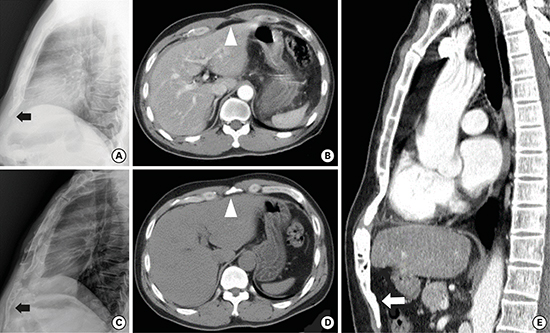
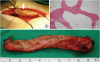
A 53-year-old male visited our hospital due to discomfort in the mid portion of the epigastrium, particularly during a bent or prone position. He previously underwent an abdominal surgery due to traumatic hemoperitoneum in November 2004. He was anesthetized and the peritoneal cavity opened with a long midline incision. After the abdominal surgery, he had been very well. However, 2 years after the abdominal surgery, he sometimes suffered from discomfort in the mid portion of the epigastrium. On physical examination, there was stiff, palpable mass from the epigastrium to the umbilicus in the midline of the abdomen, suggesting a bony structure. However, there was no tenderness, redness and edematous change. A chest X-ray showed the elongation of the xiphoid process (Fig. 1B), whereas, it was not elongated in a chest X-ray at the time of the abdominal surgery (Fig. 1A). When compared at the same level in coronal plane of chest computed tomography (CT) (Fig. 2A), the follow-up chest CT in 2012 showed the new radio opacity which indicated elongation of the xiphoid process (Fig. 2B). A sagittal plane in chest CT showed a protruded and elongated xiphoid process (Fig. 2C). The patient underwent surgical extirpation of the elongated mass of the xiphoid process under general anesthesia in July 2012. The location of the mass was between the linea alba and peritoneal adipose tissue with peritoneal adhesion underlying the abdominal structure. The cartilaginous portion of the mass was ligated and separated with the original xiphoid process (Fig. 3A). The extracted distal xiphoid process was an elongated, approximately 10-cm length cartilaginous mass (Fig. 3B). Histological findings of the resected xiphoid process revealed exostoses (Fig. 3C). He recovered uneventfully and the symptom disappeared immediately after the operation.
Fig. 1
Lateral chest X-ray findings of before (A) and after (B) surgery for traumatic hemoperitoneum. (A) Normal length of the xiphoid process (arrow). (B) Elongation of the xiphoid process (arrow heads).
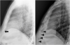
Fig. 2
CT findings of before (A) and after (B) surgery for traumatic hemoperitoneum. (A) There is no radio opacity in horizontal section (arrow). (B) There is new radio opacity (arrow head). (C) Elongated xiphoid process in axial plane (arrows).
CT = computed tomography.
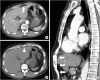
Fig. 3
Macroscopic and microscopic findings of the resected specimen. (A) Intraoperative finding of distal portion of xiphoid process which was cartilaginous (arrow). (B) Histological finding of the resected xiphoid process revealed multiple exostoses. (C) The extracted distal xiphoid process was an elongated, approximately 10-cm length cartilaginous mass.
DISCUSSION
Heterotrophic ossification is a rare phenomenon, first described in 1889 as a complication after spinal cord injury.1 Heterotrophic ossification was also reported after cervical total disc replacement and following cervical arthroplasty.23
It is a well-known fact that the ossification of the cartilaginous xiphoid process progresses with age.4 However, the patient is 53 years old and middle-aged. Furthermore, the patient had a history of abdominal surgery in the past. A postoperative heterotrophic elongation of the xiphoid process is extremely rare, with only 3 cases previously reported.567 Two cases were the postoperative elongation of the xiphoid process after thoracic surgery with median sternotomy.56 One case was the postoperative elongation of the xiphoid process after abdominal surgery with midline abdominal incision.7 Enomoto et al.6 reported postoperative elongation of the xiphoid process after median sternotomy for the operation of mitral valve replacement, and, described that the cause of the elongation of the xiphoid process is believed to be a distraction osteogenesis. In our case, the cause of the elongation of the xiphoid process is believed to be the heterotrophic ossification of the cartilaginous xiphoid process by direct or indirect injury of the xiphoid process during abdominal surgery using long midline incision or minimal fracture because of trauma 8 years before. The xiphoid process, which fractured by the indirect injury or trauma, might stimulate the ossification of the cartilaginous xiphoid process, following which the xiphoid process elongated and reconnected to the sternum. The mechanism is the same as distraction osteogenesis for limb lengthening in orthopedic surgery.8
In conclusion, a postoperative elongation of the xiphoid process is extremely rare. The surgical pathology for postoperative elongation of the xiphoid process after abdominal surgery has not yet been reported. We report an extremely rare case of the postoperative heterotopic ossification of the xiphoid process after abdominal surgery for traumatic hemoperitoneum.
 XML Download