

 PDF
PDF Citation
 Print
Print


INTRODUCTION
Sparganosis is a larval cestodiasis (metacestodiasis) triggered by infection with the plerocercoid of Spirometra spp. of which adults thrive in the small intestine of canine or feline host (https://www.cdc.gov/dpdx/sparganosis/index.html). Humans mostly serve as an intermediate host and are infected with larval worms (plerocercoid: sparganum, pl. spargana). The sparganum might grow into an adult in human intestine in extremely rare cases.1 Drinking unfiltered water that contains cyclops infected with procercoid and/or ingesting raw or inadequately cooked flesh of frogs/snakes or other paratenic hosts are important sources of human infections. Application of raw flesh/skin of poikilothermic intermediate hosts to subside sore edema and ophthalmitis is also a risk factor.234
Sparganosis has been sporadically detected globally, but the majority of cases occurred in East and Southeast Asian countries in relation to their eating habits and traditional medical practice.4 The disease is principally characterized by a slowly migrating subcutaneous nodule(s), but infection may occur in any part of the body. Infection of the worm(s) in vital organs often results in serious illness. The parasite might invoke symptoms as early as two months postinfection.2 Sometimes patients could remain asymptomatic for approximately 17 years.3 Different Spirometra spp. can cause human infections. Spirometra mansoni in Europe, Spirometra mansonoides in America, and Spirometra erinaceieuropaei in Asia including Korea.5 A recent morphological and molecular study has demonstrated that Spirometra decipiens plerocercoid is involved in Korean sparganosis.6
Sparganosis has not been uncommon in Korea. The following factors might be associated with the development of disease. Farmers working in rice paddies or in the forest might drink unfiltered water when they are thirsty during work as a long traditional lifestyle of aquaculture or forestry activity. Some people used to consume flesh of frog and/or snake to treat tuberculosis, arthralgia, and neuralgia.3 Previous seroepidemiological surveys of apparently healthy persons in Korea have demonstrated seropositive rates of 1.7%–3.3% for sparganosis. Large proportions (40.4%–62.5%) of seropositive people were old men aged over sixth decades,78 suggesting that such traditional habits might be linked to hidden infections, although non-specific serological cross-reactions by some other underlying factors could not be ruled out.9
In Korea, an authentic case of sparganosis in the lower leg was firstly found in 1917 which was described in 1924.10 Several hundreds of cases were thenceforward reported. However, systematic approaches of literature survey of human sparganosis in Korea have rarely appeared.2311 In the present study, we provide an overview and perspectives of human sparganosis in Korea by mining the publicly available databases during the period of 1924 to 2015.
METHODS
Literature survey
Published reports were searched from publicly available databases which included PubMed (www.ncbi.nlm.nih.gov/pubmed), Research Information Sharing Service (RISS; https://www.riss.kr), and Korea Medical Citation Index (KoMCI; https://komci.org/). We used terms “sparganosis,” “Spirometra,” and “Korea” in conjunction with associated Medical Subject Headings (MeSH) in the National Library of Medicine (https://www.nlm.nih.gov/mesh/).
Collection of sparganum from Korean terrestrial snakes
We examined 81 terrestrial snakes that had been caught in the southern part of Korea (Gyeongsangnam-do). Those snakes were donated by the Association of Wild Animals Protection for research purpose, including 38 Agkistrodon brevicaudus, 25 Rhabdophis tigrinus, and 15 Elaphe rufodorsata. The snakes were individually anesthetized with ethyl ether and sacrificed. Each snake was examined for the presence of sparganum.
RESULTS
Survey of Korean sparganosis cases in publicly available domains
We were able to collect information on 438 human sparganosis cases from 159 articles published in 51 journals. We found 55 papers in PubMed and 104 papers in two Korean paper search sites (RISS and KoMCI). Twenty-six and 75 articles were independently listed in PubMed and Korean search sites, respectively, among which 29 articles could be found in all search conditions. Out of the 51 Journals, the largest number of articles dealt with human sparganosis had been found in the Korean Journal of Parasitology, which contained 156 case reports. A total of 48 cases of human sparganosis were reported in the Journal of the Korean Society of Radiology. In addition, 35 and 12 cases were retrieved from the Journal of Korean Neurosurgical Society and the Korean Journal of Urology, respectively. The other 187 cases were reported in Radiology, Neuroradiology, Urology, and other international journals (We only listed three major journals that published the most Korean sparganosis).
Epidemiological characteristics
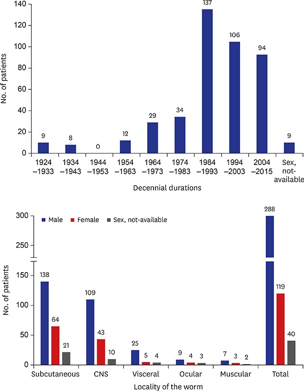
We analyzed the number of sparganosis cases reported in Korea during the period of 1924 to 2015 (Fig. 1). A previous study reported a total of 63 cases during the period of 1924–1974: 58 cases from 1924 to 1973 and additional five cases in 1975.3 At that time, preoperative diagnosis of sparganosis was almost impossible. Moreover, academic capacity to preserve medical literature between 1944 and 1953 had been chaos due to social crisis. Social and medical infrastructures were very poor in Korea, which might have hampered appropriate evaluation of medical situations including those of sparganosis.23
Fig. 1
Decennial distribution patterns of human sparganosis reported in Korea during 1924–2015. At least 438 sparganosis cases were reported in Korea. Cases reported between 1924 and 1953 referred to cases recorded in Korean peninsula and those after 1954 represented sparganosis reported in the Republic of Korea. Duplicated publications were excluded by reviewing individual papers.
aMost medical records in Korea were not preserved during 1944–1953 due to the Pacific War (1941–1945), post-war chaos (1945–1948) and the Korean War (1950–1953).3
Since the late 1970s, human sparganosis continue to occur in Korea. Min11 found additional 56 cases through literature survey between 1975 and 1989. Our literature search revealed that a total of 243 cases were described between 1984 and 2003 (Fig. 1) including those listed by Cho et al.3 and Min.11 Thereafter, cases seemed to be decreased to some extent. Nevertheless, sparganosis cases have been continually reported during the last 15 years (94 cases between 2004 and 2015).
A drastic increase of sparganosis cases from the early 1980s might be due to the development and popularization of diagnostic modalities which allow easier and earlier detection of preoperative patients. Imaging diagnostics including computed tomography (CT)/magnetic resonance imaging (MRI)/ultrasonography (US) have become widely applied for the diagnosis of sparganosis, through which pathognomonic signs for sparganosis are established (see also later sections).1213 Serological tests using crude extracts of the sparganum as antigen has yielded sensitivity of 85.7%–90% and specificity of 87%–97.5% and purified protein fractions of 31 and 36 kDa have exhibited sensitivity and specificity over 95%.91415 By these means, preoperative diagnosis of sparganosis cases, especially those of the central nervous system (CNS) and subcutaneous tissues including breast, has been substantially improved.
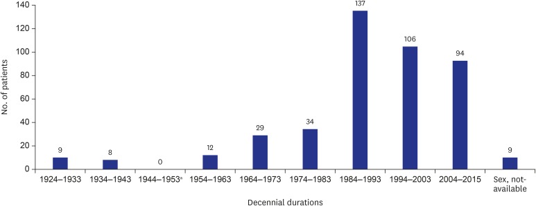
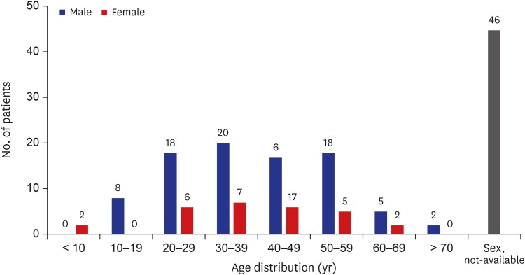
Fig. 2 shows age and gender distribution of patients. Patients were largely diagnosed between their third and sixth decades. However, 10 cases (2.5%) were detected in their early ages (under 10 years), suggesting that people might have been exposed to infection sources from childhood. Among cases with available demographic data (398 cases), the incidence of sparganosis was 2.5 times higher in men (284 cases) than that in women (114 patients).
Fig. 2
Age and sex distribution of sparganosis cases in Korea. Among 438 cases, 284 patients were men and 114 patients were women. We could not trace personal information for 40 cases.
Drinking unfiltered water contaminated with cyclops harboring procercoid and/or contraction with snakes/frogs infected with plerocercoid might be intimately involved in human sparganosis.23 We analyzed such behavioral habits of patients. Ingestion of snake and/or frog was recognized in 63.4% (203 patients) of patients whose eating history was available (320 cases). Drinking unfiltered water was also observed in 16.9% (54 cases) of these patients. However, 19.7% (63 cases) of patients denied exposure to any possible infection source (Table 1).
Table 1
Behavioral habits of Korean sparganosis casesa
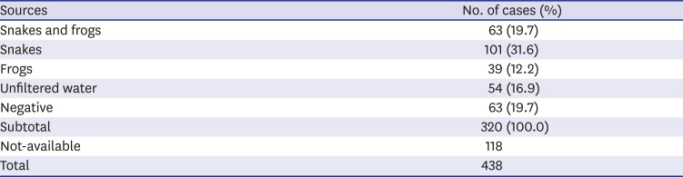
| Sources | No. of cases (%) |
|---|---|
| Snakes and frogs | 63 (19.7) |
| Snakes | 101 (31.6) |
| Frogs | 39 (12.2) |
| Unfiltered water | 54 (16.9) |
| Negative | 63 (19.7) |
| Subtotal | 320 (100.0) |
| Not-available | 118 |
| Total | 438 |
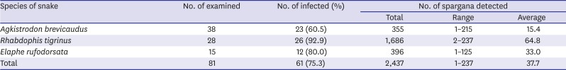
Many studies have observed infection status of the sparganum in frogs and snakes in Korea from the mid-1920s to the mid-1980s.11161718 Examination of snakes and frogs revealed variable infection rates of 3.1%–100%. Infection status of snakes was higher than that of frogs.11 In 2003, we examined 81 Korean terrestrial snakes including 38 Agkistrodon brevicaudus, 25 Rhabdophis tigrinus and 15 Elaphe rufodorsata with an aid from the Association of Wild Animals Protection because those snakes were illegally caught. We observed that the infection rate reached 60.5%–92.5%, with mean worm density of 37.7 worms per snake (Table 2). This result confirmed that snakes might be an important intermediate/paratenic host related to transmission of sparganosis to humans.
Table 2
Infection status of sparganum in terrestrial snakes collected from Gyeongsangnam-do, Korea (2003)
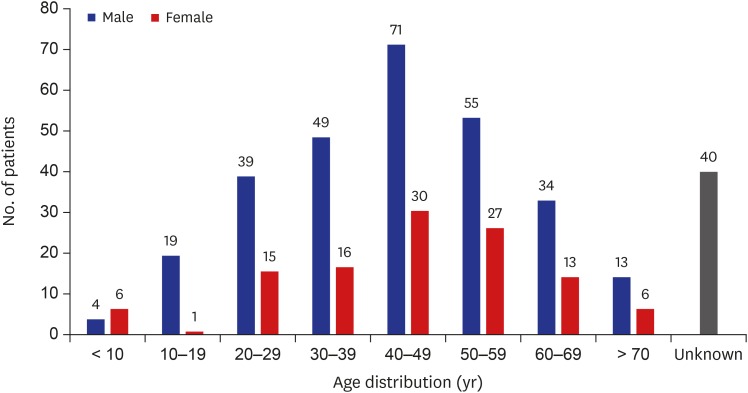
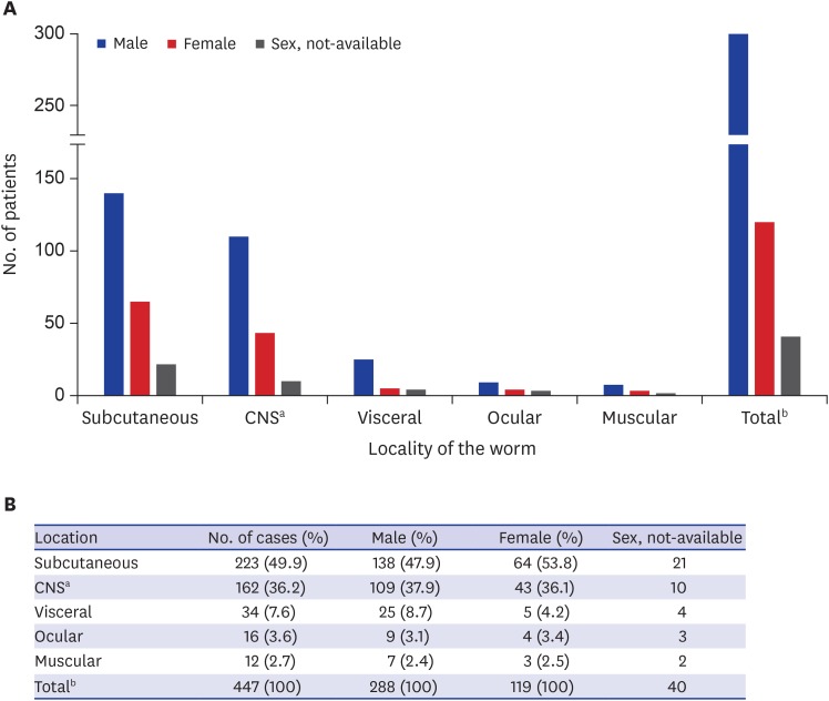
When we analyzed infection sites of sparganosis cases in the human body (438 cases), subcutaneous tissues were the most commonly affected sites (49.9%; 223 cases), followed by the CNS (36.2%; 162 patients) including the spinal cord. Infection of visceral organs was recognized in 7.6% of patients (34 cases). Ocular cases were observed in 16 patients (3.6%). Muscular sparganosis was recorded in 12 patients (2.7%) (Fig. 3). A total number of affected sites exceeded 438 due to infections at multiple sites in seven patients (four cases of subcutaneous and muscular infections, two each case with subcutaneous and visceral infections, and one case of systemic infection).
Fig. 3
Number and sex distribution of cases according to infection sites.
CNS = central nervous system.
aCNS sparganosis included spinal cord infection; bTotal number of cases exceeded 438 due to infections at multiple sites in 7 cases (4 cases of subcutaneous and muscular infections, 2 cases with subcutaneous and visceral infections, and 1 case of systemic sparganosis).
Subcutaneous sparganosis
Sparganosis of subcutaneous tissues was most commonly described in Korea, similar to that observed in other countries.4192021
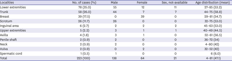
Table 3 depicts localities of lesions in the body, number of cases and gender/age distributions among 218 subcutaneous sparganosis reported in Korea (total number was 223 due to multiple infections). We were able to trace medical records of 202 patients, in which men constituted 68.3% (138/202 cases). The most frequently affected site was the lower extremity (35%), followed by trunk (26%). Involvement of the scrotum was observed in 11.7% of men. In women, breast was the most predilection site (34.2%). Clinical and imaging findings of breast sparganosis have previously been summarized.22 Major symptoms of subcutaneous sparganosis included painless mass (91 cases) and painful mass (39 patients). Other symptoms included itching sensation of the infected sites (23 cases) and hemorrhagic mass (5 patients). Some cases with scrotal sparganosis were associated with Fournier's gangrene (19.2%; 5/26 patients). Eleven patients did not complain of any symptom. We could not trace clinical information for 44 patients. Cases with subcutaneous sparganosis were largely detected in the sixth decade (age distribution, 4–85 years). However, a 4-year-old boy and a 6-year-old boy who visited a clinic with chief complaint of a painful neck mass and a painless cystic mass on the scrotum, respectively, were found to be infected with sparganum. The boy who had a neck mass had no history of exposing to any possible infection source while the other boy with scrotal mass had a history of drinking water from mineral springs.2324
Table 3
Number of subcutaneous sparganosis patients in each body compartment along with sex and age distribution
Plain radiographs of subcutaneous sparganosis often show linear or elongated calcifications. Nodular elongated soft tissue mass shadows might also be observed, but not always.25 US findings include poorly-/well-defined, heterogeneous hypoechoic mass in affected sites and multiple elongated, folded, band-like hypoechoic tracts with serpiginous tubular echogenicity.2526 On CT/MRI, multiple serpiginous tubular tracts having peripheral rim enhancement with perilesional soft tissue edema have been recognized.2728
Sparganosis of the CNS and spinal cord
In Korea, at least 162 cases of CNS involvement including the spinal cord were reported. One case was infected at both CNS and the spinal cord29 while 13 cases were spinal cord infections (Table 4). The proportion of CNS cases was remarkably high in Korea compared to that in other countries.4192021 Since the early 1980s', most cerebral cases were diagnosed preoperatively by specific serological tests together with neuroimaging modalities.912303132 CNS involvement was significantly higher in men (75.9%) than that in women (24.1%) among 116 patients whose demographical information was available. Age of these patients ranged from 6 to 72 years. The third (20.7%), fourth (23.2%), fifth (19.8%), and sixth (19.8%) decades constituted major populations (Fig. 4).
Table 4
Number of cerebral sparganosis cases in Korea
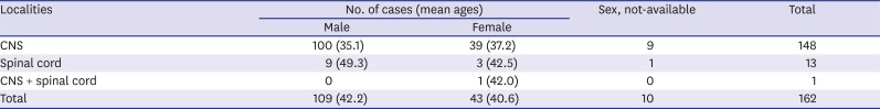
Fig. 4
Age and sex distribution of patients with cerebral sparganosis whose demographic information is available (n = 116).
Neuroimaging findings have demonstrated pathognomonic features of cerebral sparganosis, including white matter hypodensity with adjacent ventricular dilatation, irregular or nodular enhancing lesion, and small punctate calcifications. Cortical atrophy has also been recognized in many patients.30 Contrast images of MRI have better resolution than CT in detecting degenerated tissues.31 On neuroimaging scans taken at follow-up monitoring, location or shape of the enhancing lesion is typically altered, suggesting possible wondering of the worm in the hemisphere. This finding is also highly specific for cerebral sparganosis.31
Major symptoms of 148 CNS patients included seizure (20.9%), headache (12.8%), and hemiparesis (6.8%). Altered mental functions such as cognition difficulty, decreased memory and calculation, and intellectual deterioration were observed in 17 cases (11.5%). In addition, some patients manifested with motor weakness (4.7%). These symptoms were shown to be progressed with wax and wane patterns along with the course of the disease. Two patients (1.4%) underwent status epilepticus. Principal symptoms of spinal sparganosis included voiding difficulty (38.5%), back pain (38.5%), and paresis (23.1%). The patient who had concomitant infections in the CNS and spinal cord presented seizure, signs and symptoms of increased intracranial pressure, facial palsy, hearing loss, voiding difficulty, and paresis of the lower extremities. The patient's symptoms were significantly improved after surgical removal of worms in the cerebellar-pontine angle and in the 3rd–5th lumbar spinal cord together with decompression of communicating hydrocephalus by ventriculoperitoneal shunt operation.29
Visceral sparganosis
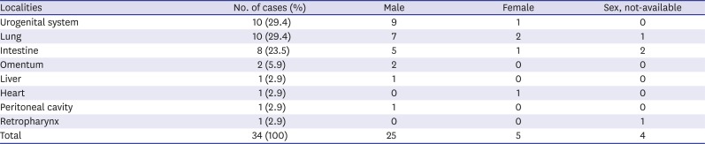
Sparganosis of visceral organs were relatively infrequent in Korea (7.8%; 34/438 patients). Patients' ages were between 23 and 70 years (mean: 50.6 years). Ratio of man and woman patients was 4.8:1. As shown in Table 5, worms were found in several different organs/tissues, among which the urogenital system (29.4%), the lung (29.4%), and the intestine (23.5%) were mainly affected organs. In the urogenital system, bladder wall, epididymis, and penile shaft infections were described. A ureteral stone associated with sparganum has been reported.32 Pleural sparganosis could provoke chest pain, pleural effusion, dyspnea, and/or empyema.33 Sparganum infected in the intestinal wall caused obstruction, perforation, or abscess formation. An intraperitoneal mass causing abdominal pain has been reported.34 Imaging scans of patients have revealed poorly defined, heterogeneous mass with increased echogenicity, and/or cavitating mass. In addition, double hypoechoic rings surrounding the mass, i.e., “tubule-in-a-tubule” appearance could be observed.35 One case of pericardial mass presenting recurrent pericardiopleural effusion, chest pain, and dyspnea was reported. The patient consumed raw frog flesh more than 30 times for treatment of thyroid disease. She was treated with three consecutive doses of praziquantel (75 mg/kg/day), after which symptoms and pericardial effusion gradually disappeared. However, follow-up surveillance of the patient's anti-sparganum specific antibody level remained to be strongly positive.36 A sparganosis causing retropharyngeal abscess has been recorded in the literature, while detailed history was unavailable.3
Table 5
Number of patients and lesions of visceral sparganosis reported in Korea
Ocular sparganosis
In Southeast Asian countries including southern China, large numbers of ocular sparganosis have occurred due mainly to application of traditional poultice to subside sores or edema of inflamed eyes and/or skins.420 However, such types of traditional therapy have not been practiced in Korea,3 which might be responsible for the low incidence of ocular sparganosis in this country. A total of 16 ocular cases (3.6%; 16/438 cases) has been reported (Table 6). Patients' age ranged from 11 to 67 years (man:woman = 3:1). Subconjunctival area was the most predilection site (68.8%), followed by eyelid (25%). Their main clinical symptoms were painless mass with/without itching. One case of intraorbital sparganosis was described in an 11-year-old girl who complained of exophthalmos and itching sensation for a year. Other symptoms such as foreign body sensation, decreased visual acuity, or visual field defect were not observed. The patient was initially diagnosed as intraocular malignancy. During operation, a hard mass tightly adhered to the upper posterior intraorbital wall was resected. A live sparganum was extracted from the lesion. Her mother admitted that she had eaten cooked frog for 2–3 times.37 The worm might have directly invaded into the orbit from the periorbital soft tissue. However, how the sparganum penetrated through the orbital wall surrounded by bony structure remains unknown.

Muscular sparganosis
Skeletal muscular tissues are relatively reluctant to allow penetration and migration of the sparganum due to their compact architectural structure. Sparganum was often found between muscles and fascial layers surrounded by fibrous capsule. Sparganosis of the muscular system was barely reported in Korea. A total of 12 cases were recorded, including seven men and three women (mean age, 61 years). Personal information for the remaining two patients was unavailable. Four cases had multiple infections in both subcutaneous tissues and muscles. Those muscular sparganosis cases revealed US findings of poorly defined intramuscular mass with heterogeneous echotextures in affected sites,27 resembling those seen in subcutaneous sparganosis.26 Most patients had a painless lump on the affected site (8 cases). The worm was discovered in the chest wall muscles in two cases and in the neck and trunk muscle in one case each.
Systemic sparganosis
An extremely uncommon case of systemic infections with multiple worms was described in Korea. A woman who complained of palpable mass on the breast visited a hospital. During evaluation of the breast mass, subcutaneous masses on the elbow and thigh were also noticed. Whole body MRI revealed multiple nodules on the subcutaneous fat layer of several organs, including the breast, elbow, thighs, calf, chest walls, abdominal wall, inguinal area, and suprapubic area. Masses in the psoas and gluteus muscles were additionally detected. A total of 16 worms were removed from respective tissues and muscles. She had a history of eating snakes to relieve leg pain 40 years ago.38
Sparganosis of immunocompromised patients
Sparganosis or proliferative sparganosis associated with immunocompromised patients, such as those with acquired immune deficiency syndrome (AIDS), cancer, or allograft recipients have been sporadically detected worldwide. Proliferative sparganosis in immunocompromised patients often result in serious outcomes.394041 We found that at least five sparganosis cases in Korea were associated with immunosuppressed conditions. All these cases were reported in recent 10 years.
The first case was identified in a woman patient. The patient who suffered from invasive ductal carcinoma of the right breast suddenly developed a mass on the ipsilateral breast. US findings compatible to sparganosis were observed. A live sparganum was removed via excisional biopsy.42 The next case was noticed in a man who underwent allogeneic hematopoietic stem cell transplantation for treatment of myelodysplastic syndrome. The patient was treated with a nucleic acid synthase inhibitor (decitabine), after which a hard and movable mass was developed on the left scrotum. Multiple enlargements of regional lymph nodes were also observed. Pelvic MRI suggested a myeloid sarcoma in the subcutaneous fat layer. However, histopathological specimen obtained from biopsy revealed a characteristic feature of sparganosis.43 Two patients who received chemotherapy for treatment of metastatic rectal cancer or B cell lymphoma abruptly developed hemorrhagic plaques on abdomen or growing subcutaneous masses of the chest and abdomen. Biopsied specimen exhibited histological features of sparganosis. Both patients agreed that they ingested cooked snakes several times.4445
Two sparganosis cases were misdiagnosed as tumor metastasis. A patient was treated for metastatic gastric cancer. After third rounds of anticancer medication, a painless mass was detected on the left lower extremity which was suspected to be a skin metastasis. Tissue biopsy demonstrated a sparganum.46 Another case was seen in a breast cancer patient. She received neoadjuvant treatment, after which an axillary nodule was additionally recognized. During curative surgery for breast cancer, axillary lesion was also resected and a viable sparganum was recovered.47
DISCUSSION
Sparganosis has been continuously detected in Korea during the past 90 years, although annual incidence seemed to be decreased in the last 20 years. However, whether the prevalence of the sparganosis has actually decreased remains unclear because most clinicians in Korea now regard sparganosis as no longer reportable disease unless unusual complications/sequelae have been combined. In this respect, the authors identified more than 15 sparganosis cases affecting various organs/tissues, including thyroid gland from samples referred from several hospitals during recent 10 years in Korea (Kong Y., unpublished observation). Some minor fractions of the elderly people still believe that snakes/frogs have special benefits for muscular tonicity and treatment of arthralgia, tuberculosis, and other chronic wasting diseases. They continue to consume snakes/frogs, although they are aware that these types of convention are illegal with a risk of parasitic infections including sparganosis. Many people who like hiking, trekking and climbing sometimes drink unfiltered water. Those cultural habits and social behaviors might be the major limiting factors for terminating sparganosis transmission in this country.
In this study, we analyzed sparganosis cases reported in Korea by mining publicly available databases. During the period between 1924 and 2015, at least 438 cases were described in the literature (Fig. 1). We surmise that many more neglected cases are not reported in the literature. Most patients (80.3%) have a history of ingesting frogs/snakes and/or unfiltered water. Patients were largely detected in their middle ages between the fifth and sixth decades. The disease was more common in men than in women (man:woman = 2.5:1). Subcutaneous tissues of the lower extremities were the most frequently affected sites (Fig. 3). These epidemiological situations were similar to those observed in other countries.4192021 However, Korean sparganosis shows some differences in terms of clinical and epidemiological aspects.
Since the 1980s, most patients were preoperatively diagnosed by imaging modalities combined with serological tests. Many cases of cerebral sparganosis have been detected in a relatively young age group (Fig. 4). Detection of a large number of cerebral sparganosis patients in relatively young age groups between third and fourth decades (43.1%) suggested that they might have severe diseases that led to early visit to clinics. Histological characteristics of the brain parenchyma might be related to early and deleterious symptoms/signs of cerebral sparganosis. Sparganum might migrate more vigorously within the brain parenchyma compared to other body compartments because brain parenchyma is mainly composed of soft tissue that is vulnerable to mechanical damage triggered by the invasive wondering worm. Bioactive molecules secreted by the sparganum including tissue hydrolyzing enzymes might also contribute to further damage to friable brain parenchyma.48 A previous study has indicated that the frontoparietal lobes are frequently affected sites.30 Infection of the occipital lobe alone was indeed rarely observed in this retrospective survey. However, involvement of parietal/parietooccipital/parietotemporal lobes was noticed to some extent.123031 The sparganum might randomly invade the whole hemisphere. It might not have a specifically favored site, but it depends on the size of the lobe.
Ocular sparganosis is relatively infrequent in Korea. Large proportions of woman patients are infected with the sparganum on the breast. Interestingly, we recognized one extremely rare case of systemic sparganosis who had index lesion on the breast. Most cases of sparganosis were caused by a single worm. Sometimes two or more worms were found simultaneously in different localities of the body. However, systemic infections with multiple worms are very rare. Whether multiple worms might have infected the patient at once or accumulation of worms by repeated infection with a single worm during long periods could not be properly evaluated in this patient.38
Sparganosis associated with immunocompromised patients has recently been reported in Korea. It is not certain whether immune status of patients truly affects exacerbation of sparganosis lesions and symptoms/signs. However, under immunocompromised conditions, mechanical activity of the sparganum and bioactive molecules secreted by the worm might accelerate and aggravate local damages. Several species of proteolytic enzymes might actively operate in the course of infection. These molecules might additionally contribute to the deterioration of lesions.4849 Although authentic case of fatal sparganosis related to immune suppression has not been reported in this country, those unfavorable conditions should be kept in mind.
We could not identify genuine proliferative sparganosis cases in Korea, although one case of multiple subcutaneous sparganosis was highly suspected to be infected with proliferative sparganum.50 However, proliferative sparganosis has been described in several other countries.51525354 Proliferative sparganum (Sparganum proliferum) is a peculiar type of budding sparganum. It is genetically distinct from Spirometra species.55 The parasite may proliferate asexually, branching, and breaking down into numerous segments, each of which has capacity to further develop. Some cases of proliferative sparganosis are associated with immunosuppressed conditions.394041 Occurrence of proliferative sparganosis should be carefully monitored in Korea.
Recent molecular phylogenetic analysis employing mitochondrial cytochrome c1 (cox1) gene has demonstrated that plerocercoid of S. decipiens can trigger human infections in Korea, in addition to plerocercoid of S. erinaceieuropaei.6 The plerocercoid of S. decipiens has also been recovered from Korean terrestrial snakes. Interestingly, all sparganum (n = 904) recovered from snakes were identified as plerocercoid of S. decipiens, but not that of S. erinaceieuropaei.56 When we consider the fact that S. erinaceieuropaei has not been discovered in naturally infected carnivorous hosts,57
S. decipiens might be the responsible species found in the natural sylvatic cycle. These results suggest strongly that sparganum found in human cases should be carefully revisited for their species identification in the near future.
Currently, drugs effective for treating sparganosis are unavailable.58 In this series, we observed that some patients were treated with praziquantel. A few patients seemed to respond to the drug because their symptoms subsided after medication. However, follow-up monitoring of anti-sparganum specific antibody levels demonstrated strong positive reactions,36 suggesting that the sparganum might become dormant in the body, but not eradicated. This situation should be carefully considered when managing patients. Sparganosis of vital organs and/or related to immune suppressions may result in serious consequences. Public health education with regular interview by local health authorities, periodic check-up of suspected cases, continuous monitoring of the prevalence and medical surveillance system through patient-doctor rapport network might be needed in Korea.
 XML Download