

 PDF
PDF ePub
ePub Citation
 Print
Print


INTRODUCTION
Gout is one of the most common types of inflammatory arthritis, and its incidence and prevalence is increasing (1). Gouty arthritis is characterized by hyperuricemia, which results from overproduction or impaired renal excretion of uric acid (2). It was shown recently that gout-associated monosodium urate (MSU) crystals activate the NALP3 inflammasome (3), leading to processing of procaspase-1 to caspase-1, and the production and secretion of active IL-1β and IL-18 (4).
In a recent study of multiple sclerosis, caspase-1 was measured in biological body fluids, and could be used as a marker of inflammation (5). In that study, the concentration of caspase-1 in synovial fluid (SF) was found to be higher in patients with juvenile idiopathic arthritis (JIA) than in patients with other inflammatory diseases.
We hypothesized that caspase-1 would be more active, both within cells and in the synovial fluid, in crystal-induced arthritis (CIA) than in other arthritides such as osteoarthritis (OA), spondyloarthropathy (SpA), and inflammatory arthritides including rheumatoid arthritis (RA). In this study, we evaluated levels of caspase-1 in synovial fluid (SF) which might be useful in the differential diagnosis of CIA from other arthritides.
MATERIALS AND METHODS
Subjects
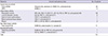
A total of 112 SF samples were obtained from patients with CIA, inflammatory arthritis, OA, and SpA. The patients met the American College of Rheumatology criteria for acute gout (6), RA (7), systemic lupus erythematosus (SLE) (8), and knee OA, and the 1984 New York criteria for ankylosing spondylitis (9). Arthrocentesis was carried out to diagnose and/or to attempt to treat arthritis flares such as acute gouty attacks. The presence of MSU crystals was proven in 22 patients with gout, and calcium pyrophosphate dihydrate (CPPD) crystals were identified in two patients. Only two gout patients were diagnosed clinically. Table 1 gives the diagnoses and current treatments of patients enrolled in the study.
Sampling and assays
Samples of SF were collected from patients requiring knee or ankle arthrocentesis for diagnostic or therapeutic reasons, from September 2010 to March 2012. Plain tubes were used to collect SF for routine examination. Cells were removed by centrifugation at 2,500 RPM for 15 min for ELISAs and the SF was stored at -20℃. Levels of caspase-1, IL-1β, and IL-18 were measured with commercial ELISA kits (caspase-1: Wuhan EIAab Science Co., Ltd., Wuhan, China; IL-1β and IL-18: Quantikine, R&D Systems Inc., Minneapolis, MN, USA; IL-1RA: Invitrogen, UK). The detection range for the caspase-1 ELISA kit was 15.6-1,000 pg/mL. Uric acid in SF was measured with a quantitative colorimetric assay kit as described by the manufacturer (BioAssay Systems, Hayward, CA, USA). C-reactive protein (CRP) and erythrocyte sedimentation rate (ESR) were determined using the data recorded in the medical records within ± 1 day of the study day.
Statistical analysis
All statistical analyses were performed using SAS software, V.9.2 (SAS Institute, Cary, NC, USA). Results are presented as means ± SDs unless specified otherwise. The non-parametric Kruskal-Wallis test was used for between-group comparisons of arthritides and between-phase comparisons of gout. Pearson's and Spearman's correlation coefficients were used to examine the relationships between concentrations of biomarkers and laboratory parameters. The Bonferroni correction was applied to multiple comparisons within each category of variables. Fisher's exact test was used to assess the frequencies of high levels (≥ 125 pg/mL) of caspase-1 in between-group comparisons. P values less than 0.05 was considered significant.
RESULTS
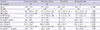
The clinical and laboratory characteristics of the various patients are summarized in Table 2. As expected, age and gender were disease-specific. CRP was highest in patients with CIA and inflammatory arthritis. The white blood cell (WBC) count in SF was highest in CIA, followed by inflammatory arthritis, SpA, and OA in descending order.
Contrary to our hypothesis, caspase-1 in SF from patients with CIA, mostly with gout, was not higher than in the other arthritides (35.9 ± 86.7, 49.7 ± 107.7, 2.1 ± 7.0, and 152.6 ± 155.7 pg/mL for CIA, inflammatory arthritis, OA, and SpA patients, respectively). In fact, caspase-1 in SpA was significantly higher than in the others, even after Bonferroni correction. The frequency of caspase-1 levels ≥ 125 pg/mL was also higher in SpA (62.5%) than in CIA (7.1%), inflammatory arthritis (13.9%), and OA (0%) patients. When the patients with gout were divided into three groups according to the acute gout phases proposed by Scanu et al. (10), the level of caspase-1 in phase I (41.2 ± 48.3 pg/mL, n = 13, 0-48 hr after onset of attack) was higher than in phase II (6.5 ± 11.3 pg/mL, n = 3, days 3-4) or phase III (0 pg/mL, n = 3, days 5-7), but the difference between groups was not statistically significant.
IL-1β and IL-18 were highest in CIA and lowest in OA. Moreover, the ratio of IL-1β to caspase-1 differed significantly between groups. Caspase-1 in SF as a whole, and in CIA or inflammatory arthritis in particular, was not correlated with levels of WBC, IL-1β, or IL-18 in SF (data not shown).
DISCUSSION
We hypothesized that since intracellular caspase-1 is processed from procaspase-1 during MSU-induced acute inflammation and is released into the extracellular environment, as shown by Franciotta et al. (5), there should be a high concentration of caspase-1 in SF in acute gouty arthritis. Unfortunately for this hypothesis, however, caspase-1 was not higher in CIA than in the other arthritides. Moreover, the highest mean level of caspase-1 was found, unexpectedly, in the SF of patients with SpA, and the frequency of high level caspase-1 (≥ 125 pg/mL) was also highest in SpA. In the SF of 18 patients with juvenile idiopathic arthritis (JIA), the level of caspase-1 was 945.5 ± 126.6 pg/mL. It remains to be determined whether caspase-1 is inherently high in the SF from patients with JIA or whether our findings differ from those of other studies because of differences in the methods used. Meanwhile, the levels of IL-1β and IL-18 in this study were compatible with previous results in OA and RA (11-14).
Unlike those of caspase-1, levels of IL-1β and IL-18 were highest in the SF of CIA. This difference between the levels of caspase-1 and IL-1β in CIA may be due in part to caspase-1-independent activation of IL-1β in neutrophil-predominant arthritis (15, 16).
The legitimacy of estimating intracellular pro-inflammatory caspase-1 from measurements of caspase-1 in extracellular body fluids has not been established, but parallelism between the concentration of caspase-1 in cell lysates and in supernatants of THP-1 cells has been demonstrated in vitro (5). In that study the highest mean caspase-1 levels were found in the SF of patients with JIA, which correlated with ESR. These workers did not mention a correlation between caspase-1 levels in serum and SF, although they measured caspase-1 in paired SF and serum samples from patients with JIA. Therefore, they may not have found any correlation.
NALP3 mRNA levels were found to be higher in synovial tissue in RA than in OA, but this was thought to be due to a higher number of infiltrating macrophages rather than synovial fibroblasts in RA synovium (17). Kolly et al. (18) showed that synovial fibroblasts lack NALP3, and therefore are unable to activate caspase-1, and that the main sources of inflammasome-associated IL-1β secretion in RA synovium are infiltrating myeloid cells and endothelial cells. With respect to the histopathological features of the synovial membrane in SpA and RA, the expression of CD163 (a scavenger receptor expressed on mature tissue macrophages) on the lining and sublining layers was reported to be significantly higher in SpA than in RA (19). Judging from observations on the activation of NALP3 in infiltrating macrophages in RA, and the histopathologic characteristics of SpA synovium, the unexpectedly high concentration of caspase-1 in the SF of SpA seems to be associated with the elevated number of infiltrating macrophages. Meanwhile, resident macrophages are very important in initiating MSU crystal-induced inflammation in acute gouty arthritis, whereas neutrophils are the main cells recruited in the acute attack phase of gout (20). The fact that macrophages are abundant in the invaded tissues of patients with ankylosing spondylitis, or undifferentiated spondyloarthropathy (19), not only in synovial tissues (21) but also in sacroiliac joint tissues (22), and even in colonic mucosal tissues (21, 23), has been observed through many studies. Furthermore, in a recent reviews, similar information was mentioned in articles regarding synovial immunopathologic studies for rheumatoid arthritis and spondyloarthropathy (24). Therefore, the increased number of macrophages in peripheral joint lesions in spondyloarthropathy can explain our study results, and caspase-1 is not high in acute gout, even in phase I.
Our study has some limitations. As shown in Table 1, almost all the patients had been taking disease-specific medications. Although arthrocentesis was carried out during arthritic flares, the medication may have affected the results. Since we were focusing on patients with gout, the number of patients with SpA was too small to draw any conclusions about the issue of caspase-1 in SpA.
In conclusion, contrary to our hypothesis, caspase-1 in the SF from patients with gout is not higher than in that from other arthritides. The high level of caspase-1 in SpA could be helpful in differentiating it from other arthritides.
 XML Download