

 PDF
PDF ePub
ePub Citation
 Print
Print


INTRODUCTION
Intracerebral hemorrhage (ICH) is a severe type of stroke and comprises about 15% of total acute stroke. The worldwide incidence of ICH ranges from 10 to 20 cases per 100,000 population and increases with age (1-4). The mortality rate at 6 months after ICH ranges from 23% to 58% (5-7). Given the high mortality and economic burden of ICH, strategies to improve outcome are important. Chronic hypertension is reported to be a major risk factor for ICH. Elevated blood pressure (BP) is observed in 78% to 88% of the patients with ICH (6). Another study estimated that about 45%-55% of non-lobar ICH would be prevented if the effect of hypertension was eliminated (8).
Persistent marked elevation of BP can predispose patients to hematoma enlargement. Factors commonly reported to be associated with poor outcome include hematoma enlargement. Lowering BP is commonly practiced to prevent hematoma enlargement in patients with ICH (2, 3, 9-14). However, the association between elevated BP and hematoma enlargement remains unclear. There has therefore been considerable controversy regarding the initial control of BP after the onset of ICH. On the other hand, there may be areas of disturbed cerebral autoregulation resulting in increased intracranial pressure and focal ischemia adjacent to a hematoma, and reduction of BP has been assumed to promote hypoperfusion and further ischemia. Elevated BP could also be a protective Cushing-Kocher response to preserve cerebral perfusion (2, 3, 8, 10, 13, 15-22).
Nicardipine hydrochloride is a dihydropyridine-derivative calcium channel entry blocker, a potent coronary and cerebral vasodilator, and is water-soluble, making it suitable for intravenous administration; it also has a less negative inotropic effect than other calcium channel entry blockers. A previous report demonstrated that intravenous nicardipine is effective and safe for the rapid lowering of BP. They also showed that continuous intravenous infusion of nicardipine HCl did not affect cerebral arterial flow velocity, intracranial pressure, and cerebral perfusion pressure (18, 23-27). Nevertheless, a few studies assessing the results of nicardipine in patients with acute ICH have been undertaken, and optimal management and its feasibility and effectiveness have yet to be determined.
The primary objectives of this prospective multi-center study were to determine the feasibility of initiating intravenous nicardipine treatment using a standardized BP management protocol within 24 hr after symptom onset and to determine the outcomes of neurological deterioration and hematoma enlargement among patients in whom blood pressure is reduced using intravenous nicardipine medication.
MATERIALS AND METHODS
The study was performed with 88 patients in 5 medical centers with dedicated stroke services. The same protocols were applied in all of the centers as standard practice. The study collected clinical data on all patients treated using the antihypertensive nicardipine protocol as part of their routine care during the study period.
Patient selection
The 88 patients in this study represent a consecutive cohort of patients over 18 months who were treated within 6 hr of symptom onset. This study was performed between August 2008 and November 2010. In patients for whom time of onset could not be determined, the time when patients were last seen intact was taken as the time of onset. Patients were included in whom 1) surgical hematoma evacuation was not performed; 2) supratentorial ICH that was defined as the sudden occurrence of bleeding into the parenchyma of the brain that may extend into the ventricles and, in rare cases, the subarachnoid space, confirmed by clinical history and computed tomography (CT) scan; 3) neurological state was more than 8 points by Glasgow coma scale; 4) volume of hematoma was less than 60 mL in CT scan calculated by length × width × height/2 (volume of hematoma in ventricle was excluded); and 5) BP at admission was more than 140 mmHg. Patients were excluded in whom 1) time of symptom onset could not be reliably assessed; 2) progressive onset or fluctuating characteristics were observed; 3) previously known neoplasms, arteriovenous malformation, intracranial aneurysms, trauma were documented; or 4) location of ICH was infratentorial such as cerebellum, brain stem, or ventricle only; 5) any history of bleeding diathesis or coagulopathy or medication that promote the coagulopathy was present; and 6) any history of congestive heart failure, renal failure, myocardial infarction, or blood glucose level was less than 50 mg/dL or more than 300 mg/dL was reported. The study included patients who were evaluated as requiring antihypertensive intravenous nicardipine medications.
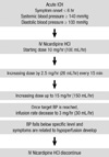
Treatment of Acute Hypertension Protocol (Fig. 1)
The goal in the treatment group was to maintain systolic BP < 140 mmHg and diastolic BP < 100 mmHg within 24 hr from onset of symptoms. Initial treatment was started using intravenous nicardipine (10 mg/hr) if required as follows: if target blood pressure was not achieved, the dose was increased by 2.5 mg/hr every 15 min up to the maximum tolerable dose (15 mg/hr), or until side effects of nicardipine limited the use of the regimen prior to reaching maximum dose.
The protocol was made available as preprinted order sheets at each center and incorporated as routine orders in the chart after initial evaluation. BP was monitored using an intra-arterial catheter or automated cuff inflation device at the discretion of the physician. The lower end of systolic BP and diastolic BP was 110 mmHg and 70 mmHg, respectively. If the BP fell below the specified levels and symptoms related to or possibly exacerbated by hypoperfusion developed, antihypertensive medications were discontinued and further management was performed according to the direction of the treating physician.
Outcome data
From a standard questionnaire, data were recorded on all patients who were treated using the present protocol for antihypertensive medication. We collected information from each eligible patient's medical record, that is, age, sex, and the presence of any of the following risk factors before onset of hemorrhage: hypertension, diabetes mellitus, previous cerebral stroke, and heart disease. The information was acquired on presentation from either the patient or family or obtained from previous medical records. Neurological status at presentation was determined using the initial Glasgow Coma Scale (GCS) score as documented by initial examination. Any episodes of neurological deterioration, defined by a decline in GCS by 2 points or greater that is not explained by use of sedatives or hypnotics, were recorded.
The patients were routinely evaluated for neurological state at admission within the first 24 hr using the GCS score. Based on these investigations, neurological deterioration was further categorized as related to hematoma expansion, ventricular blood, and hydrocephalus. Outcomes at 3 months were graded according to the modified Rankin scale. Outcome for discharged patients was determined at the time of an outpatient clinic visit.
Measurement of hematoma volume
The hematoma volume was determined from the baseline CT scan for all patients and from a 24-hr CT scan using a bedside method by the participating physicians. The location of ICH in this study was basal ganglia (49 patients), thalamus (29 patients) and subcortical cortex (10 patients). An attempt was made to perform measurements without knowledge of the patient's clinical condition. For the bedside length × width × height/2 method, the CT slice with the largest area of hemorrhage was identified. The largest diameter (A) of the hemorrhage on this slice was measured. The largest diameter perpendicular to A on the same slice was measured next (B). Finally, the approximate number of 10 or 5 mm slices in which the ICH was visualized was determined (C). These CT hemorrhage slice values were then added to determine the value for C. All measurements for A and B were made with the use of the centimeter scale on the CT scan to the nearest 0.5 cm. A, B, and C were multiplied and the product divided by 2 to yield the volume of hemorrhage in cubic centimeters. Broderick et al. (28) and Kothari et al. (29) demonstrated excellent agreement between hematoma volumes measured by the bedside technique and computerized planimetric method.
Hematoma enlargement was defined as an increase in the volume of intraparenchymal hemorrhage of > 33% on the 24-hr CT compared with the baseline CT scan. The cutoff for hematoma enlargement is based on the definition provided by Brott et al. (30). They recommended a cutoff of 33% because a 33% change in the volume of a sphere corresponds to a 10% increase in diameter, a clear difference to the naked eye of a physician viewing serial CT scans of a patient with ICH. Second, measurement of serial CTs in patients with ICH indicated that some patients can have up to a third less volume of hemorrhage on the 1-hr CT than on the baseline CT owing to different positioning and angles of the CT slice images rather than to an actual decrease in hemorrhage volume. This observation was particularly true for small hemorrhages. Therefore, the present CT definition of hematoma enlargement is most likely to represent true hemorrhage growth and not variability in CT imaging.
RESULTS
A total of 88 patients were included in this registry. They included 53 men (60.2%) and 35 women (39.7%) with a mean age was 58.3 yr (range 26-87 yr). There were preexisting known risk factors such as hypertension in 44 patients who required antihypertensive medication; other risk factors included diabetes mellitus (8 patients), previous cerebral stroke (3 patients), coronary heart disease (2 patients), and end-stage renal disease (1 patient). No significant abnormality was observed in laboratory data including the total protein, serum glucose, liver function, and platelet function.
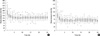
Hourly BP recordings for the first 24 hr of treatment are presented for all of the treated patients. The mean initial SBP/DBP at admission was 175.4 ± 33.7 mmHg and 100.8 ± 22 mmHg, respectively. Administration of nicardipine by protocol resulted in a decrease in mean SBP/DBP during the first 2 hr after infusion. However, after 2 hr, there were no changes in mean SBP/DBP up to 24 hr (Fig. 2). The mean SBP/DBP at 6 or 24 hr were 127.4 ± 16.7 mmHg and 67.2 ± 12.9 mmHg or 130.7 ± 14.3 mmHg and 67.7 ± 9.2 mmHg after infusion, respectively (P < 0.001, the mixed-effect linear models).
All patients underwent follow-up CT at 24 hr. Among them, hematoma expansion (more than 33% increase in hematoma size) at 24 hr was observed in 3 (3.4%) of 88 patients. Initial mean hematoma volume was 19.83 ± 20.8 mL. After administration of nicardipine by protocol, there was no significant change of hematoma volume at 24 hr (17.6 ± 20 mL, P < 0.05).
Neurological deterioration (defined as a decrease in initial Glasgow coma scale ≥ 2) was observed in 2 (2.2%) of 88 patients during treatment. Initial mean Glasgow coma scale (GCS) was 13.6, and GCS at 3 months was 14.3 (P < 0.05). Likely functional independence (modified Rankin scale ≤ 2) at 3 months was observed in 3 (3.4%) of 88 patients.
Hydrocephalus was found in 10 patients and ventricle blood in CT was found in 26 patients. However, these data were not related to results such as a decrease of BP, change of hematoma volume, and neurological state. Adverse drug reaction to nicardipine was found during treatment in 12 (13.6%) of 88 patients. These were hypotension (11 patients), creatinine elevation of unknown etiology (1 patient), and scalded skin syndrome (1 patient). Patients with hypotension fell below the specified levels, and no symptoms related to exacerbated by hypoperfusion developed; therefore, discontinuation of nicardipine and management were performed under the direction of the treating physician. The other patients were treated with proper management without any residual symptoms.
DISCUSSION
In terms of treatment of acute hypertension in patients with ICH, the appropriate control of BP is still controversial. Many previous studies have identified hypertension as a major risk factor for the development of primary and recurrent ICH. Persistent uncontrolled hypertension, especially systolic BP over 200 mmHg in acute ICH, may increase the risk of ongoing or recurrent bleeding leading to hematoma enlargement. The strength of association between BP and ICH appears to be greater than that observed for ischemic stroke. Furthermore, because of chronic hypertension, large proportions of patients with ICH have cardiac hypertrophy and diastolic dysfunction and decompensate in the presence of high afterload. Therefore, investigators are recently recommending treatment of acute hypertension using antihypertensive medication in patients with ICH (2, 3, 9-17, 21, 31, 32).
Untreated severe hypertension may also cause excessive perfusion pressure, which may promote or worsen existing edema, increase intracranial pressure and ischemia, and cause failure of cerebral perfusion. Worsening cerebral edema has been implicated in neurological deterioration that occurs within 24-48 hr after the onset of hemorrhage (2, 11, 15, 28, 30-34). Several previous studies have suggested the presence of transient reduction in regional cerebral blood flow in regions both surrounding and distant from the hematoma, presumably induced by compression of adjacent microvasculature. It is further hypothesized that autoregulation is impaired in the perihematoma region due to local ischemia and acidosis. Therefore, reduction in systemic BP may theoretically further impair blood flow regions with reduced regional cerebral blood flow and provoke ischemia. In addition, reducing systemic BP may lead to autoregulatory vasodilatation of cerebral vessels and adversely affect intracranial pressure (2, 3, 8, 10, 13, 15-22). Powers et al. (26) evaluated the effect of BP reduction in 14 patients with supratentorial ICH 6 to 22 hr after onset. Regional cerebral blood flow was measured using positron emission tomographic scan. Pharmacologic reduction of mean arterial pressure with nicardipine by up to about 17% from baseline (mean arterial pressure 143-119 mmHg) produced no change in global or peri-clot regional cerebral blood flow while maintaining the cerebral perfusion pressure at > 65 mmHg. Autoregulation was not impaired and there was no evidence of cerebral ischemia. Another factor that determines the pathologic significance of regional cerebral blood flow change on the tissue is the metabolic demand of the regions involved.
Nishiyama et al. (23) determined the effect of intravenous nicardipine infusion (initiated at 1 µg/kg/min and titrated to maintain systolic BP between 120 mmHg and 160 mmHg) on intracranial pressure, middle cerebral artery flow velocity, and CT findings of rebleeding and edema in 22 patients with putaminal ICH after surgical evacuation. Intracranial pressure decreased during the infusion with no change in mean middle cerebral artery flow velocity or evidence of rebleeding or exacerbation of edema.
In this prospective multi-center study, we did not find any evidence to suggest excessively high rates of hematoma expansion and neurological deterioration in patients with ICH who received intravenous nicardipine antihypertensive medication within 6 hr of symptom onset. Among patients who underwent follow-up by CT, hematoma expansion at 24 hr (more than 33% increase in hematoma size at 24 hr) was observed in 3 (3.4%) of 88 patients. Neurological deterioration (defined as a decrease in initial Glasgow coma scale ≥ 2) was observed in 2 (2.2%) of 88 patients during treatment. Likely functional independence (modified Rankin scale ≤ 2) at 3 months was observed in 3 (3.4%) of 88 patients.
The rates of hematoma expansion and neurological deterioration were compared favorably with those described in previous studies (12, 14, 25, 30, 34). Also, we did not find any case of transient reduction in regional cerebral blood flow and impairment of autoregulation to provoke ischemia.
Kazui et al. (12) reported that poorly controlled diabetic patients with high systolic BP at admission were at high risk of hematoma enlargement. Rapid expansion of ICH in putamen and thalamus occurred in 19.4% and 8.3%, respectively (30, 34). Other studies have reported a rate of neurological deterioration ranging from 23% to 33% (14, 25). The rates of hematoma enlargement range from 20% to 38% (12, 30). These findings are consistent with other previous studies that found no detrimental effects of antihypertensive medication on cerebral blood flow, cerebral edema, or intracranial pressure (23, 26).
There are some limitations to our study in design. The first limitation relates to adequacy of BP control. Target BP in our study (SBP < 140 mmHg, DBP < 100 mmHg) was relatively lower than that in other studies (9, 10, 24), given that baseline BP tends to be high in chronic hypertension patients, especially in old age group. In chronic hypertension, it is reported that there is an upward shift in the lower limit at which autoregulation of cerebral blood flow is maintained, although the amount of such a shift is unpredictable. Therefore, the study does not specify the exact threshold of tolerance for reduction in blood pressure after ICH. The threshold of tolerance is also expected to vary based on presence of chronic hypertension. A history of hypertension was documented in most but not all patients, which may reflect a certain heterogeneity in response. Second, the selection criteria of patients in this study were small ICH volume (less than 60 mL) and relatively good GCS score. These findings may not be an accurate reflection of ICH in exclusion of large-sized ICH. Although target systolic BP might have tended to be set high in patients of advanced age or with a large volume of hematoma, hematoma enlargement occurred more often in younger patients with smaller hematomas. And, those with large ICH may have increased intracranial pressure associated with a secondary rise in systemic BP. As cerebral arterial perfusion is a function of the difference between systemic BP and intracranial pressure, a decrease in systemic BP may compromise cerebral perfusion. This problem may be exacerbated in patients with chronic hypertension. Third, our study with a relatively small number of cases (88 patients) with no control group is not sufficient to definitively conclude the feasibility and safety of acute ICH treatment with nicardipine. We cannot rule out the presence of unknown, confounding variables, not accounted for in the final analysis. This study provides preliminary estimates of outcomes in a series of patients with ICH who were treated using intravenous nicardipine within 6 hr of symptom onset. A low rate of neurological deterioration and hematoma expansion was observed in the treated patients. Further clinical trials with a large, well-designed, randomized, controlled trial are required to confirm our results. It is suggested that intravenous therapy with nicardipine is effective and safe in ICH patients with hypertension.
 XML Download