

 PDF
PDF ePub
ePub Citation
 Print
Print


INTRODUCTION
Although the exact pathogenesis of systemic lupus erythematosus (SLE) is unknown, several drugs cause drug-induced lupus erythematosus (DILE) (1). DILE is a rare disease that is diagnosed when the following features are present: 1) exposure to a drug suspected to induce DILE; 2) no history of SLE prior to drug therapy; 3) detection of positive antinuclear antibodies (ANA) with at least one clinical sign of SLE; 4) rapid improvement and a gradual fall in ANA, and other serologic findings, upon withdrawal of the drug (1).
Bullous SLE is a kind of LE-non-specific bullous skin disease in which autoantibody-mediated subepidermal blistering occurs. Histopathological analysis showed marked neutrophil infiltration with papillary microabscess formation (2). To date, the only known drugs reported to induce bullous SLE are hydralazine and penicillamine.
We present here the first case of bullous SLE triggered by methimazole, which was treated with prednisolone, dapsone, hydroxychloroquine, and methotrexate, finally progressed to SLE nephritis.
CASE DESCRIPTION
A 31-yr-old woman presented with generalized erythematous to brownish patches and multiple bullae. She was diagnosed with Graves' disease in July 2008, with a decreased TSH level (0.05 µIU/mL; reference range [RR] 0.15-5.0 µIU/mL) and increased free T4 level (21.2 µg/dL; RR 4.6-14.0 µg/dL). Propylthiouracil was prescribed for 6 months, but was changed to methimazole in December 2008 due to inadequate control of hyperthyroidism. One month after the introduction of methimazole, generalized erythematous maculopapular rash developed over her whole body but responded to topical steroids. The woman developed arthralgia of both knees and elbows in early July 2009, which resolved spontaneously within 1 month. At the end of July, intra-oral blisters developed with erythematous patches on both extremities accompanied with pruritus. The skin lesions then spread to the whole body. In early August, multiple 0.5-2.0 cm diameter bullous lesions developed from some of the previous patches. The bullous lesions usually developed on the sites where pruritus was present. Methimazole was discontinued on suspicion of drug allergy on August 10, and 131I therapy was administered on August 19, 2009. Prednisolone at a dosage of 15 mg/d was initiated on August 25 for the skin lesions but was not effective. Hydroxychloroquine, which was initiated on September 25, was not effective either. Only dexamethasone brought temporary relief. Fever developed (38.5℃) from September 23, 2009, and the woman was referred to the rheumatology division for further evaluation. She had no past medical history or personal familial history of bullous disease.
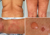
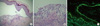
On physical examination, her blood pressure was 130/94 mmHg; pulse rate, 90/min; and body temperature, 38.5℃. Her eyes had reddish conjunctivae. Her voice was husky, the oral cavity was filled with multiple ulcerations, and both lips were swollen. Multiple dark-reddish annular lesions and dark-brownish patches were observed on her trunk, extremities, palms, and soles. Several blisters that varied in size (0.5-2.0 cm) were found on both extremities, and on her palms and soles (Fig. 1). However, neither lymph node enlargement nor hepatosplenomegaly was detected. Chest auscultation revealed no evidence of abnormal sound or friction rubs. Joint examination revealed no evidence of arthritis. Laryngoscopic examination revealed severe inflammation and erosion of the larynx. Ophthalmology examination showed bilateral conjunctivitis. Laboratory test results were as follows: white blood cell count, 5,070/µL; hemoglobin, 9.5 g/dL; and platelet count, 221,000/µL; reticulocyte count, 5.56%; haptoglobin, 10 mg/dL (RR 30-180 mg/dL). The result for Coombs' test was positive, suggesting hemolytic anemia. Viral cultures for Herpes simplex, Varicella zoster, and cytomegalovirus were all negative. Her ANA was over 1:320, homogeneous pattern, anti-histone antibody (47.0, RR < 40.0 U/mL), and anti-ds DNA were positive (56.5, RR 0-10 IU/mL). Also, anti-cardiolipin IgM and Anti-neutrophil cytoplasmic antibody (ANCA) were positive. Rheumatic factor, anti-Sm, lupus anticoagulant, anti-Ro, and anti-La antibodies were negative. Complement levels were decreased with C3 being 70 mg/dL (RR 70-150 mg/dL) and C4, 7 mg/dL (RR 10-35 mg/dL). The bulla on her left thigh was biopsied, and histopathologic examination showed a subepidermal blister with abundant neutrophils (Fig. 2). Direct immunofluorescence examination showed linear deposition of lgG, lgA, C3, fibrinogen, and C1q at the dermo-epidermal junction (Fig. 2). There was no evidence of vasculitis.
Because bullous SLE was suspected, 40 mg/d prednisolone and 300 mg/d hydroxychloroquine were started on September 28, which alleviated the fever, and relief provided relief from the annular skin and patch lesions. The bullous skin lesions, which were refractory to prednisolone, were partially relieved with the introduction of 125 mg bid dapsone on October 1. Methotrexate at 15 mg/week was required for complete relief of the bullous lesions. The redness of the eyes disappeared slowly, and her voice returned to normal. After discharge, at 12 months, lupus nephritis occurred and she under the treatment with prednisolone, mycophenolate mofetil.
DISCUSSION
In this case, SLE developed after the administration of methimazole, with multiple oral ulcerations, arthritis, hemolytic anemia, low complement levels, and anti-nuclear, anti-cardiolipin, and anti-ds DNA antibodies. Anti-histone antibodies were also present. In addition to fulfilling the classification criteria for SLE (3), this case satisfied the diagnostic criteria for bullous SLE proposed by Yell: 1) subepidermal blistering in SLE, and 2) IgG, IgA, C3, fibrinogen, and C1q deposits at the basement membrane zone on direct immunofluorescence examination (4).
DILE is a lupus-like disease caused by exposure to drugs. Like idiopathic SLE, DILE can be classified into systemic, subacute, and chronic forms depending on the characteristics of the cutaneous manifestations. The drugs frequently implicated in systemic DILE are hydralazine, procainamide, isoniazid, methyldopa, chlorpromazine, quinidine, and minocycline (5). For subacute forms, calcium channel blockers, angiotensin-converting enzyme inhibitors (6), docetaxel (7) and thiazide diuretics are reported to be frequent triggers. Antithyroid drugs such as propylthiouracil and methimazole have also been reported to cause systemic DILE (8-12). Among the 8 cases of methimazole-induced SLE reported to date, none had bullous SLE (13). One case in which vesiculobullous skin lesions developed after administration of methimazole was due to antineutrophil cytoplasmic antibody-positive vasculitis, and did not satisfy the criteria for SLE (14). Drug-induced bullous SLE has been reported for hydralazine and penicillamine (15, 16).
Our case showed peculiar autoantibody profiles. Drug-induced lupus is generally characterized by anti-histone antibodies, but subacute forms, which include annular papulosquamous and bullous forms, are reported to show anti-Ro and anti-La antibodies without anti-histone antibodies (8). However, our patient showed anti-dsDNA and anti-histone antibodies without anti-Ro or anti-La antibody. These autoantibody profiles suggest that the disease status in our patients was between the systemic and subacute forms of lupus. At the time of initial presentation, drug-induced bullous SLE was the more favored diagnosis than idiopathic SLE, based on the reasons that follow. First, it developed 1 month after the use of methimazole. This is consistent with a previous report that DILE develops 2 weeks to 3.2 yr after use of a drug (8, 17). Second, the rash was distributed over the woman's whole body, including the lower extremities. In idiopathic SLE, rash usually develops on the sun-exposed upper body, but it can develop over the whole body, including the lower extremities, in DILE cases (5, 18). Third, systemic signs were not prominent in our case. There was a fever, but other common systemic symptoms in DILE, such as arthritis and serositis, were absent (19). But, after 1 yr, she presented with lupus nephritis, which means methimazole triggered SLE later.
For bullous SLE, dapsone is the cornerstone treatment; it induces a dramatic response within a week (8). In some cases where an adequate response is not achieved with dapsone, other immunosuppressants, such as prednisolone, methotrexate, and azathioprine, can be tried (20). In our case, dapsone alone was not sufficient to control the symptoms, and prednisolone, methotrexate, and hydroxychloroquine were also required for control of the symptoms. The combined features of systemic and subacute forms in our case could have necessitated combined treatment.
In conclusion, we report the first case of bullous SLE triggered by methimazole. The findings in our case suggest that SLE should be considered as a differential diagnosis when bullous skin lesions develop in patients treated with methimazole for hyperthyroidism.
 XML Download