

 PDF
PDF ePub
ePub Citation
 Print
Print


INTRODUCTION
Acupuncture is being increasingly used to treat a variety of conditions (1). Acupuncture is known to be a safe procedure. However, various adverse events do occur regarding its misplacement in the body. There has been no report documenting the whole acupuncture needle that migrated to the pleural cavity or lung parenchyma in published English journals. We experienced five patients who had acupuncture needles in their thoracic cavity during last 10 yr. Thoracotomy was performed in the earlier three patients to remove the acupuncture needles, but video-assisted thoracoscopic surgery was performed in two patients later as the development of minimally invasive surgery. Herein we introduce our diagnostic and therapeutic experience for the migration of acupuncture needle to the pleural cavity and or lung parenchyma.
MATERIALS AND METHODS
We analyzed retrospectively the medical records of the patients who were diagnosed with migration of acupuncture needles to the pleural cavity and or lung parenchyma from January 2000 to September 2009 in the Department of Thoracic and Cardiovascular Surgery, College of Medicine, Kyung Hee University, Seoul, Korea.
The diagnosis was made by the chest radiography and chest computed tomography which revealed straight metallic materials in the pleural cavity and or lung parenchyma in the patients who had been treated with acupuncture. Treatment consisted of the administration of antibiotics empirically followed by early removal of the acupuncture needles via thoracotomy or thoracoscopic procedures.
RESULTS
There were 5 patients diagnosed with migration of acupuncture needles to the pleural cavity and or lung parenchyma during last 10 yr. Mean age of the patients was 55.8 yr old. Three patients were male and two patients were female. All patients suffered from the sequela of the previous cerebrovascular accident. Mental status was alert in one patient and drowsy in four patients. Motor status was hemiplegia in two patients and quadriplegia in three patients. All the patients had been treated with acupuncture as a conservative therapy to the sequela of the cerebrovascular accident. Three patients had dyspnea and fever, and other two patients had dyspnea or fever. Four patients had one acupuncture needle in hemithorax and one patient had two acupuncture needles, one in right-side and another in left-side hemithorax (Table 1).
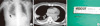
Treatment strategy was administration of antibiotics empirically followed by early surgical removal of the acupuncture needle with or without pleural decortications. All the six needles from the five patients were located in the thoracic cavity. Four needles were located in both pleural cavity and lung parenchyma, one needle in pleural cavity, and one needle in lung parenchyma. Pleural decortications were needed in the four patients because empyema was also presented. One patient who had two needles in both sides of thoracic cavity was treated with removal of the two needles and evacuation of pleural fluid in left side and adhesiolysis only in right side because pleural cavity of the right side had adhesion only without pleural effusion. Wedge resection of the lung was performed in only one patient who had necrotized lung around the acupuncture needle (Fig. 1). Surgical approaches were thoracotomy in the first three patients and thoracoscopic procedure in the last two patients. The last patient, who had two migrated needles, was treated with bilateral thoracoscopic procededures in one-stage operation (Table 2, Fig. 2).
The total length of the removed acupuncture needles was 5-6 cm; handles of 2 cm and needles of 3-4 cm. Handles consisted of the spiral coil and its thickness was only 1 mm. Postoperative courses were uneventful in all the patients and were discharged without fever or respiratory symptoms.
DISCUSSION
Acupuncture is performed to manage various symptoms such as paresis following a stroke, neuralgia, myalgia, headache, dyspepsia, sprain and so forth. The risk of serious events occurring in association with acupuncture is low when compased to other common medical treatment, but the risk for any individual patient may vary with the practitioner (2). In acupuncture, from only a few to hundreds of needles are used and the tip of the needle is placed at subcutaneous or muscle layer. If the needle is placed too deep in thorax, the tip may be wrongly positioned at thoracic cavity and may penetrate lungs or heart and lead to serious adverse events.
Common acupuncture-related adverse events are pneumothorax and injury to the central nervous system (2). The needle used in acupuncture is so fine that the symptoms of pneumothorax due to acupuncture are generally mild, and some cases show spontaneous recovery, but there has been a such case involving bilateral tension pneumothorax after acupuncture, which led to the mortality of the patient (3). There was a report that thoracoscopic removal of the migrated embedded acupuncture needle caused pneumothorax. In that patient, only tip of the needle was located in the pleural cavity (4). Also, there was a report of multiloculated empyema following acupuncture. In that patient, acupuncture needle was not presented in the pleural cavity (5). Regarding to the heart, there were cases of cardiac tamponade caused by acupuncture needle penetrating the right ventricular free wall (6, 7).
To the best of our knowledge, there was no report of empyema caused by fully migrated acupuncture needle into the thoracic cavity in published English journals. After acupuncture treatment on the chest wall, not removed needles may penetrate the chest wall and place itself into the pleural cavity. In our cases, the whole needle including its handle was placed in the thoracic cavity. To our knowledge, all of our patients had been received acupuncture treatment from doctors of oriental medicine. The exact time of migration into the pleural cavity was obscure but the patient was referred soon after the detection of the foreign body on chest radiography which was taken during routine evaluation of high fever. We thought that because the patient was quadriplegic and unable to communicate, the remaining needle traveled itself along the subcutaneous and muscle layers during his and her motion.
If an acupuncture needle was lost during acupuncture therapy, or if symptoms of high fever or dyspnea develops during or after acupuncture therapy, simple radiography or other imaging studies should be performed to investigate the possibility of a needle being placed in the body. In particular, exceptional care should be provided to quadriplegic patients, who are not able to communicate. If the acupuncture needle migrates to the pleural cavity, the occurrence rate of empyema is very high. Pleural decortication was needed in the four out of five patients and drainage of pleural effusion was needed in one patient in our series. The tip of needle was penetrated to the lung parenchyma in the four out of five patients but wedge resection of the lung was necessary only in one patient who had focal severe necrotizing pneumonia around the needle.
Additionally, if a needle is found in the thoracic cavity, administration of empiric antibiotics with early surgical removal is the best management option. As the development of minimally invasive surgery, we performed thoracoscopic surgery in the last two patients. Although we experienced only five patients who have migrated acupuncture needles in their thoracic cavity, we suggest that thoracoscopic removal of the acupuncture needle with or without pleural decortication is the most optimal modality of treatment in those patients.


 XML Download