

 PDF
PDF ePub
ePub Citation
 Print
Print


INTRODUCTION
The incidence of endometrial carcinoma in Korea has increased rapidly in the last ten years possibly due to an increasingly westernized life style, decreased number of pregnancy, and increase of postmenopausal hormone therapy (1). Surgery is the treatment of choice and total hysterectomy with bilateral salpingo-oophorectomy is the cornerstone of both the staging and treatment of endometrial carcinoma. However, although the International Federation of Gynecology and Obstetrics (FIGO) adopted surgical staging in 1988, and the National Comprehensive Cancer Network (2) also recommended standard surgical procedures, there is a significant variety in the actual surgical procedures employed in the treatment of patients with endometrial cancer. The management of patients with cervical involvement remains controversial and treatment options range from extrafascial hysterectomy (EH) to radical hysterectomy (RH), with or without radiation (3, 4). However, the literature regarding the treatment of these patients is limited by its low frequency.
Cervical extension of endometrial carcinoma represents about 10% to 15% of cases at the time of diagnosis (5). Such cervical invasion is considered to increase the risk of nodal metastasis; and those patients are reported to have a worse prognosis than those without it (6-9). The rationale for performing RH in patients with cervical involvement is to eradicate all possible parametrial involvement (PMI). However, the preoperative detection of cervical involvement or parametrial disease is not entirely reliable despite the progress in imaging techniques such as magnetic resonance imaging (10, 11). Thus, although the only definitive method of ensuring the status and extent of disease in the parametria is through radical excision and a histological study of the operative specimen, the unreliability of preoperative prediction, and the relative rarity of PMI may make it unwise to resort to RH as a matter of routine in patients with cervical invasion (12).
We reviewed the medical records of endometrial cancer patients with cervical involvement to determine the relation of parametrial spread with other histopathological variables and to evaluate whether the type of primary surgery using EH or RH alters the patients' outcome from a histological perspective.
MATERIALS AND METHODS
The cancer database from five different institutes were reviewed; patients surgically diagnosed with stage II and a part of stage III or IVa endometrial cancer with cervical involvement during the period of 1993-2005 were identified. The recorded data were then extracted by a medically qualified gynecology trainee in each institute. The original pathology reports and operation reports of 133 patients were reviewed for the following information: age at diagnosis, pre-operative data, operative procedure, adjuvant treatment, follow-up data including date of recurrence, the patient status at last visit. A pathologic data such as FIGO stage, histologic type, tumor grade, depth of myometrial invasion, and the status of parametrium, and retroperitoneal lymph node were also reviewed. Patients with mixed Müllerian tumor, including carcinosarcoma or endometrial stromal sarcoma, were excluded from the review. When the tumor grade varied between pre-operative biopsy and final pathologic reports, the higher grade was recorded.
All surgeries were performed by board-certified gynecologic oncologists. Confirmation of cervical invasion and staging was made by reviewing pathology reports, although pathology slides review was not performed. The term 'Radical hysterectomy' included modified hysterectomy (MRH) that could not be exactly differentiated in operating records.
Patients with surgically staged IIa cancer who had undergone EH were selectively treated with adjuvant radiation according to the degree of depth of myometrial invasion. Otherwise, most patients with stage IIb disease or above underwent post-operative pelvic irradiation or brachytherapy. If sufficient follow-up information regarding survival and recurrence was unavailable through medical records, we referred to death certificates to obtain the required information.
Statistical analysis was performed using SPSS version 12.0 (SPSS, Inc., Chicago, IL, USA). Logistic regression analysis was done to identify the risk factors of PMI. We finally analyzed the follow-up results including the recurrence rates of patients.
RESULTS
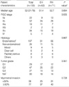
One hundred thirty-three patients with endometrial cancer stage IIa to IVa were found to have cervical invasion. Patients' characteristics are summarized in Table 1. Sixty two women underwent EH and 71 underwent RH or MRH. Ninety-three percent (124/133) of the patients underwent complete surgical staging with or without paraaortic lymph node dissection. We found no significant difference in age, body mass index (BMI), distribution of FIGO stage, histology, and tumor grade between the EH and RH groups.
Preoperative evaluations for disease extent including cervical involvement were mostly done by magnetic resonance imaging (MRI) and partly by computed tomography (CT) or endocervical curettage. Pre-operative MRI was performed in 63.9% (85/133) of the patients and sensitivity for cervical invasion was 44.7% (38/85). When the cases with stromal invasion were analyzed, the sensitivity was 53.3% (24/45).
Table 2 presents the data for the choice of the extent of hysterectomy according to the results of preoperative evaluation. EH was performed in 12/62 (19.4%) patients who were preoperatively suspected of cervical involvement, and more than half (37/71, 52.1%) of the patients who underwent RH had no preoperative evidence of cervical invasion. The results show that many surgeons did not strictly follow the results of preoperative evaluation in choosing the type of hysterectomy and the decision regarding EH or RH was made at their own discretion.
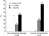
Fig. 1 shows the relationship between the depth of myometrial invasion and that of cervical stromal invasion. A total of 121 cases had available information about both about the depth of myometrial and presence of cervical stromal invasion. In patients with disease limited to the endometrium, no case showed cervical stromal invasion. On the other hand, a majority of patients with disease involving cervical stroma had deep myometrial invasion (P=0.001).
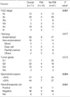
Table 3 showed the relationship between prognostic factors and parametrial involvement in 71 patients undergoing RH. Ten patients (14.1%) had pathologic parametrial involvement. All the patients with PMI were stage III or above. Eight of ten patients (80.0%) had pelvic node metastasis, 1 patient had grossly positive adnexal involvement, and 3 had tumor spread outside the pelvis (2 of 3 had coexisting pelvic lymph node metastasis). Depth of myometrial invasion and pelvic or paraaortic lymph node positivity were significantly correlated with paramatrial involvement. Of the 19 patients with pelvic lymph node metastasis, 8 patients (42.1%) had concomitant PMI. Conversely, of the 10 patients with PMI, 8 (80.0%) had lymph node metastasis.
In 74 patients with surgical stage II, i.e., disease showing no demonstrable evidence of tumor spread in the pelvic cavity, RH was performed in 41 cases and no patients showed parametrial involvement. Sixty six of 74 (89%) patients underwent adjuvant radiation therapy (35 whole pelvic irradiation, 7 vaginal brachytherapy, 23 both, 1 unknown) and there were 3 patients who developed recurrent disease in the RH and none in the EH group (mean follow-up: 51 months, Table 4).
DISCUSSION
Our results show that a large number of Korean gynecologic surgeons have a tendency to choose the surgical extent of hysterectomy through their own disposition and do not strictly adhere the results of pre-operative evaluation. Actually, the type of hysterectomy procedure selected for endometrial cancer varies in each nation and in each institution. Watanabe et al. (13) reported the results of a survey of a Japanese group showing that more than 70% of institutes never perform RH without regarding the preoperative status of cervical involvement. Moreover, according to a survey in North America, nearly 30% of centers never performed class II or III extended hysterectomy for the treatment of endometrial cancer (14). As shown in Table 2, surgeons who overuse RH without regarding the pre-operative results may believe that the current pre-operative evaluation method is not sensitive enough to detect cervical invasion and thus, worry about possible PMI or post-operative stump recurrence. On the other hand, those who perform only EH despite the positive findings from pre-operative evaluation may believe that cervical stromal invasion should be followed by adjuvant radiotherapy and thus, the prognosis would not be changed by performing a 'high morbidity producing surgery' considering the low incidence of PMI. This inconsistent treatment policy, in fact, necessitates the need for an evidenced-based reformed guideline to which most clinicians can conform, although the definition of RH or EH is not standardized among surgeons and thus, the actual extent of surgery investigated as RH or EH by medical records might be different for each patient and this is one of the limitations of this study.
Our results also showed that the PMI rate in patients with cervical invasion is about 14% in the RH group. This is within the range reported by other investigators, and is not significantly lower than reported values (15-17). However, parametrial spread was not found in any of the patients with FIGO stage II, i.e., patients showing no intraoperative tumor spread, but was identified in 8 patients with positive pelvic lymph nodes, and two showed grossly frank extrauterine spread around the adnexa and another site in the pelvic cavity.
These findings indicate that a risk of leaving an occult metastasis in parametrial tissue when performing only EH in patients without evidence of extrauterine tumor spread seems to be very low, even though all lymph node metastases may not be completely screened by intraoperative frozen section.
In contrast to the present results, Yura et al. (17) reported 11.5% of PMI in FIGO stage II patients. Although they reported a relatively high rate of parametrial involvement, the points we focused on is not the crude value of the PMI itself, but the necessity of parametrial resection. Undoubtedly, cervical stromal invasion is known to increase the risk of PMI. However, the pattern of spread in the invaded cervix of endometrial cancer is quite different from that of primary cervical carcinoma. As shown in Fig. 1, the pattern of cervical spread of endometrial cancer seems to follow myometrial invasion and originate from the endocervical glandular region, and as it advances, it invades the stroma. Therefore, we can speculate that before the tumor of the endometrium reaches the deep myometrium, a chance to detect direct PMI in the patients with suspicious cervical invasion seems to be low. However, PMI in endometrial cancer cannot be completely excluded only by the absence of cervical stromal invasion because PMI is reported to consist histologically of 2 patterns of spread: direct invasion of cancer cells to the parametrial connective tissues like that seen in cervical cancer, and parametrial lymph-vascular space invasion frequently seen in patients with deep myometrial involvement without cervical involvement (17-19). Current guidelines for endometrial cancer show that cervical stromal invasion is regarded as one of the major indications of adjuvant radiotherapy. We might guess that adjuvant radiation is required due to the implication that cervical stromal invasion raises the risk of recurrence not just by direct PMI from the cervix, but also by increasing the risk of occult parametrial involvement from the preceding deep myometrial invasion or myometrial lymph-vascular space invasion.
Suffice it to say, this suggest that performing RH only in patients with cervical involvement cannot guarantee the complete eradication of possible parametrial involvement. In addition, it is very likely that we may have 'cut through' the invaded parametrium in stage II patients who underwent EH even though it was not revealed by pathology because there was no direct connection between the invaded cervical stroma and parametrium itself. Even acknowledging that occult parametrial spread had existed, our data showed that no recurrences occurred in patients who only underwent EH followed by adjuvant radiation and thus, it suggests that occult parametrial metastasis in patients with no gross extrauterine spread can be successfully controlled by radiation therapy.
We could not evaluate the value of RH in patients with stage III or over because their survival outcome is much likely to be influenced by variables other than parametrial involvement, i.e., lymph node status, extent of tumor spread, different treatment strategy to the recurred patients, etc. We did not overcome the limitations of retrospective analysis, i.e., lack of verifying the actually performed surgical extent, lack of centralization of pathologic review, and limited number of cases. Nonetheless, we cautiously suggest that routine RH in patients with cervical invasion and having no grossly positive extrauterine spread should be reappraised and that these patients are worthy of consideration for less radical surgery performed in conjunction with pelvic and paraaortic lymphadenectomy considering the low rate of parametrial involvement, inaccurate preoperative prediction, high post operative morbidity, and successful control through adjuvant radiation.
Since this is an area of continued debate and a principal shift from our current practice and attitudes, a randomized study would be required to define the low-risk population that would have a modification of the extent of parametrial resection performed. In this regards, we can conclude that the real value of RH in endometrial cancer patients with suspicious cervical involvement can be evaluated by performing a large scaled randomized controlled study comparing the survival and quality of life between the group who underwent RH only or followed by tailored adjuvant radiation and the group with EH and routine adjuvant radiation therapy.


 XML Download