

 PDF
PDF ePub
ePub Citation
 Print
Print


INTRODUCTION
Hookworm is one of important soil-transmitted helminthes (STH) for humans around the world. About 740 million people are estimated to be infected by hookworm (1). Human infection by hookworms induces blood loss, iron-deficiency anemia, and other anemia associated-symptoms and signs. Human hookworm includes two species, Necator americanus and Ancylostoma duodenale. Necator is rather prevalent in tropics and southern subtropics while Ancylostoma is in subtropic or northern temperate zones. Worldwidely, N. americanus accounts for the predominant etiology of human hookworm infection, whereas A. duodenale occurs in more scattered focal environments (2).
Until the 1970s, STHs were highly prevalent in Korea, and the population of infected people far outweighed those uninfected. Infections of STH were decreased, however, by a national mass control program in the 1970s to 1990s (3). Only clonorchiasis and sporadic tissue parasitic helminthiases are recognized at present in Korea (4). Among STHs, hookworm disappeared first, and thus anemia and malnutrition by hookworm infection became negligible in Korea in 1980s. The hookworm egg positive rate in Korea was 10.7% in 1971, but decreased to 3.7% in 1978, 0.08% in 1985, and 0% in 2004 (3, 5). Of human hookworms, A. duodenale was predominant in Korea but N. americanus was recognized in a few localities (6-8). The last case of N. americanus infection in Korea was reported in 1978 (8). Later, in 1992, an imported case of A. duodenale was reported (9).
Herein, we describe a case of an 82-yr-old woman who suffered from severe chronic iron deficiency anemia by Necator americanus infection in Korea. It is a recent piece of evidence of hookworm transmission in Korea.
CASE REPORT
An 82-yr-old woman was transferred to the National Police Hospital, Seoul, on October 22, 2007, complaining of shortness of breath and dizziness which lasted for years. She lived in Uljin-gun (county), a remote rural area of eastern mountain of Gyeongsangbuk-do, Korea. She had resided in Uljin-gun throughout her entire life and had no history of overseas traveling. She cultivated a small dooryard farm at home.
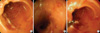
For past medial history, she had been treated for anemia including two transfusions in 2006 at a general hospital in Seoul. She also visited another general hospital complaining exertional dyspnea, and was treated for pulmonary tuberculosis. On physical examination at admission, she was pale, ill-looking, and undernourished, but her vital signs were stable. Laboratory results revealed overt anemia: RBC 1.66×10⁶/µL (normal 4.0-5.4×10⁶/µL), WBC 3.8×10³/µL (normal 4-10×103/µL), Hb 3.4 g/dL (normal 12-16 g/dL), Hct 12.4% (36-48%), MCV 74.7 fL (normal 79-95 fL), MCH 20.5 pg (normal 27-33 pg), MCHC 27.4 g/dL (normal 32-36 g/dL), platelet count, 162×103/µL (normal 130-400×103/µL), Fe 9 µg/dL (normal 37-145 mg/dL), total iron-binding capacity 318 mg/dL (normal 228-428 mg/dL), ferritin 10 ng/mL (normal 10-130 ng/mL), neutrophils 56%, lymphocytes 25%, monocytes 11%, and eosinophils 8%. RBCs were microcytic and hypochromic. Stool examination was negative for occult blood, but was not examined for parasite eggs. Other biochemistry examinations, including electrolytes, liver and renal functions were within normal limits. Serum ELISA against tissue invading helminthiases was negative for Clonorchis, Paragonimus, cysticercus, and sparganum. Gastroduodenoscopy found hyperemic mucosa and numerous 10-mm long slender reddish moving roundworms in the duodenum (Fig. 1).
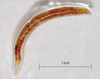
A total of 7 worms were removed by endoscopic biopsy forceps and 5 of them were transferred to the Department of Parasitology and Tropical Medicine, Seoul National University College of Medicine. The worms were round-cylindrical in shape and blood-tinged, and the 5 worms (male: 3, female: 2) measured 10.5 mm (range; 8-11 mm) long and 0.5 mm (range; 0.4-0.5 mm) wide (Fig. 2). The buccal cavity at the anterior end was characterized by one pair of cutting plates. Two male worms showed the copulatory bursa at the posterior end, the dorsal costa of which had a deep notch. SEM findings confirmed cutting plates (Fig. 3). Those gross and microscopic findings of the worms identified them as Necator americanus.
After the diagnosis, she was treated with packed RBC transfusion, iron supportive therapy and albendazole medication, 400 mg twice per day for two days. Her symptoms of dyspnea, dizziness, and severe anemia improved. She was discharged on November 5, 2007. Repeated tests two months after the treatment found her hemoglobin level to be 10.6 g/dL (12-16 g/dL) and gastroduodenoscopy found normal mucosa and no N. americanus worms in her duodenum.
DISCUSSION
The present case constitutes evidence of local transmission of hookworm infection in Korea in 2007. The patient had no history of overseas travel and stayed at home for long periods of time. The patient's home is at an isolated remote mountainous village in Uljin-gun (county), Gyeongsangbuk-do, and she cultivated a small dooryard farm there. It is presumed that she got hookworm infection at her dooryard farm. The environment of her door-yard must have been favorable for active transmission of hookworm. This transmission of N. americanus may be residual of the past endemicity of hookworm in the locality. However, it is a surprising finding for the recent epidemiology of helminthiasis in Korea because most parasitologists and physicians regard that STHs were eradicated.
The morphological characteristics of the present hookworms were clear and definite enough to identify them as N. americanus. Most human hookworms in Korea in the past have been A. duodenale, but N. americanus has also been found in remote agricultural localities in Chungcheong-do or Gyeongsang-do (6-8). Since STHs, including hookworm, heavily infected throughout Korea in the past, whole rural villages were endemic for the helminthes. Uljin-gun has been also an endemic area of those helminthes, and N. americanus must have been prevalent there. Upgrades of environmental sanitation and mass anthelminthic chemotherapy successfully eliminated STHs and their associated diseases in the whole of Korea (3, 5). Therefore, STHs are now neglected by doctors and also by patients in Korea. However, the present case allows for the possibility that some remaining endemic foci of STH transmission may be scattered in remote mountainous localities. In the village where the present patient lived, Ascaris or Trichuris are presumed remaining as well as hookworms because they were more prevalent. It is necessary to screen infection status of STHs in such remote isolated villages in the mountainous zones of Korea because other similar cases may live there.
Clinical findings of the present case included anemia, dyspnea, and undernourished condition, which are known to be associated with hookworm infection (10). The severe anemia may have been due not only to the hookworm infection, but also to undernourishment and low hematopoietic activity due to her old age. The physician observed hundreds of worms on the duodenal mucosa by endoscopy but recovered only 7 of them. Picking out all of them was impossible. In addition to endoscopy visuals, the laboratory hematological findings also suggested that the present patient was infected by at least hundreds of hookworms, because N. americanus suck less blood than A. duodenale do (1, 2). In general, infection by a few hookworms is not sufficient to induce clinical anemia. The present patient recovered greatly from anemia by albendazole therapy and supportive medical treatment. This fact confirmed that the anemia of the present case was caused by heavy infection of the hookworm, N. americanus.
Reviewing her medical records, it was unfortunate that her fecal specimen was not examined properly throughout her whole past medical history. She had visited several hospitals but no doctors had suspected hookworm anemia and her feces had not been properly checked for more than 2 yr. The present patient suffered from this disease for at least 2 yr because the possibility of hookworm anemia in this case was completely neglected by physicians.
This is the first case of hookworm anemia in Korea to be diagnosed by endoscopy. Although most of past endemic STHs have disappeared in Korea, it is still necessary for physicians to be mindful of the possibility of STHs.

 XML Download