

 PDF
PDF ePub
ePub Citation
 Print
Print


INTRODUCTION
Gastrointestinal stromal tumor (GIST) is relatively rare neoplasm occurring in the gastrointestinal tract, omentum, or mesentery, but is the most common among sarcoma of the gastrointestinal tract and accounts for 5% of all sarcoma. In general, only complete resection can lead to cure, but recurrence in the liver and peritoneum is common after surgery, and unresectable or recurrent tumor does not respond to conventional cytotoxic chemotherapy and therefore, its prognosis has been very poor. However, the identification of signal transduction pathway associated with the development of GISTs and the use of so-called molecular targeted therapy with imatinib (Glivec, Novartis Korea, Seoul, Korea) have yielded remarkable achievement. In addition, imatinib has been shown not only the prolongation of survival time but also the great effectiveness on quality of life with very mild side effects compared with conventional cytotoxic chemotherapy. Based on these results, imatinib is used as first-line therapy in metastatic GISTs, and furthermore, neoadjuvant and adjuvant treatments with imatinib are being investigated.
In the western countries, histopathologic criteria and molecular pathologic mechanism of GIST have recently standardized and the guidelines for this entity have been published by the National Comprehensive Cancer Network (NCCN) (1) and the European Society of Medical Oncology (ESMO) (2). And in Japan and Australia, the guidelines appropriate for clinical practice in each country have been also published (3, 4)
However, in Korea, no standardized guidelines for diagnosis or treatment of GISTs are available. As a result, diagnosis is not consistent between institutions and in addition to the problem associated with diagnosis, optimal treatments are sometimes not provided because of numerous uncertainties related to treatment and a lack of treatment guidelines. To recognize and to find ways to solve these problems, pathologists, surgeons, gastroenterologists, diagnostic radiologists, and medical oncologists organized a multidisciplinary study group called the Korean GIST Study Group (KGSG) in December in 2006. We made the first guideline in 2007 for diagnosis and treatment of GISTs that is suitable for clinical practice in Korea (5). In this second version of the guideline, we sought to update changes in the topics and reflect modified and added recommendations. Expert panel members of the KGSG thoroughly reviewed the relevant literature including the European Society of Medical Oncology and National Comprehensive Cancer Network guidelines and shared their experience and opinions to make a consensus on twenty topics related to pathologic diagnosis, surgical and medical treatment of GIST. The consensus was presented as the basis for a guideline of diagnosis and treatment for patients with GIST that would be used to facilitate the optimal clinical practice in Korea.
PATHOLOGIC DIAGNOSIS OF GIST
Definition of GIST
GIST is the most common mesenchymal tumor of the gastrointestinal tract (6). GISTs arise from the interstitial cells of Cajal or their common stem cell (7). GISTs range in size from tiny tumors discovered incidentally during tests for other diseases, measuring less than 1 cm to very large lesions measuring upwards of 35 cm (median 5 cm) (8). Irrespective of tumor size, GISTs share morphologic features and immunoreactivity for KIT and contain an oncogenic mutation in the KIT (80-85%) or platelet-derived growth factor receptor (PDGFRA, 5-7%) genes (9). GISTs can arise in any portion of the gastrointestinal tract, but usually occur in the stomach (60%) or the small intestine (30%) (10, 11).
Pathologic findings of GIST
On gross examination, GIST is a well circumscribed, fleshy, pink, or tan-white mass. Large tumors frequently show hemorrhage, necrosis, and cystic degeneration. Microscopically, GISTs can be divided into three different histologic subgroups. Spindle cell GISTs (70%) are composed of cells with palely eosinophilic, fibrillary cytoplasm, ovoid uniform nuclei, and ill-defined cell borders, often with a somewhat syncytial appearance, arranged in short fascicles or whorls (Fig. 1). Epithelioid GISTs (20%) are composed of rounded cells with eosinophilic to clear cytoplasm arranged in sheets and nests (Fig. 2). The final group shows mixed spindle and epithelioid cells (10%). The frequency of these histological types varies according to location. GISTs of the stomach mostly fall into one of 4 spindle cell subtypes of sclerosing, palisading-vacuolated, hypercellular, and sarcomatous or one of 4 epitheloid subtypes of sclerosing epitheloid variant, dyscohesive epithelioid, hypercellular, and sarcomatous (12). GISTs of the small intestine have great amounts of skeinoid fiber, and are most likely to become malignant if epitheloid type or mixed type is present. Many of GISTs in the large intestine are spindle cell type. GISTs developed in the omentum are similar to histological types of the stomach whereas GISTs of the mesentery are similar to histological findings of the small intestine. The diagnosis of GISTs is mainly based on clinical and histological findings, but immunohistochemical staining is needed to confirm diagnosis (13).
Immunohistochemical staining of GIST
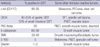
The most important immunohistochemical staining in the diagnosis of GISTs is c-kit (CD 117), and other several antibodies may be helpful in the diagnosis and differential diagnosis. Approximately ~95% of cases are positive for c-kit protein (Figs. 1, 2). c-kit negative GISTs account for ~5% of cases and can cause diagnostic difficulties, but given the rather limited choice in mesenchymal diagnostic considerations at these sites, they can often be diagnosed by excluding other potential mimics by immunohistochemical characterization (14). c-kit staining with polyclonal anti-c-kit antibody is mandatory for diagnosis. Extreme caution should always be taken to avoid false-positive or false-negative c-kit staining results by carefully observing positive control (mast cells or interstitial cells of Cajal) and negative control (smooth muscle cells or endothelial cells). Because c-kit may also be positive for other soft tissue tumors, interpretation of c-kit based on H&E findings is necessary (Table 1).
CD34 is positive in 60-80% of GISTs, and the frequency of CD34 positivity depends on location of GISTs. The frequency of positive CD34 is high in GISTs of the esophagus and colon (95%), but relatively low in the small bowel and extra-gastrointestinal sites. In the small intestine, CD34 is positive in 50% of cases while c-kit is positive in almost 100% of cases. However, GISTs of the colon can readily be misdiagnosed as other soft tissue tumors such as inflammatory fibroid polyp or inflammatory myofibroblastic tumor, which is attributable to the rare occurrence of GISTs in the colon and greater incidence of negative or focal staining of c-kit in GISTs of the colon relative to other organs. Thus, negativity of c-kit staining does not exclude a possibility of GIST, and every effort should be made to obtain the diagnosis of GISTs through proper differential diagnoses. Protein kinase C (PKC)-theta staining is positive in approximately 90% of GISTs. The quality of PKC-theta staining must be managed by observing ganglion cells of the intermyenteric plexus as an internal positive control and smooth muscle or blood vessel as a negative control. When the staining is properly performed, it can serve as an important adjunct tool in the diagnosis of c-kit negative GISTs, particularly developed in the stomach and extragastrointestinal locations (15). H-caldesmon is positive in 60-80% of GISTs, which may be helpful in the diagnosis of c-kit negative GISTs. Smooth muscle actin is positive in 30-40% of GISTs, and the frequency of the positive staining is high especially in the small bowel. S-100 and desmin is positive in 5% and 1-2% of GISTs, respectively. A recently developed antibody against DOG1 (discovered on GIST) was reported to be superior in sensitivity and specificity to c-kit and CD34. However, c-kit negative GISTs express DOG1 in only 36% of cases, limiting its use in this setting (16). Fig. 3 shows the algorithm of diagnosis in GISTs based on the immunohistochemical staining results.
Extragastrointestinal GISTs (EGISTs)
In addition to the gastrointestinal tract, GISTs are also found in extragastrointestinal sites, although rare. Caution should be taken because histological and immunohistochemical findings of EGISTs are different from those of GISTs and consequently, it may be very difficult to make a diagnosis.
c-kit negative GISTs
In GISTs, c-kit is negative in ~5% of cases. These c-kit negative GISTs are common in the stomach and omentum/mesentery. In such cases, examining other immunohistochemical markers (PKC-theta, CD34, SMA or DOG1) and mutation analyses may be useful in diagnosis. Among c-kit negative GISTs, 75% are positive for PKC-theta, 44% for CD34, 40% for SMA, and 36% for DOG1 (14, 15, 17-19).
Pathologic prognostic parameters
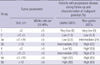
Morphologic risk assessment in GISTs provides the basis for clinical management and optimal patient care. The vast majority of studies of GISTs suggest that the two most important prognostic features to assess the risk of aggressive behavior in a primary localized GIST are mitotic activity and tumor size. These two features were the foundation of the consensus approach for risk assessment in GISTs published by Fletcher and colleagues in 2002 (13). Subsequent data collected by Miettinen and colleagues (20), analyzing large series of GISTs, confirmed that tumor size and mitotic activity are essential prognostic parameters; they proposed additional new parameter, location of tumor, in the evaluation of the clinical behavior of localized GISTs, and KGSG adopted this risk stratification with slight modification (Table 2) (12).
Pathologic reporting for GIST
It has been suggested that one block per centimeter should be examined histologically. The pathology report should include the size of the tumor, mitosis (per 50 high powered fields [HPF]), location, resection margin status, and the presence or absence of metastases. Presence of metastasis or perforation during operation leads to a diagnosis of malignant GIST. Pathologic report may include histological type including the degree of cellularity and atypia, presence of necrosis or cystic change, invasion into mucosa or adjacent structures.
Mutational analysis
At present, mutation analysis is not required for the diagnosis of GIST when tumors have a typical histology and immunohistochemical staining pattern. However, because the presence and location of mutations in KIT or PDGFRA can have implications for prognosis and management in patients with advanced disease, mutation analysis should be considered at the time of diagnosis. Mutational analysis for KIT exons 9, 11, 13, and 17 or PDGFRA exons 12, 14, and 18 can be performed with unstained slides from formalin-fixed paraffin-embedded tissue or fresh frozen tissue.
SURGICAL TREATMENT OF GIST
Surgical treatment as first-line therapy
The main treatment of resectable localized GIST is surgery. The goal is complete resection without residual tumor cells (R0).
Diagnosis
The initial diagnosis is generally made by endoscopy, endoscopic ultrasound, gastrography, or computed tomography (CT) of the abdomen due to difficulty with obtaining adequate tissues. It should be confirmed by pathologic histological findings after resection. Preoperative histological diagnosis is feasible, but it may be difficult to interpret definitively (21-24). Imaging tests to detect metastasis include chest radiography (or chest CT), triphasic CT of the abdomen and pelvis, and/or magnetic resonance imaging (MRI) if necessary (25). Positron emission tomography (PET) may be performed when evidence of metastasis may be equivocal or for clinical trials (21).
Biopsy
There is no consensus regarding the need of endoscopic ultrasound biopsy or percutaneous biopsy for preoperative diagnosis. The important part of histological diagnosis is not to cause tumor seeding during biopsy. Therefore, unless multiple metastases are present, excisional biopsy with laparotomy is suggested (21, 26). If diagnosis is unknown at the time of resection, post-operative frozen tissue examination must be performed in order to elucidate the treatment strategy for GIST as treatment varies for an adenocarcinoma or lymphoma. Biopsy is necessary when planning neoadjuvant therapy.
Indications for surgery
Due to the high potential for malignancy of GIST, resection should be the first-line treatment (21, 27). We strongly recommend resection for tumors larger than 2 cm or growing tumors (21). Smaller tumors (<2 cm) confer a lower potential for malignancy and may be observed. However, small tumor size does not exclude the potential for malignant transformation. Therefore, patients should be informed about the possibility of malignancy.
Surgical margins
The main objectives of surgical treatment are to acquire negative margins and to resect without causing tumor rupture. In case of inadvertent tumor infiltration into the surrounding organs, a complete en bloc resection with negative margins should be performed (21, 26, 27) regardless of size. Therefore, even tumors are small, endoscopic shell-out procedure or enucleation should be avoided if GIST is suspected. In many cases, wedge resection of gastric GIST and segmental resection of small bowel GIST are appropriate treatments. Subtotal or total gastrectomy may be performed based on size and location. We recommend en bloc resection for omental or mesenteric GIST. Adjacent organs adherent to tumor should also be completely resected en bloc to avoid tumor rupture or intraabdominal seeding (26).
Laparoscopic resection
Laparoscopic resection is feasible if intraabdominal tumor rupture or seeding is unlikely. Laparoscopic resection should follow principles of oncologic surgery. Generally, it is reserved for small, favorably located gastric GISTs (28-31). Intra-operative endoscopy or laparoscopic ultrasound may be used to assist in laparoscopic resection if needed.
Lymphadenectomy
Unlike adenocarcinoma, GIST rarely metastasizes to local regional lymph nodes. Therefore, lymphadenectomy is warranted only if metastasis is suspected, i.e. enlarged lymph nodes.
Post-operative follow-up and Surveillance
For patients in the high- or intermediate-risk group, we recommend follow-up with CT of the abdomen and pelvis every 3 to 4 months for the first 3 yr after surgery and then every 6 months until 5 yr; then annually thereafter (21, 26). For patients in the low or very low risk, we recommend follow-up with CT every 6 months for 5 yr. Ultrasonography may replace CT once a year (21, 26).
The role of PET for this purpose is not established, and clinical studies on the role of PET are ongoing. Most recurrences occur within 2 yr after surgery and the liver and peritoneum are the most common sites of recurrence (32). Due to the high incidence of gastric cancer in Korea, the National Cancer Screening program recommends biennial stomach-cancer screening for men and women older than 40 yr with endoscopy or upper-gastrointestinal series.
Post-imatinib resection in metastatic GIST
Medical treatment with imatinib alone usually does not result in complete response in metastatic GIST. And, responses are not usually maintained indefinitely. It is now well recognized that clones of tumor cells resistant to imatinib develop continuously over time after start of imatinib. Therefore, in cases where partial or stable responses are shown after adequate duration of imatinib therapy (usually 4 to 12 months of treatment), complete surgical resection of residual tumor may be considered to reduce the risk of development of resistant clones by eradicating the residual viable tumor cells. Several retrospective studies suggested that resection of residual lesions could prolong progression-free survival if it is done during the tumors are under control with imatinib (either in PR or SD) (33, 34). It is also emphasized that imatinib should be continued after resection (21). However, the role of resection of residual tumors after imatinib therapy has not been established, and several phase III clinical trials to investigate the role of surgical resection in this setting are ongoing or planned worldwide. Hepatic or peritoneal metastases may be locally treated with radiofrequency ablation (RFA) or chemoembolization. Importantly, management of GIST should be coordinated by a multidisciplinary team of experienced medical and surgical specialists.
MEDICAL TREATMENT OF GIST
Adjuvant treatment
Adjuvant treatment with imatinib is given to enhance the possibility of cure by eradicating microscopic lesions that might still be present after complete resection of visual tumors. Given the great efficacy of imatinib on metastatic or recurrent GIST, imatinib appears to have a sufficient potential as adjuvant treatment.
In recently published ACOSOG Z9001 study of patients with tumor diameter ≥3 cm who received imatinib for 1 yr following complete resection, imatinib demonstrated a significant increase in recurrence-free survival (RFS) compared with placebo although improvement in overall survival (OS) not observed (35). The benefit in RFS appears to be related to tumor size, with the most marked improvement in patients who had large tumor (≥10 cm) in the study.
A Korean phase II study, recently presented in an abstract, evaluated the efficacy of 2-yr imatinib adjuvant therapy in patients who were at high risk of recurrence (tumor size ≥5 cm and mitotic index ≥5/50 HPF, tumor size ≥10 cm, or mitotic index ≥10/50 HPF) based on the NIH risk criteria and had KIT exon 11 mutation which had been recognized as an independent poor risk factor of recurrence in a Korean retrospective study (36, 37). The recurrence-free survival of these patients looked much improved compared with historic data.
These results strongly suggest that adjuvant imatinib therapy should be recommended to reduce the risk of recurrence after curative resection of localized GISTs. However, there are several issues to solve with regard to adjuvant treatment with imatinib. In the ACOSOG Z9001 trial, rapid disease recurrence was observed after stopping 1 yr of imatinib adjuvant treatment. The study did not demonstrate a benefit of adjuvant imatinib in terms of overall survival. Adjuvant treatment with imatinib may delay recurrence. But, it may not increase the cure rate or eliminate the disease in patients with metastatic GISTs; therefore the majority of them eventually develop resistance to imatinib, leading to treatment failure despite its great efficacy. Although currently it remains yet to be determined whether adjuvant imatinib improves cure rate or just delays recurrence, adjuvant imatinib can be proposed as an option for patients at a substantial risk of relapse. Only tumor size and mitotic count were included in the 2002 NIH Consensus risk classification. More recent risk classifications incorporate primary tumor site and/or tumor rupture in addition to the tumor size and mitotic count. Current consensus of the experts is to recommend adjuvant imatinib for patients at high risk of relapse and not for those at low risk of relapse. But, there is no consensus for the patients at intermediate risk (20, 38). Patients with tumor rupture should be considered as having high likelihood of micrometastasis and treated with imatinib. In addition to the risk assessment, mutational analysis may be helpful for the selection of patients who are not likely to get benefit from the treatment. For example, GIST with D842V mutation in PDGFRα exon 18 is known to be not responsive to imatinib. With the currently available data, it is recommended to use imatinib as an adjuvant treatment for at least one year. And, most of the experts agree that 1 yr of adjuvant imatinib is not long enough for especially patients at high risk. However, optimal duration of adjuvant imatinib remains yet to be determined. Phase III trials comparing one year versus three years and no treatment versus two years of treatment duration are ongoing.
Neoadjuvant treatment
Outside of a clinical trial, neoadjuvant treatment with imatinib is not recommended unless there are clinically significant grounds to improve results of surgery by downsizing tumors with neoadjuvant therapy at the initial diagnosis (21). However, neoadjuvant treatment with imatinib may be considered if R0 resection is not feasible, for the purpose of preserving organ functions in GISTs of the rectum, esophagus, and duodenum, or for complete resection of gastric GISTs accompanied by severe, local infiltration into the pancreas or duodenum (39-41). When such a neoadjuvant treatment is considered, progression and response of tumors before and during the treatment should be assessed very carefully by CT and/or PET scan, and it should be done by an experienced multidisciplinary team. Early assessment of tumor response by CT and/or PET is recommended so that surgery is not delayed in the case of non-responding tumors. Duration of neoadjuvant therapy with imatinib may vary according to response to the treatment, but surgery should be performed after sufficient shrinkage of tumors is observed (typically after 4 to 6 months and within 12 months of imatinib treatment) (21). Mutational analysis might be helpful to exclude GISTs not sensitive to imatinib.
Advanced disease
Initiation of imatinib
Once an advanced GIST is diagnosed, imatinib should be immediately initiated regardless of the presence or absence of symptoms. It is optimal to administer imatinib in patients with liver metastasis or localized metastasis to the peritoneum because cure is hard to achieve even if tumors are completely resected visually and histologically (32, 42). The concept of adjuvant therapy does not apply in this setting.
Optimal dosage of imatinib
The optimal initial dose of imatinib is 400 mg per day. In a large European phase III study that compared 400 mg daily with 800 mg daily, the group with 800 mg daily did not achieve an increased survival rate while side effects were increased (43, 44). However, in subgroup analysis, a clinically significant improvement of progression-free survival was observed in the group with KIT exon 9 mutations that received 800 mg per day. So, high dose imatinib is now recommended in the Western countries as the initial treatment for patients with KIT exon 9 mutant GIST (45). However, it is still unclear if this recommendation is valid for Asian patients as well. In recent two retrospective studies of Korean and Taiwanese patients with GIST, there was no difference in the treatment outcomes according to the KIT genotype with imatinib administered at a dose of 400 mg per day (37), which suggest that 400 mg/day of imatinib might be enough for KIT exon 9 mutant GIST patients in Asia. A larger scale retrospective study is ongoing to address this issue.
Duration of imatinib treatment and surgical resection of responding tumors
Treatment with imatinib should be continued indefinitely unless disease progresses, intolerable adverse events occur, or a patient refuses the treatment. If imatinib is discontinued after tumor response with imatinib is achieved, the disease progresses in most cases. Many patients show responses to the reintroduction of imatinib when the disease progresses following imatinib discontinuation, but imatinib treatment should not be interrupted outside of a clinical trial or unless clinically indicated. Surgical resection of stabilized tumor lesions can be considered optionally with enough discussion and with a multidisciplinary approach.
Standard tests for tumor response
CT is the most useful tool to date to determine response to treatments. We recommend dynamic or triphasic CT scanning through arteries and veins after contrast enhancement (25). Fluourodeoxyglucose (FDG) PET is highly sensitive in early tumor response, but given its cost and availability, it is not easy to include it in basic imaging tests (46-48). Intervals to determine response may be various according to clinical situations, and tumor response is usually determined every 3 to 4 months after the initial response is confirmed.
Criteria for determination of response
Determining tumor response or continuation of treatment solely based on tumor size should be avoided because tumors may enlarge due to intratumoral hemorrhage or myxoid degeneration despite therapeutic effects during the beginning of treatment (25, 49-51). Because GIST, hypervascular neoplasm, exhibits hypoattenuating findings resulting from reduced vascularity, hyaline degeneration, and occasional cystic changes following imatinib treatment, the strict application of the response evaluation criteria in solid tumors (RECIST) or WHO criteria requires caution, and the development of new criteria is warranted. Particularly for liver metastasis, when tumor response is determined solely based on the portal-venous phase CT, small and new lesions with hypodensity may be seen, which are most likely the resulting findings of clear margins secondary to necrosis of preexisting tumors that were present before the treatment with the same radiodensity as the hepatic parenchyma, and they require differential diagnosis (25, 51). Both tumor size and tumor density should be considered for the response evaluation. Improvement of patients' symptoms or degree of a reduction in CT attenuation coefficient (Hounsefield Units, HU) or maximum standardized uptake value (SUVmax) on PET may be used to determine tumor response (25, 49-51).
Recurrence and progression
Recurrence or progression includes appearance of a new lesion at the surgery site, development of a new metastasis, or an increase in tumor size. In some cases, a new intratumoral nodule or an increased solid tissue in hypodense tumors previously responded is identified as recurrence or progression. The patterns of the progression cannot be determined based on the RECIST or WHO criteria, and thus inside and cell walls of each lesion should be examined very carefully (25, 52).
Treatment of disease progression during imatinib therapy
Resistance is classified into primary resistance and secondary resistance. Primary resistance is defined as progression within the first 6 months of imatinib therapy, and most of the cases progress multifocally (21). Secondary resistance is defined as progression after 6 months of the initiation of imatinib therapy, and generally two types of progression are seen (21, 25).
Focal progression: It is called when one or a limited number of multiple lesions exhibits intratumoral nodules or lesions become larger, which results in an increased FDG uptake on PET scan, and the remainder of the lesions are relatively well controlled. Treatment for the focal progression requires multidisciplinary approaches. Local treatment such as resection of localized metastasis to the liver or peritoneum, radiofrequency ablation, as well as chemoembolization can be considered to control the focal progressing lesions. But, systemic treatment should be also continued to control hidden micrometastatic lesions. If local progressing lesion(s) was removed, standard dose of imatinib can be continued. But, if those lesions were not removed, dose escalation of imatinib or switch to sunitinib should be considered. No prospective study on the efficacy of local treatment on focal progression has been conducted. Some studies suggest that a part of patients with focal progression may get benefit from local treatment on focal progression, but others eventually show general progression within short time after local treatment (33, 34, 53).
General progression (multifocal resistance): It is called when the majority of multiple lesions simultaneously progress. The efficacy of local treatment on the multifocal resistance is extremely limited and mostly negative (33). Therefore, local treatment for general progression is not recommended except for palliation of symptoms. Administration of imatinib at increased doses or second-line drug such as sunitinib should be considered.
Increase of imatinib dose during disease progression: When disease progresses at the dose of 400 mg per day, an increase of imatinib to 800 mg per day has been known to show partial responses or control of tumors for a certain period in about 30-40% of patients (43, 54). With the daily dose of 800 mg, adverse events associated with imatinib are not increased except malaise and anemia, but if an intolerable side effect occurs, it may be reduced to 600 mg per day (54). When severe adverse events are expected with direct dose escalation to 800 mg per day, imatinib can be first escalated to 600 mg per day, and then sequentially to 800 mg per day. The median progression-free survival is about 3 months and a 12-month progression-free survival rate is 18-30% with the dose escalation of imatinib (55).
Use of sunitinib during disease progression: Sunitinib is a new tyrosine kinase inhibitor which can confer antitumor activity by both direct antitumor activity and antiangiogenic activity because it can inhibit VEGFR as well as kit or pdgfra (56). Sunitinib was approved for the treatment of patients with advanced GISTs after failure of the 1st line imatinib. A phase III pivotal study results showed that sunitinib at a dose of 50 mg daily with 4 weeks on and 2 weeks off schedule was significantly superior in time to progression over placebo (median 27.3 weeks vs 6.4 weeks, P<0.001) (56). Since progression during the rest period was occasionally observed, a continuous dosing schedule with a lower daily dose (37.5 mg per day) was also developed and proven to be effective and well tolerated, although no randomized trials have been performed to compare the intermittent and the continuous dosing schedule. So, the continuous dosing schedule with a lower daily dose can now be considered as an option on an individualized basis.
Use of conventional cytotoxic chemotherapy during disease progression: No conventional cytotoxic chemotherapy has ever been reported to be effective in GISTs. Thus, we do not recommend the use of such drugs except in clinical studies (21).
Continuous use of imatinib or sunitinib after failure of both imatinib and sunitinib: There is no effective systemic treatment option after failure of both imatinib and sunitinib. And, general oncology principles indicate that the use of the same agent that already failed is not beneficial or recommended. However, in spite of resistance to these agents in a majority of tumor cells in this situation, at least a fraction of tumor cells may remain responsive to these agents. So, it is allowed and recommended in many countries to continue one of these agents to slow down the progression of the disease even after the tumor is determined resistant according to the RECIST criteria. For this indication, imatinib may be preferred to sunitinib and daily 400 mg imatinib may be appropriate. However, this issue remains to be addressed in a well designed clinical trial.
CONCLUSION
Although several clinical practice guidelines for GIST based on clinical practice of each country have recently been published by the study group of each country (NCCN, ESMO, the Japan Society of Clinical Oncology, and in Australia), there was no standardized guideline for diagnosis or treatment of GISTs in Korea. To solve this problem, pathologists, surgeons, gastroenterologists, diagnostic radiologists, and medical oncologists organized a multidisciplinary study group called the Korean GIST Study Group (KGSG). We made the first guideline in 2007 for diagnosis and treatment of GISTs that is suitable for clinical practice in Korea. This study is the second version of the guideline for Korean GIST patients. Through series of workshops to review and discuss evolving new evidences, we have updated recent clinical recommendations and reflected changes in diagnosis, surgical and medical treatment for more optimal clinical practice for GIST in Korea. We hope the guideline can be of help in enhancing the quality of diagnosis by members of the Korean associate of physicians involving in GIST patients' care and subsequently in achieving optimal efficacy of treatment.


 XML Download