

 PDF
PDF ePub
ePub Citation
 Print
Print


INTRODUCTION
Solitary or multiple lipomas can occur almost anywhere in the trunk, extremities, mediastinum, pelvis, and retroperitoneum, but they rarely originate in the intestinal mesentery (1-4). A search of the English literature has revealed less than 30 documented cases. In the literature review, mesenteric lipomas do not cause any intestinal symptoms in most cases as they usually allow the passage of intestinal contents (1, 5), and those causing abdominal pain make up only a small percentage (2, 6). Mesenteric lipomas may cause abdominal pain by complete intestinal obstruction as a result of torsion or volvulus (3, 7-9) or partial intestinal obstruction associated with compression of the intestine.
In the present report, we describe a case of a giant mesenteric lipoma causing abdominal pain by entrapment of an ileal loop and review the cases with mesenteric lipomas causing abdominal pain.
CASE REPORT
A 29-yr-old woman presented with colicky lower abdominal pain. Her abdominal pain had developed 3 yr before intermittently, but it had worsened over the past 6 months. She was unable to sleep well due to her abdominal pain, which aggravated after eating meals. She also experienced episodes of abdominal distension, constipation, and urinary frequency or urgency. Other symptoms, as well as her past medical history and family history, were otherwise unremarkable.
The patient had a height of 164 cm and weight of 59.6 kg, for a body mass index of 22.2. Physical examination revealed a slightly distended abdomen, but the remainder of the examination was unremarkable. Routine blood tests, including renal function and urine analysis, were normal.
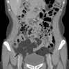
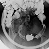
Computed tomography (CT) of the abdomen and pelvis revealed a homogenous fatty mass in the lower abdominal cavity (Fig. 1). The entire small bowel loop was displaced upward by the mass, and one segment of the ileal loop was slightly straightened by entrapment of the bowel. Small bowel series revealed ileal loop entrapped in fatty tissue and associated ileal diverticula (Fig. 2).
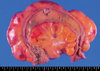
Laparoscopic surgical resection was recommended because of her symptoms. The laparoscopic surgery showed an encapsulated, yellowish mass originating from the ileal mesentery in the lower abdominal cavity. The mass extended to the antimesenteric border of the small bowel and was free of any adhesions or signs of necrosis. Some segment of the ileum appeared to be "stretched" over the fatty mesenteric mass with edema, however, its luminal patency was preserved in spite of luminal narrowing by compression. Lymphadenopathy or fluid collection was not observed. Surgical removal of the fatty mass and the affected ileal loop was performed without difficulty under a laparoscopic magnified view. The resected mesenteric mass was a soft, yellow mass and 19×10×5.1 cm in size (Fig. 3). Microscopically, the mass was characterized by homogenous mature adipose tissue without cellular atypia, which was compatible with the diagnosis of a mesenteric lipoma (Fig. 4). Any evidence of necrosis or malignancy was not observed.
She had no postoperative complications and was free of any signs of disease recurrence 15 months after the operation.
DISCUSSION
The exact prevalence of lipomas is unknown, however, it is likely far higher than reported because most cases with lipomas may be ignored due to their silent nature. Lipomas may show a familial tendency and an increased incidence in people who are obese, have diabetes mellitus or hypercholesterolemia, and experience a traumatic injury (4); however, none of these factors were applicable to our patient. Although benign lipomas can arise in any location in which fat is normally present, reported mesenteric lipomas have been very rare (1-9). They generally form as a slow-growing, non-lobulated, soft, mobile mass that does not penetrate into surrounding organs (1, 4). Although many lipomas are an incidental finding, they may cause variable symptoms when the tumor grows very large. Their uncommon symptoms include anorexia, abdominal distension, weight loss, abdominal pain, constipation, and sensation of fullness, especially after meals, as our case. When the tumor is near the intestinal lumen and far from the mesenteric root, it may cause abdominal pain by pressing upon the intestinal loops, however, passage of the intestinal contents may be allowed as our case due to the consistency of the lipoma and liquid nature of small intestinal contents (4). Our patient, who suffered from chronic intermittent abdominal pain and constipation, has been examined at the primary clinic several times during the last 3 yr without receiving a definite diagnosis. Even though pelvic ultrasonographic examination was performed by primary physician, it was misdiagnosed as mesenteric fat. Mesenteric lipomas may pose diagnostic difficulties due to absence of abnormal laboratory findings and its vague non-specific symptoms as our case. Therefore, physicians should be aware of the various manifestations of mesenteric lipomas and avoid inappropriate managements.
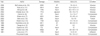
We reviewed reports of mesenteric lipomas identified using a computerized search of Pubmed (articles in English between 1965 and 2007), and thirteen cases of which causing abdominal pain (Table 1). The incidence of this tumor is equal for both sexes, and seven of the cases have been reported in children (2, 8, 11-13, 15). All tumors are located in the mesentery of the small intestine, mostly the ileum, as our case. The main sequelae of mesenteric lipoma are complete intestinal obstruction as a result of torsion or volvulus (3, 7, 9-13) or partial intestinal obstruction due to compression of intestine by giant lipomas (2, 8, 14-16). Abdominal pain may also be related to altered peristalsis secondary to adhered small bowel loops. In terms of size, mesenteric lipomas have a tendency to produce symptoms dependent on their size. The size of the mesenteric lipoma in our patient was one of the largest reported in the literature.
Generally, the size of mesenteric lipomas with obstruction by compression of intestinal loop (mean=21 cm) was larger than those of mesenteric lipomas with obstruction caused by torsion or volvulus (mean=13 cm).
Radiological examination suggests the possible diagnosis, and the best definitive diagnostic procedure is CT, which demonstrates a proliferation of homogenous adipose tissue in the lower abdominal cavity (18, 19). The CT number reading provides information about the nature of the lesion, and the density of the fatty mass with attenuation values between -80 and -120 HU, which is identical to that of subcutaneous fat, may lead to its diagnosis (19). When a large mesenteric lipoma extends lower in the abdominal or even pelvic cavity due to its gravity as our case, CT findings of mesenteric lipoma may mimic pelvic lipomatosis, which is a rare condition defined as a nonmalignant overgrowth of normal fat in the perivesical and perirectal spaces (20-22). Ultrasound can detect mesenteric lipoma as a homogenous echogenic mass (7), however, it may be misdiagnosed as mesenteric fat as our case because the mesentery is rich in fat. Therefore, primary physicians should keep in mind the possibility of this rare disease when faced with the prominent fatty tissue in the abdominal cavity during ultrasonographic examination.
Definitive treatment for mesenteric lipoma has not been established. Entire resection with, or if possible without, the affected intestinal loop may be the treatment of choice for the large mesenteric lipomas due to the risk of partial or complete intestinal obstruction by compression or volvulus. As reported by Tsushimi et al. (23), laparoscopic surgery has gained wide acceptance as the treatment of choice for benign tumors of the abdomen because it is less invasive than conventional open surgery and is economically, cosmetically, and physically beneficial. In addition, it may provide better surgical planes under magnified views than conventional laparotomy. In this regard, laparoscopic surgery may be favored for the removal of a large mesenteric lipoma despite limited experiences for this rare disease.
Mesenteric lipomas are rare, however, they may be considered in the differential diagnosis of unusual abdominal pain. Despite the benign nature of this tumor, total excision with or without the affected intestinal loop by laparoscopic approach may be considered if intestinal symptoms such as abdominal pain are present.

 XML Download