

 PDF
PDF ePub
ePub Citation
 Print
Print


INTRODUCTION
Anti-Müllerian hormone (AMH) and inhibin B are members of the transforming growth factor β superfamily and both are expressed by the granulosa cells. Circulating levels of AMH and inhibin B are correlated with primordial follicle recruitment, suggesting that they may be potential clinical markers for ovarian reserve. Numerous studies have reported that basal serum levels of AMH (1-5) or inhibin B (5-7) are good predictor of ovarian response in patients undergoing ovarian hyperstimulation and in vitro fertilization (IVF). Several reports also indicate that basal serum levels of AMH is a more discriminatory marker of ovarian response than basal follicle stimulating hormone (FSH), inhibin B, or estradiol (8-10).
However, there were few reports addressing the clinical significance of AMH and/or inhibin B levels measured at late follicular phase during ovarian hyperstimulation.
Serum and follicular AMH levels at the time of oocyte retrieval are positively correlated with the number of mature follicles and oocytes retrieved (11). A recent report indicated that serum AMH values at the time of human chorionic gonadotropin (hCG) administration are also correlated significantly with the number of mature follicles, number of oocytes retrieved and serum estradiol levels (12). Moreover, AMH levels are correlated significantly with a greater number of 6-cell embryos and better embryo morphology score.
Serum inhibin B levels at ovulation triggering day and the levels in follicular fluids at the time of oocyte retrieval are strongly correlated with the number of oocytes retrieved (13). Serum (14) and follicular fluid (15) levels of inhibin B at ovum pick up are also correlated positively with the number of oocytes collected and were predictive of clinical pregnancy. Serum inhibin B levels measured around late follicular phase during ovarian hyperstimulation could predict the number of oocytes retrieved both in normal and poor responder (16).
Since AMH is secreted mainly in preantral and early small antral follicle (17, 18), the circulating AMH level decreases through maturing follicle in normal menstrual cycle, and further decreases in FSH-treated cycles (19-21). In ovarian hyperstimulation and IVF cycles, however, continued recruitment of additional antral follicles during the stimulatory phase results in higher AMH levels, thus it may be related with the number of mature follicles and number of oocytes retrieved.
In contrast to AMH, inhibin B rises from early follicular phase to reach a peak during mid-follicular phase (22) but continues to increase during ovarian hyperstimulation (16). This suggests that inhibin B is secreted by the developing cohort of antral follicles (23).
Follicular pool at late follicular phase encompasses various stages of follicular development during ovarian hyperstimulation. Therefore, AMH and inhibin B levels at late follicular phase could reflect overall ovarian pool, including small and large antral and mature follicles. However, follicular AMH levels were reported to be three times higher in small follicles (<12 mm) than large follicles (>16 mm) and serum AMH levels were tightly correlated with the follicular levels (11). These findings suggest that serum AMH levels more reflect small antral follicular pool, thus we postulated serum AMH levels at ovulation triggering day could be related with the number of immature oocytes obtained. Although previous studies by other researchers demonstrated that serum AMH (12) or inhibin B (13, 16) values at the time of hCG administration correlated significantly with the number of oocytes retrieved, they described only the number of total or mature oocytes. The aim of this study was to investigate whether serum levels of AMH and/or inhibin B at ovulation triggering day correlate with the number of immature oocytes obtained from stimulated IVF cycles.
MATERIALS AND METHODS
Fifty-nine IVF cycles from 45 infertile couples were recruited in which at least one oocyte was obtained between May 2005 and November 2006. The characteristics of the study subjects were presented in Table 1. Only women with tubal (n=18) or unexplained infertility (n=27) were included. Ovulatory factor such as polycystic ovary syndrome (PCOS) was entirely excluded since women with PCOS may have unusually higher serum AMH levels during ovarian hyperstimulation (21). Patients with present or past history of ovarian endometriomas were also excluded. We obtained an approval from Institutional Review Board of Seoul National University Bundang Hospital for the measurement of AMH and inhibin B from patients' frozen sera.
Ovarian hyperstimulation was performed using recombinant FSH (rFSH, Gonal-F®, Serono, Geneva, Switzerland) (31 cycles) or combination of rFSH with urinary hMG (Menogon®, Ferring, Malmo, Sweden in 11 cycles, and Pergonal®, Serono in 15 cycles). Urinary hMG only were used in two cycles. The pituitary was suppressed only by GnRH antagonist. When mature leading follicle reached 14 mm in diameter, GnRH antagonist (Cetrotide®, Serono) 0.25 mg/d was started until HCG administration. When mature leading follicle(s) reached 19 mm in diameter, recombinant hCG (Ovidrel®, Serono) 0.25 mg was given for final ovulation triggering and then oocytes were retrieved 35-36 hr later. During the study period, almost all follicles over 10 mm in diameter were aspirated strictly.
In conventional IVF cycles (n=25), the maturity of oocytes was initially assessed by their morphological criteria in reference of cumulus cells. When immature oocytes were suspected, they were treated with phosphated buffered saline (PBS) solution including 0.1% hyaluronidase (Cook, Brisbane, Australia), and then, the cumulus cells were removed mechanically by pipetting. In intracytoplasmic sperm injection (ICSI) cycles (n=34), cumulus-enclosed oocytes were treated with 0.1% hyaluronidase, and the cumulus cells were removed mechanically by pipetting. Finally, immature oocytes were defined by the absence of the first polar body, and then classified as germinal vesicle (GV) or metaphase I stage depending on visible GV. The mature oocyte was determined as a presence of the first polar body under the stereomicroscope.
Mature oocytes were inseminated by conventional method or ICSI. Immature oocytes were cultured (MI oocytes for 4 hr and GV up to 44 hr) in organ culture dish containing 1 mL of each G2 medium (Vitrolife, Kungsbacka, Sweden) at 37℃, 5% CO2 with high humidity. If matured, they were fertilized by ICSI. Normal fertilization was confirmed when two distinct pronuclei were present 16-18 hr later. The cleaved embryos with good quality, either from in vivo or in vitro matured oocytes, were transferred transcervically 3 days after insemination. Pregnancies were followed by serial serum β-hCG and ultrasonogram (USG).
Blood samples were collected on the day of hCG administration, and serum aliquots were immediately separated, then frozen at -80℃ till assay. The concentrations of serum estradiol were measured using a radioimmunoassay kit (TKE21, Diagnostic Products Corporation, U.S.A.). AMH and inhibin B concentrations were measured by sensitive enzyme-linked immunosorbent assay (ELISA) (MIS/AMH; DSL-10-14400, Inhibin B; DSL-10-84100i, Diagnostic Systems Laboratories, Webster, Texas, U.S.A.). The sensitivity of AMH was <0.017 ng/mL and the intra-assay coefficient of variation (CV) was 4.6% at 0.144 ng/mL. The sensitivity of inhibin B was <7 pg/mL and the intra-assay CV was 5.6% at 472 pg/mL.
Statistical analysis was performed using MedCalc (ver. 4.15, MedCalc Software, Mariakerke, Belgium). A p value <0.05 was considered statistically significant.
RESULTS
Approximately one third of the retrieved oocytes were immature oocyte in our series (33.7%, 144/427). The overall maturation and fertilization rate for immature oocyte was 56.9% (82/144) and 61.0% (50/82), respectively. Embryo transfer was possible in all study subjects except one cycle. Up to five embryos were transferred and the resultant pregnancy rate was 25.9% per transfer (15/58). Between pregnant and non-pregnant cycles, there were no differences in oocytes number and serum levels of AMH, inhibin B or estradiol.
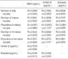
Univariate analysis revealed that the number of immature as well as mature oocytes was positively correlated with serum AMH, inhibin B and estradiol levels, respectively (Table 2). In serum AMH and inhibin B, the correlation coefficients for immature oocyte were slightly higher than those for mature oocyte. AMH levels had a strong positive correlation with inhibin B. Either AMH or inhibin B levels were correlated positively with estradiol levels. There was no relation between mature and immature oocyte count.
Since serum levels of AMH, inhibin B and estradiol may reflect overall oocyte pool including mature (MO) and immature oocytes (IM), we tested multiple regressions and the following equations were generated.
AMH (ng/mL)=0.1312+0.0874 [MO]+0.1417 [IM] (r=0.5424, p=0.008)
Inhibin B (pg/mL)=-188.7851+130.5844 [MO]+235.8635 [IM] (r=0.7318, p=0.022)
Estradiol (pg/mL)=234.1401+118.9406 [MO]+87.0733 [IM] (r=0.6593, p=0.014)
Multiple regressions showed that numbers of mature and immature oocytes were still independently correlated with three markers. Multiple regressions also indicated that all of three markers could reflect the combined pool of mature and immature oocytes with a statistical significance. These equations also indicate serum AMH and inhibin B levels measured on triggering day were more closely related with the immature oocyte count than mature oocyte count in IVF cycle.
DISCUSSION
Our results clearly demonstrated that the number of immature as well as mature oocytes is independently correlated with serum AMH and inhibin B levels measured on ovulation triggering day. Very interestingly, the correlation coefficients from univariate analysis were higher in the number of immature oocytes than number of mature oocytes. This means that the number of immature oocytes have a better correlation with serum AMH and inhibin B levels. We could generate multiple regression equations in the hypothesis that number of retrieved oocytes represents follicular pool at the time of oocyte retrieval. From the equations, we found again that number of immature oocytes could be a better contributor to determine serum levels of AMH and inhibin B.
Although previous studies by other researchers demonstrated that serum values of AMH (12) or inhibin B (13, 16) at the time of ovulation triggering correlate significantly with the number of oocytes retrieved, they did not describe the number of immature oocytes separately. Our findings are in agreement with those of the previous studies in that serum AMH or inhibin B at ovulation triggering day have a significant positive correlation with the number of total or mature oocytes collected. Moreover, we found for the first time that serum levels of AMH and inhibin B could be good predictors to determine the number of immature oocytes retrieved in IVF cycles.
At the time of ovulation triggering, follicular pool with various sizes exists within ovary. During ovarian stimulation, the oocyte population at the time of hCG may be heterogeneous and this leads to retrieval of oocytes at different stages of maturation. Immature oocytes at the time of oocyte retrieval may be originated from small antral follicles or from large preovulatory follicles unresponsive to hCG. In one study, 61.8% of immature oocytes are obtained from small antral follicles (7-12 mm) and the remaining from larger follicles (>12 mm) (24).
In IVF cycles, 10-15% of oocytes are generally obtained as immature form. Although they represent a compromised group with low developmental competence, those are valuable source for surplus oocytes if they can be matured in vitro. The clinical use of oocytes matured in vitro for IVF is more important especially in poor responders; thereby the needs for development of in vitro maturation are increasing (25). In the present study, 33.7% of oocytes were retrieved as immature form. This higher rate might be principally attributed to our strict policy to puncture all follicles over 10 mm in diameter during the period of study. The selection of those who were used GnRH antagonist could be another factor. A slightly higher immaturity was reported in GnRH antagonist cycles compared to GnRH agonist cycles (24).
We also found a strong positive correlation between serum concentrations of AMH and inhibin B at ovulation triggering day. Serum estradiol levels also correlated positively with either serum AMH or inhibin B levels. These strong correlations indicate that growing follicular pool produces AMH, inhibin B as well as estradiol into the circulation, and thus suggest that serum AMH and inhibin B give no additive information over conventional measurement of serum estradiol levels. Serum AMH and inhibin B may be valuable in that these protein markers represent preferentially immature oocyte count, as shown by our study.
In the present study, the mean serum level of AMH at ovulation triggering day was 0.90 ng/mL (range: 0.05-2.99 ng/mL) and this was quite lower than 2.7 ng/mL (range: 0-28.5 ng/mL) reported by Silberstein et al. (12) However, it cannot be directly compared since the method of measurement was different. We used ELISA kits purchased from Diagnostic Systems Laboratories (DSL), and it was reported that AMH levels are almost 4.6-fold lower with the DSL kit compared to the Beckman Coulter kit (26). One study using the Beckman Coulter kit reported the median serum level of AMH was 1.44 ng/mL on the day of oocyte retrieval (range: 0.38-4.96 ng/mL) (11). Serum AMH levels on the day of oocyte retrieval may be lower than those in ovulation triggering day because hCG-induced luteinization curtails follicular AMH production (27). Therefore, serum AMH values may be different according to the time of measurement and the ELISA kits used.
In conclusion, our study demonstrated that the number of immature as well as mature oocytes retrieved in stimulated IVF cycles is independently correlated with serum AMH and inhibin B levels measured on ovulation triggering day. Our results also indicate that serum AMH and inhibin B levels on triggering day are more closely related with immature oocyte count, thus serum levels of AMH and inhibin B seem to be good predictors to determine the immature oocyte count in IVF cycle.
 XML Download