

 PDF
PDF ePub
ePub Citation
 Print
Print


INTRODUCTION
In addition to hematopoietic stem cells (HSCs), the bone marrow contains a second type of stem cells that are capable of giving rise to multiple mesenchymal cell lineages, namely, mesenchymal stem cells (MSCs) (1, 2). Bone marrow stromal cells have been shown to include MSCs, which are thought to support hematopoiesis by creating an optimal microenvironment (the marrow matrix), and by providing cytokines and other regulatory factors that stimulate and enhance proliferation of the hematopoietic elements (3). Moreover, multipotent human MSCs can be isolated from bone marrow and expanded more than 1-billion-fold in cell culture without loss of stem cell capacity (4). These cells are capable of differentiating into osteoblasts, adipocytes, chondrocytes, myocytes, neural elements, and stromal fibroblasts under defined in vitro or in vivo conditions, and show an identical phenotype when grown from a single cell clone (5-7). In the area of hematopoiesis, MSCs are being used clinically to support hematopoietic recovery by replacing stroma. Moreover, a growing body of clinical evidence suggests the existence of a link between the efficiency of hematopoietic recovery and the degree of stromal damage that occurs as a result of myeloablative cancer therapy (8). As a result, therapy-ablated marrow may not be able to sustain hematopoietic stem cell maintenance or their differentiation into specific lineages, such as, megakaryocytes and platelets. In addition, a prolonged stromal defect in growth factor production, like early-stage cytokines, such as, interleukin-6, leukemia inhibitory factor, stem cell factor and granulocyte-colony stimulating factor, after autologous bone marrow transplantation has been observed for as long as a year in patients with hematologic disorders (9).
Independent laboratories have shown that recipients of unmanipulated allogeneic bone marrow transplants have only host-type marrow stromal cells and MSCs in bone marrow (10, 11). These results are attributed to the inability of the conditioning regimen to ablate host marrow stroma and/or the inability of stromal progenitors of donor origin to engraft. In addition, the number of MSCs has been estimated to be low in an average bone marrow graft (400-1,000 MSCs/kg). On the other hand, a recent report noted that allogeneic osteoblasts could be detected in recipients with osteogenesis imperfecta (OI) after sibling-matched allogeneic bone marrow transplantation (12). These findings suggest that osteoblasts may be transplanted successfully under certain conditions. Stromal chimerism has been achieved using infusions of cultureexpanded stromal progenitors in murine models, and xenotransplantation models have been developed to determine the homing of human MSCs in animals. Human MSCs were detected predominantly in bone marrow, and less frequently in other organs of immunodeficient NOD/SCID mice 8 weeks after infusion. Moreover, recent data indicate that MSC coinfusion enhances human hematopoiesis in NOD/SCID mice (13, 14). Based on encouraging data obtained in both rodent and large animal models, clinical studies of MSC transplantation are underway in both autologous and allogeneic settings. Koc et al. (15) found that the co-infusion of autologous MSCs together with autologous HSCs is free of toxicity and that it is associated with rapid hematopoietic recovery in 28 women receiving high dose chemotherapy for advanced breast carcinoma. In this report, we demonstrate that co-transplantation with human MSCs can enhance the engraftment of human hematopoietic stem cells in NOD/SCID mice, and that this engraftment-enhancing effect increases according to the number of infused MSCs. We also show that an increased dose of MSCs enhances the tissue distribution of human MSCs in multiple tissues in NOD/SCID mice. Our results show that MSCs are capable of enhancing hematopoietic engraftment and distribution to multiple organs in a dose-dependent fashion.
MATERIALS AND METHODS
Collection and isolation of CD34+ cells from umbilical cord blood (UCB)
UCB was obtained at the time of full-term deliveries, after receiving informed consent for the study protocol by our hospital's ethics committee. UCB was collected after clamping and cutting of cords by draining blood into sterile collection tubes containing the anticoagulant citrate-phosphate dextrose. Mononuclear cells (MNCs) were isolated from UCB using Ficoll-Hypaque (1.077 g/cm3, Sigma, St Louis, MO) density centrifugation (400 g for 25 min). CD34+ cells were isolated using a magnetic cell sorting system, Mini-MACS (Miltenyi Biotec, Auburn, CA), according to the manufacturer's instructions. Purity was determined by flow cytometry using a FITC-conjugated anti-CD34 (anti-HPCA-2, Becton-Dickinson, Mountain View, CA), and was found to be 90 to 95%.
Human MSCs isolation and ex vivo culture
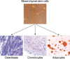
Ten to twenty mililiters of bone marrow aspirate was obtained under sterile conditions by posterior iliac crest puncture from bone marrow transplantation donors with given informed consent. Human MSCs were isolated and culture-expanded according to the method described by Pittenger et al. (1). MNCs were isolated from bone marrow, and cells were cultured in human MSC medium composed of Dulbecco's modified Eagle's low glucose medium (DMEM-LG; GibcoBRL, Grand Island, NY) containing 10% fetal bovine serum (FBS; GibcoBRL) and 1% antibiotic-antimycotic solution (GibcoBRL). Human MSCs were confirmed to be negative for hematopoietic markers by flow cytometry and to be capable of differentiating into osteocytes, chondrocytes, and adipocytes in vitro (Fig. 1, 2).
Differentiation of MSCs
MSCs were induced to differentiate into adipocytes by treating confluent second passage monolayer cultures with 1 µM dexamethasone (Sigma, St. Louis, U.S.A.), 0.5 mM methylisobutylxanthine (Sigma), 10 µg/mL insulin (Sigma), and 100 mM indomethacin (Sigma) in DMEM containing 10% FBS. Adipogenic differentiation was demonstrated by the accumulation of lipid vesicles stained red by Oil red O (Sigma). The osteogenic differentiation of MSCs was induced by culturing confluent monolayers in DMEM containing 10% FBS, 0.1 µM dexamethasone, 50 µM ascorbate (Sigma), and 10 µM β-glycerol- phosphate (Sigma). MSC osteogenic differentiation over 21 days was demonstrated by increases in alkaline phosphatase and calcium accumulation. Alkaline phosphatase was detected histologically, and calcium was stained by the Kossa's method. The chondrogenic differentiation of MSCs was in- duced by pellet culture in 500 µL of DMEM containing 10% FBS, 0.1 µM dexamethasone, 50 µM ascorbic acid 2-phosphate (Sigma), and 1 µg/mL transforming growth factor-β (βTGF-β R&D System, Minneapolis, MN). Under low speed centrifugation (1,000 g for 5 min), a dense mass of cells formed at the bottom of conical centrifuge tubes. Chondrogenic differentiation was demonstrated by toluidine blue staining after 3 weeks of culture.
Mice
Female NOD/SCID mice (5-6 weeks old), purchased from Charles-River Laboratory (Tokyo), were housed in micro-isolator cages on laminar flow racks at the animal facilities of the Catholic University of Korea. Mice were provided a sterile diet and autoclaved acidified water.
Xenotransplantation of human cord blood CD34+ cells and MSCs into mice
NOD/SCID mice were sublethally irradiated (3.5 Gy) 4 to 6 hr before transplantation. Human female cord blood CD34+ cells were injected at a final volume of 100 µL Iscove's Modified Dulbecco's Media (IMDM; GibcoBRL) per mouse and human male MSCs were injected with IMDM at a volume of 100 µL/106 MSCs per mouse. Both CD34+ cells and MSCs were injected via a tail vein. Two mice were assigned to each experimental group. In three separate experiments, mice were assigned to receive 1×10⁵ CD34+ cells, CD34+ cells plus 1×10⁶ MSCs, or CD34+ cells plus 5×10⁶ MSCs. One mouse treated with 5×10⁶ MSCs in the first experiment died after MSCs infusion due to myocardial infarction (pathologically confirmed, data not shown).
Detection of human hematopoietic cells in NOD/SCID mice by flow cytometry
Four weeks after transplantation, mice were sacrificed by carbon dioxide inhalation, and bone marrow (BM) cells were collected by flushing both femurs with RPMI medium (Gibco BRL). Cell suspensions were stained with mouse anti-human monoclonal antibodies, fluorescein isothiocyanate (FITC)-conjugated CD45 antibody and phycoerythrin (PE)-conjugated CD34, CD13, CD33, CD3, and CD19 antibodies (Becton-Dickinson, Mountain View, CA). Flow cytometry to detect human CD45+ and CD34+, CD13+, CD33+, CD3+, or CD19+ cells was performed on a FACScan flow cytometer (Becton-Dickinson). FITC- and PE-conjugated mouse isotype control antibodies were used for each culture, and 5,000-10,000 events were counted for each analysis. Differences in engraftment percentages were calculated using the Student's t-test. p values of <0.05 were considered statistically significant.
Detection of human MSCs in NOD/SCID mice by fluorescence in situ hybridization
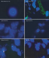
Fluorescence in situ hybridization (FISH) for the human Y chromosome was used to detect transplanted MSCs in BM, spleen, liver, lung, kidney, heart, skeletal muscle, intestine, and skin of NOD/SCID mice at four weeks after transplantation in the second and third experiments. Total numbers of Y chromosome positive cells in ten×high-power fields were counted.
RESULTS
Differential potential of MSCs
Culture of bone marrow MNCs produced a monomorphic confluent adherent layer of elongated fibroblast-like cells that survived multiple passages under mesenchymal culture conditions. At the end of the second passage, bone marrow derived MSCs had successfully differentiated along osteogenic, chondrogenic, and adipogenic lineages, using methods described above (Fig. 2).
Effect of MSCs on human hematopoietic cell engraftment in NOD/SCID mice
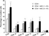
Transplantation of UCB CD34+ cells (1×10⁵/mouse) in the presence of MSCs (1×10⁶ and 5×10⁶/mouse) resulted in significantly higher engraftment levels in the BM of NOD/SCID mice than was observed after transplanting UCB CD34+ cells alone. The mean percentage of CD45+ cells in the BM of NOD/SCID mice according to the MSCs number, i.e., 1×10⁶ or 5×10⁶, increased to 41.05±5.62 and 55.58±8.26, respectively, compared with 29.18±1.00 for transplantation with CD34+ cells without MSCs (p<0.05). The difference between the group infused with 1×10⁶ MSCs and the group infused with 5×10⁶ MSCs was statistically significant (p<0.05) (Fig. 3). The mean percentage of CD45+-CD34+ cells increased to 13.78±1.50 and 17.43±2.05 at these MSC dosages, compared with 7.76±0.37 for transplantation with CD34+ cells without MSCs (p<0.05). In addition, the groups infused with 1×10⁶ or 5×10⁶ were significantly different (p<0.05) (Fig. 3).
Effects of MSCs on human myeloid cell engraftment in NOD/SCID mice
Co-transplantation of MSCs significantly increased the level of human myeloid cell engraftment in NOD/SCID mouse BM. The mean percentage of CD45+-CD33+ cells in NOD/SCID mouse BM at MSC doses of 1×10⁶ and 5×10⁶ were 19.62±1.18 and 24.38±3.05, respectively, compared with 6.54±0.17 for transplantation with CD34+ cells alone (p< 0.05). Moreover, the level of myeloid cell engraftment in the group infused with a high dose of MSCs was significant higher than in the group infused with a lower dose (p<0.05) (Fig. 3). Mean percentages of CD45+-CD13+ cells increased to 21.64±2.04 and 24.45±3.41 for high and low MSC doses, compared with 10.34±0.57 for transplantation with CD34+ cells alone (Fig. 3). Although there was a distinct difference between the group co-transplanted with MSCs and the group transplanted with HSC alone (p<0.05), we did not notice a difference between levels of HSC engraftment according to the number of MSCs co-transplanted.
Effects of MSCs on human lymphoid cell engraftment in NOD/SCID mice
The mean percentages of CD45+-CD19+ cells in NOD/SCID mouse BM according to the MSC dose were significantly increased to 9.93±4.77 and 24.39±11.22, respectively, compared with 7.08±1.46 of CD34+ cells alone (p<0.05). Co-transplantation of MSCs at 5×10⁶ significantly increased the level of human B lymphoid cell engraftment in NOD/SCID mouse BM (p<0.05). Mean percentages of CD45+-CD3+ cells engrafted by low and high MSC doses were 2.89±1.90 and 4.43±1.64, respectively, compared with 3.02±1.17 for CD34+ cells alone. Co-transplantation of MSCs did not increase the level of human T lymphoid cell engraftment in NOD/SCID mouse BM (Fig. 3).
Engraftment of human MSCs in NOD/SCID mice
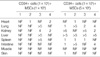
Human Y chromosome positive cells were found in BM, spleen, liver, lung, kidney, heart, intestine, and skin of NOD/SCID mice at four weeks after transplantation but not in skeletal muscle. The distribution of human Y chromosome positive cells differed by tissue; Y chromosome positive cells were found more frequently in the liver, lung, and kidney than in other tissues (Fig. 4, Table 1). In addition, numbers of these cells were higher in mice that received a higher dose of MSCs in same tissues.
DISCUSSION
We infused relatively low doses of CD34+ cells (1×10⁵ cells) into NOD/SCID mice in this study. We tried to evaluate the dose-dependent effect of MSCs on HSC engraftment, and thus, we used relatively high doses of MSCs (5×10⁶/animal) as compared with the doses (1×10⁶/animal) usually applied. We observed that the high-dose MSC group showed significantly higher levels of HSC engraftment than the lowdose (1×10⁶/animal) MSC group, thus confirming that MSCs have a dose-dependent effect on HSC engraftment.
Noort et al. (13) demonstrated in NOD/SCID mice that engraftment levels were higher in the co-transplantation of MSCs (1×10⁶) and UCB CD34+ cells than in transplantation with UCB CD34+ cells alone. They also demonstrated that the effects of MSC co-transplantation on BM engraftment were more pronounced after transplantation with relatively low doses of CD34+ cells (0.03-0.1×10⁶). The engraftment level increased three- to four-fold with MSCs cotransplantation with low doses of UCB CD34+ cells. However, no further additive effect was observed at higher doses of UCB CD34+ cells. Angelopoulos et al. (14) reported that MSC co-transplantation enhanced human cell engraftment when CD34+ cell infusion was limited, and that the efficacy of engraftment was blunted when the CD34+ cell dose was increased to 1.5×10⁶. These data suggested that MSC co-transplantation can enhance the efficacy of engraftment if only a low dose of CD34+ cells is available. We observed similar effects in the present study.
Few reports have evaluated whether the engraftment-enhancing effect of MSCs is related to the MSC dose, which is why we decided to use a higher dose in this study. However, problems occur when animal models are administered higher doses. Initially, we wondered whether higher doses increase the risk of lethal vascular complications, such as, pulmonary embolism. In fact, the frequency of sudden death within 24 hr after MSC infusion did tend to increase slightly, but this was not significant (not shown data). Additionally, the use of a high cell dose could be criticized for not being clinically relevant. However, the objective of this study was to determine the effect that MSCs have on HSC engraftment, and therefore we chose a somewhat extreme model.
The effect of MSCs on HSC engraftment is not lineage-restricted, with grafts being comprised B lymphoid cells as well as myeloid cells. However, T lymphoid cell levels were not increased by co-transplanting HSCs with MSCs. This supportive activity may have been derived from the specific characteristics of MSCs, which have a suppressive effect on the proliferation of T lymphocytes and a modulating effect on the immune system (16, 17). However, no mechanism explaining the immunoregulatory effects of MSCs has been elucidated (16-20).
Several studies in animal models have demonstrated that stromal cells not only seed the bone marrow but also enhance hematopoietic recovery (13, 21, 22). However, the transplantation ability of bone marrow stromal elements still remains an open issue in humans (23). Several independent clinical studies have shown that recipients transplanted with unmanipulated allogeneic bone marrow have only host-type marrow stromal cells and MSCs in bone marrow (11, 24). This finding suggests that neither stromal progenitors nor MSCs are able to engraft into bone marrow because a "normal" bone marrow graft contains too few MSCs to support engraftment. However, others have reported contrary results (25, 26). This discordance may originate from either methodological differences concerning the detection of donor-derived mesenchymal cells, or from differences between doses of reinfused stromal cells. To detect engrafted donor derived cells in various tissue types, green fluorescence protein (GFP) is most widely used as a reporter. However, even though the GFP method has a higher sensitivity than the monoclonal antibody method for membrane antigen, it is difficult to determine whether GFP expressing cells are quiescent. Therefore, we decided to use FISH for the human X and Y chromosomes to detect engrafted donor derived cells in the organs of NOD/SCID mice. The data obtained provide evidence that human HSCs can be engrafted into various organs in NOD/SCID mice, and that the number of engrafted cells increases in proportion to the number of MSCs infused.
Our results reveal that MSCs have an accelerating effect on HSC engraftment and that this is dose-dependent. It is also suggested that a graft engineered to increase the number of MSCs may pave the way for further applications in the field of transplantation with respect to hematopoietic support and efficient engraftment.
 XML Download