

 PDF
PDF ePub
ePub Citation
 Print
Print


INTRODUCTION
Electrical stimulation of acupoint (ESA; electroacupuncture) has been used to treat a wide range of musculoskeletal disorders and reported to relieve pain and inflammation, strengthen muscle, and reduce abnormal muscle tone (1-5). Its mechanisms of action are not fully elucidated, but some of its action on the endogenous opioids system through multiple neuronal pathways have been identified in experimental studies.
Previous studies have revealed that ESA with low (2-10 Hz) and high (≥100 Hz) frequencies has different mechanisms with selective release of β-endorphins, enkephalins or dynorphins (6-8). ESA at different frequencies activates the distinct different regions in the spinal cord and the central nervous system (9, 10), and evokes the different responses of autonomic nervous system (11, 12). Based on these findings, ESA with low and high frequencies seems to produce the different therapeutic effect.
Ankle sprain is a very common condition of acute musculoskeletal injuries and is induced by accidental overextension of ligaments in ankle joint, resulting in pain, reduction of weight bearing during walking, and edema around ankle. Early weight bearing, bracing, and functional rehabilitation are the generally accepted management of mild and moderate ankle sprain (13, 14). In addition, cyclo-oxygenase inhibitors and opioids are known to be useful in reducing pain and edema and to achieve earlier recovery of normal function (15, 16).
The ankle sprain model in rats was demonstrated to generate similar clinical symptoms of mild degree of ankle sprain (3). There have been few studies on the effect of ESA for ankle sprain (3), and moreover, discrete effects of ESA at low and high frequencies on pain and edema induced by ankle sprain have not been reported.
Therefore, we aimed to investigate whether ESA with two frequencies, 2 Hz and 100 Hz, can reduce pain and edema induced by ankle sprain and whether there is a difference in therapeutic effects between 2 Hz and 100 Hz ESA in a recently developed ankle sprain model in rats.
MATERIALS AND METHODS
Animal preparation
Male Sprague-Dawley rats were housed in separate cages and allowed to acclimate for 7 days by using a 12/12 hr day/night cycle. The experiments were performed on rats weighing 210-270 g. The experimental protocol was approved by our Institutional Animal Care and Committee.
Procedure for ankle sprain
Rats were anesthetized with 2% enflurane in O2 via a mask. The ankle sprain model was made according to the method described by Koo et al. (3). Ankle sprain was produced by manually overextending the lateral ligament.
Electrical stimulation
Under general anesthesia, electrical stimulation was delivered to contralateral SI6 point (Yangno) through a pair of bipolar needles at 24 hr after ankle sprain. It was controlled by Grass S88 electrical stimulator (Grass Telefactor, West Warwick, RI, U.S.A.) equipped with SIU5 isolation unit (Grass Telefactor, West Warwick, RI, U.S.A.). The SI6 acupoint is known as an effective analgesic point for pain induced by ankle sprain in rats (3) and located at the posterior distal end of the forearm between the radius and ulna. Electric current (1 ms pulse duration, 5 times intensity as muscle twitch) was delivered for 30 min. Rats were allocated randomly into three groups (2 Hz ESA, 100 Hz ESA or control groups: n=9/group). In the control group, needles were not inserted and electrical current was not delivered.
Measurement of pain
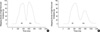
The pain was determined by the stepping force in the paw of the sprained ankle during walking (3). Rats were allowed to acclimate within plastic walking channel (10 cm width, 10 cm high, 60 cm long) for 10 min and then allowed to walk spontaneously. The stepping force in the paw of the sprained ankle was recorded by Pocket Pro 250-B electronic balance (Acculab, Newton, PA, U.S.A.), which is placed on the floor at the midway area of the right walking path. The signal of the balance was converted to digital signal by an AD converter (Physiolab, Seoul, Korea), which was fed into a computer monitor. The waves of stepping force were observed, and the peak amplitude was measured during walking. To estimate the level of pain produced by ankle sprain, stepping forces were measured before ankle sprain and at 24 hr after ankle sprain. To evaluate the analgesic effect of ESA, stepping forces were measured at 1, 2, 4 hr after the termination of ESA. The rats with a weight bearing ratio less than 20% or more than 40% were excluded from the study. The data were converted by the following formula: % recovery=(improvement of stepping force by EA in gram/reduction of stepping force by sprain in gram)×100.
Measurement of paw volume
The volume of the right hindpaw was measured before and 24 hr after ankle sprain. To evaluate the anti-edematous effect of ESA, the volume of the right hindpaw was measured at pre-ESA and at 2, 4, 6, 12 and 24 hr after the termination of ESA. The volume of paw was measured by a water replacement plethysmometer (Ugo Basile, Comerio, Italy). The position at about 5 mm proximal to the ankle is clearly divided by dense long hairs and sparse thin hairs. This clearly demarcated boundary has been used as a convenient landmark (3). The paw was immersed into water up to the marked line. The volume of paw edema was calculated from experimental paw volume minus pre-sprained paw volume. The data were converted by the following formula: decreased paw edema (%)=(volume of paw edema after ESA in mL/volume of paw edema at 24 hr after sprain in mL)×100.
RESULTS
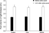
The sprain injury of right hindpaw produced a marked decrease (p<0.001) in stepping force during walking (Fig. 1) and a significant increase (p<0.001) in foot volume (Fig. 2). The weight-bearing ratios were decreased significantly (p<0.001) from 53.1±0.5% (2 Hz ESA), 53.5±0.6% (100 Hz ESA) and 52.9±0.3% (control) to 33.4±0.9%, 34.2±1.0% and 32.3±1.5%, respectively. The foot volumes at 24 hr after sprain were increased (p<0.001) to 170.4±5.33% (2 Hz ESA), 174±5.03% (100 Hz ESA), 174±5.03% (control), compared to the values at pre-sprain.
In 2 and 100 Hz ESA groups, the % recovery of stepping force was significantly (p<0.05) increased at 1 and 2 hr after post-ESA compared to the values at pre-ESA. In the control group, no differences between pre-ESA and post-ESA were observed (Fig. 3).
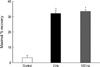
The maximal % recovery at 2 Hz ESA and 100 Hz ESA were 32% and 33%, respectively, showing no differences between two groups, but showed a significant (p<0.05) increase compared to the values in the control group (Fig. 4).
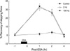
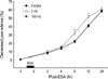
In three groups, the edema volumes were reduced as time elapsed. The edema volumes in the 2 Hz ESA group were reduced more significantly (p<0.05) than the values in 100 Hz ESA and control groups at 6 and 12 hr after ESA (Fig. 5).
DISCUSSION
In the present study, ESA at 2 and 100 Hz showed improvement in stepping force and produced a similar degree of analgesic effects, but only ESA at 2 Hz facilitated the reduction of edema in ankle sprain.
The analgesic effect of ESA observed in the current study is consistent with several studies in which ESA shown to be effective in several pain models (8, 17, 18). It has been also reported that ESA effectively alleviated ankle sprain pain in rats (3). However, they used the electrical current with train of four pulses (100 Hz of intra-train frequency) repeated at 2 Hz, which was different from continuous electrical stimulation used in the present study and did not specifically examine the analgesic and anti-edema effects of low and high frequency ESA.
In our study, analgesic effectiveness of ESA at 2 Hz was similar to that at 100 Hz. In the previous studies, ESA at low and high frequencies produced different analgesic effects in various pain conditions. Some studies have reported that high frequency was more effective against inflammatory hyperalgesia (19) and post-operative pain (20). Another study has reported that low frequency ESA produced superior analgesic effects in response to noxious radiant heat stimuli (10). Moreover, it has been reported that ESA at 100 Hz reduced mechanical, but did not thermal inflammatory hyperalgesia (21). These inconsistent results reflect that ESA of specific frequencies may produce different effects to the several types of pain and hyperalgesia.
The reasons of similar analgesic effect of both frequencies on ankle sprain in the present study might be due to the specific nature of ankle sprain itself or the measurement method of pain. Ankle sprain has a combined property of mechanical and inflammatory pain. We measured the stepping force during walking as an index of ankle pain. This index has been also widely used in several arthritic pain models (3, 22, 23). However, the changes in stepping forces during walking may be affected by ankle sprain pain as well as muscle activity and joint movement. Also, ESA can strengthen muscle power and improve the range of motion of joint (4, 5). Therefore, improvement of motor activity induced by ESA at 2 and 100 Hz may affect positively the changes in the amount of stepping forces.
In this study, ESA at 2 Hz produced more rapid reduction of edema compared to spontaneous progression of edema reduction, but ESA at 100 Hz failed to show any effects on edema reduction. This is consistent with a previous report that showed only low frequency ESA inhibited paw edema in the complete Freund's adjuvant-induced inflammation model (2). This finding, together with the previous data, suggests that ESA at 2 and 100 Hz have different therapeutic effects on edema resulting from ankle sprain.
The anti-inflammatory effect of low frequency ESA has been observed in several studies. ESA at low frequency produced significant inhibition of paw edema in capsaicin or carrageenan-induced inflammation (24-26) and suppressed the activity of macrophages induced by lipopolysaccharide (27). The anti-inflammatory effect of low frequency ESA is considered to be related to µ opioid receptor agonist. Its effect was reduced or suppressed by low doses of naloxone (25, 27). These findings are in line with the observation that morphine suppressed carrageenan-induced edema and anti-edema effect of morphine was reversed by low doses of naloxone (28). On the other hand, some studies showed that low frequency ESA induced the release of ACTH from the pituitary gland and elevated the levels of peripheral cortisols, which can suppress inflammation by shutting off the production of inflammatory mediator at the sites of injury (2, 29, 30). In addition, some investigators showed that anti-inflammatory effects of low frequency ESA were not affected by naloxone (31), suggesting that a non-opioid mechanism may be involved in anti-inflammatory effect by low frequency ESA. This is different from previous findings that naloxone inhibited antiinflammatory effect of low frequency ESA (25, 27). They assumed that the discrepancy might be due to different inflammation-induced agents (carrageenan vs. capsaicin).
Although the mechanism underlying ESA is not clear, it has been demonstrated that ESA is mediated by the endogenous opioids system and ESA at 2 Hz facilitates the release of β-endorphin, enkephalin, or endomorphin, whereas ESA at 100 Hz releases dynorphin. The effect produced by 2 Hz ESA was naloxone-reversible, while high frequency stimulation was not (6). In a cross-tolerance study, high frequency ESA tolerant rats showed cross-tolerance to κ opioid receptor agonist (dynorphin) but did not show cross-tolerance to a specific agonist for δ opioid receptor (7). Also, endomorphin-1 was involved in 2 Hz, but not 100 Hz, ESA-induced analgesia (8). These results suggest that ESA with low and high frequencies have different mechanisms, and optimization of stimulation frequency is important in determining the therapeutic effect in various pathological conditions.
Interestingly, 2 Hz ESA reduced ankle sprain pain at 1 and 2 hr after the electrical stimulation, but inhibited edema at 6 and 12 hr after the electrical stimulation. This time dissociation between analgesic effect and anti-edema effect of ESA is in agreement with the previous observation that low frequency ESA attenuated inflammatory hyperalgesia at 2.5 hr, but inhibited edema at 24 hr (24). These results, together with present data, suggest that the analgesic effect of ESA is independent of an effect on peripheral inflammation, and the anti-inflammatory mechanisms of ESA may be different from analgesic mechanism. Although the endogenous opioids systems are closely involved in mechanisms of action of ESA, further studies are required to clarify the mechanisms underlying ESA-induced anti-inflammatory effects.
In conclusion, the present study shows that 2 and 100 Hz ESA are similarly effective against ankle sprain pain, but only 2 Hz facilitates the reduction of ankle sprain edema. This result suggests that there is different therapeutic effects between ESA at 2 Hz and 100 Hz, and 2 Hz ESA may be considered as a preferred therapeutic method for ankle sprain.
 XML Download