

 PDF
PDF ePub
ePub Citation
 Print
Print


INTRODUCTION
Despite continuous improvements in intensive care medicine and antibiotic therapy, bacterial meningitis remains a serious disease at any age, and is still accompanied by significantly high mortality and neurologic morbidity in survivors. The prognosis is particularly poor in neonates, with mortality rates of 20-40% and long-term neurologic sequelae in up to 50% of survivors (1). Although the precise mechanism of neuronal injury has not been completely delineated, host inflammatory responses to bacterial components in the subarachnoid space including increased intracranial pressure (ICP), increase in cytokines, leukocytes, and lactate in the cerebrospinal fluid (CSF) (2, 3), and cerebral ischemia resulting from reduced cerebral perfusion pressure (CPP) due to intracranial hypertension and/or systemic hypotension (2, 4, 5), are presumed to be responsible for much of brain injury during bacterial meningitis. Therefore, the development of new adjunctive therapy to antibiotics aimed at attenuating acute inflammatory responses and reducing cerebral ischemia would be necessary to improve the prognosis of this disease.
Hypertonic saline (HTS) has been primarily used as an initial therapy in severe hypovolemic shock associated with trauma in context with small-volume resuscitation (6), however, its use is being considered in various conditions including perioperative setting (7), burn injury (8), sepsis syndrome (9), and brain injury (10). In animal studies, HTS immediately increases mean arterial pressure (MAP) (11), enhances cardiac function (12), and restores microvascular perfusion (13). HTS also decreases the rising of ICP and improves CPP and regional CBF in various animal studies (14, 15). According to recent in vitro studies, HTS also possesses significant anti-inflammatory capabilities (16-18). HTS can interfere with neutrophil-endothelial cell interaction (16), inhibits lipopolysaccharide-induced nitric oxide synthase expression by inhibiting nuclear factor kappa B (17), and attenuate the neutrophil cytotoxic response by interfering with intracellular signal transduction (18). These features make HTS an attractive therapeutic agent to attenuate neuronal injury by improving CPP and down-regulating acute inflammatory responses in bacterial meningitis.
In the present study, we evaluated the efficacy of HTS as an adjunctive therapy in attenuating brain damage in experimental neonatal bacterial meningitis. We tested the hypothesis that HTS attenuates brain injury by improving CPP and down-regulating acute inflammatory responses in experimental Escherichia coli meningitis in the newborn piglet.
MATERIALS AND METHODS
Animal preparation and surgical procedures
The experiments described herein were reviewed and approved by the Institutional Animal Care and Use Committee of the Samsung Biomedical Research Center (Seoul, Korea). We also followed the institutional and National Institutes of Health guidelines for laboratory animal care in this study.
Newborn piglets of mixed strain (Yorkshire, conventional breed, purchased from Paju Farm, Paju, Gyeonggi-Do, Korea) less than 3 days old, weighing 1.0-1.6 kg were used in this study. Animals inhaled ether for sedation, and anesthesia was induced with 5 mg/kg intravenous sodium thiopental. After local injection with lidocaine (1%), tracheostomy was performed and the piglet was paralyzed with 0.1 mg/kg intravenous pancuronium and ventilated with a neonatal pressurelimited, time-cycled mechanical ventilator (Sechrist Infant Ventilator, IV-100B; Sechrist Industries Co., Anaheim, CA, U.S.A.). Ventilator settings were adjusted to keep PaO2 at 80-100 mmHg and PaCO2 at 35-45 mmHg. Supplemental doses of sodium thiopental and pancuronium were given every 2, 3 hr when spontaneous movement of the animal including self-respiration was observed to maintain anesthesia. Femoral arteries and veins were cannulated for blood pressure monitoring, blood sampling, and medication and fluid infusion, respectively. Electrocardiogram, oxygen saturation, ICP, and blood pressure were continuously monitored using a Hewlett-Packard neonatal monitoring system (Hewlett-Packard model M1276A, Hewlett-Packard Co., MA, U.S.A.). Cisternal puncture was done with a 22-gauge spinal needle (Becton Dickinson, Franklin Lakes, NJ, U.S.A.), and the needle was kept in situ for continuous ICP monitoring and CSF sampling. Throughout the experiment, the piglet was placed under the servo-controlled warmer (Airshields, Hatboro, PA, U.S.A.), and rectal temperature was maintained between 38.0 and 39.0℃, which is normal for newborn piglets.
Experimental protocol
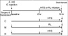
The experimental protocol was schematized in Fig. 1. In twenty five newborn piglets, preliminary study was done in the control group (C, n=6) to determine the infusion rate of HTS to maintain hypernatremia of 150-160 mEq/L, and the rest was randomly assorted into the meningitis group (M, n=10) and the meningitis with HTS infusion group (H, n=9). After surgery and a stabilization period, meningitis was induced by intracisternal injection of 108 colony forming units of E. coli in 100 µL of saline.
Continuous monitoring of heart rate, electrocardiogram, oxygen saturation, ICP, and MAP were done for 10 hr after bacterial inoculation, and CPP was calculated as MAP minus ICP. Serum electrolytes concentration, arterial blood gas analyses, and glucose and lactate concentration in the blood and CSF were measured at baseline and every 2 hr for 10 hr after bacterial inoculation. Leukocyte counts and bacterial titers in the cerebrospinal fluid (CSF) were determined at baseline, 6 and 10 hr of the experiment.
In C and H, hypernatremia was induced by giving a bolus of 10 mL/kg HTS (7%) for 30 min at 6 hr after intracisternal bacterial inoculation. After a bolus infusion of HTS, continuous infusion was begun at a rate of 4 mL/kg/hr by using infusion pump. Then, if serum sodium concentration measured every 30-45 min was between 150-160 mEq/L, infusion rate was unaltered, if serum sodium concentration was above 160 mEq/L, infusion was stopped until the next serum sodium concentration was determined, and if serum sodium concentration was below 150 mEq/L, infusion rate was increased to 8 mL/kg/hr. However, during the real experiment, only 5 piglets needed infusion rate alteration during HTS continuous infusion. They all needed infusion stop at least once during HTS continuous infusion because serum sodium concentration exceeded 160 mEq/L. There was no incidence that needed increase of infusion rate because serum concentration dropped below 150 mEq/L. The other 10 piglets received HTS at a constant rate (4 mL/kg/hr) all through the HTS continuous infusion (3.5 hr) because their serum sodium concentration was maintained between 150-160 mEq/L at all times.
To match the volume of HTS infused and thus eliminate the effect of differing volume on the outcomes, Ringer's lactate solution was infused as a bolus of 10 mL/kg and a subsequent continuous infusion at a rate of 4 mL/kg/hr to 10 piglets in M.
Separately from HTS or Ringer's lactate solution, D7.5W solution was infused continuously at a rate of 4 mL/kg/hr to the piglets in all experimental groups as the maintenance fluid during the experiment.
As we tried to settle the debate as to whether higher serum levels are not simply the result of sufficient amounts of HTS in order to restore CPP, we decided to aim for a certain serum level instead of a predetermined amount of volume in the present study. The serum sodium concentration of 150-160 mEq/L selected in the present study was assumed to be clinically acceptable and optimal for maximal clinical effects (19) and least side effects (6, 14).
CSF leukocyte counts were measured using a hemocytometer, arterial blood gases were measured on a blood gas analyzer (Ciba-Corning Diagnotics Corp., Medfield, MA, U.S.A.), and concentrations of glucose and lactate were measured using a YSI model 2,300 dual analyzer (Yellow Springs Instrument Co., Yellow Springs, OH, U.S.A.). Bacterial titers in the CSF were determined by plating 10-fold dilutions on blood agar plates overnight at 37℃ in room air. At the end of each experiment, brain cortex was harvested using a guillotine, rapidly frozen in liquid nitrogen, and stored at -80℃ for further biochemical analyses.
Biochemical analyses of brain cortex
Methods of brain cell membrane preparation and determination of cerebral cortical cell membrane Na+, K+-ATPase activity, concentration of conjugated dienes, ATP and phosphocreatine (PCr) were described in detail previously (3). Briefly, brain cell membranes were prepared according to the method described by Harik et al. (20). The activity of Na+, K+-ATPase was determined by subtracting the enzyme activity in the presence of ouabain from the total activity in the absence of ouabain (21). The concentration of conjugated dienes was determined using the method of Recknagel and Glende (22). Brain ATP and PCr concentrations were determined with a coupled enzyme assay using the method of Lamprecht et al. (23).
Statistics
Data were analyzed by Kruskal-Wallis test (nonparametric ANOVA) for intergroup comparisons at each time point. To detect significant changes over time within each group and between groups, data were compared using repeated measures ANOVA with Bonferroni correction. Statistical analyses described above were done using SPSS software program version 11.5. A p value of less than 0.05 was considered significant. Data are given as mean±standard error (S.E.).
RESULTS
Physiologic and laboratory data
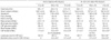
Physiologic and laboratory data from the three experimental groups at 6 hr (before HTS infusion) and 10 hr (the end of the experiment, 4 hr after HTS infusion) are summarized in Table 1. There were no significant differences in the baseline (at 0 hr) values of these variables between the three experimental groups.
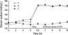
Serum sodium concentrations had risen to 150-160 mEq/L after the bolus infusion of HTS and remained at that level to the end of the experiment in C and H, while remaining at the baseline level in M (Fig. 2).
Significantly elevated lactate level and leukocyte counts in the CSF observed in M compared to C, were significantly improved in H. Elevated bacterial colony counts and decreased glucose level in the CSF observed in M and H were not significantly different during the experiment.
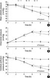
Changes in ICP, MAP, CPP
The changes in ICP, MAP, and CPP in the three experimental groups during the experiment are shown in Fig. 3. In C, there were no significant changes in ICP, MAP, and CPP throughout the experiment. In M, MAP decreased gradually during the experiment, and became significantly lower compared to C 8 hr after the induction of meningitis, and this abnormality was improved in H after HTS infusion. ICP increased progressively, and became significantly elevated 2 hr after the induction of meningitis in M and H, and this elevation was significantly attenuated after HTS infusion in H. CPP, calculated as MAP minus ICP, decreased gradually and became significantly lower compared to C 6 hr after the induction of meningitis in M and H, and this reduction was significantly attenuated in H after HTS infusion.
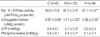
Biochemical Data in Cerebral Cortex
Decreased cerebral cortical cell membrane Na+, K+-ATPase activity and increased level of lipid peroxidation products (conjugated dienes) observed in M compared to C, indicative of meningitis-induced alterations in brain cell membrane function and structure, were slightly but significantly improved in H (Table 2). Marginally reduced concentrations of cerebral ATP and PCr observed in M compared to C failed to improve in H.
DISCUSSION
In the present study, HTS infusion significantly decreased lactate level and leukocyte count in the CSF, and increased CPP in the newborn piglet model of bacterial meningitis. HTS has been shown to possess significant anti-inflammatory capabilities in various in vitro studies (16-18, 24). In our previous studies of experimental neonatal bacterial meningitis (3, 25), increased ICP and elevated CSF lactate level were primarily attributable to acute inflammatory responses in the subarachnoid space rather than to brain edema and cerebral energy depletion from cerebral ischemia, respectively. Thus our data of significant attenuation of meningitis-induced increase in lactate level and leukocyte count in the CSF with HTS infusion suggest the possible in vivo anti-inflammatory effects of HTS. However, we adopted only leukocyte count in the CSF as an inflammatory marker in this study. Other inflammatory markers such as tumor necrosis factor-α, interleukins were not measured. Moreover, although statistically significant, the difference of leukocyte count in the CSF between H and M was small. This is a major drawback of our study. Therefore, it seems to be precipitate to conclude that HTS showed an anti-inflammatory effect in this study. Further studies will be necessary to prove the anti-inflammatory effects of HTS in bacterial meningitis.
Besides the acute inflammatory responses, cerebral ischemia resulting from the critical reduction of CPP with intracranial hypertension and systemic hypotension may have an additional and even critical role in the pathogenesis of brain injury during bacterial meningitis (2, 4, 5, 26, 27). In accordance with other studies (26, 27), our data of maintenance of adequate CPP by simultaneous improvement of reduced MAP and increased ICP with HTS infusion in this study suggest the possibility that HTS could also be an attractive adjunctive therapy to prevent cerebral ischemia and the ensuing brain injury in bacterial meningitis.
The anti-inflammatory property of HTS might not be attributable to a stabilization of hemodynamics because we have observed that maintenance of adequate CPP and cerebral blood flow with dopamine infusion did not attenuate the acute inflammatory responses in our previous study of experimental bacterial meningitis (26). Which effect of HTS, either anti-inflammatory effect or improvement of CPP, might have been primary in the attenuation of brain injury observed in the present study is a subject of intricacy, because these two effects are related to each other, in which as meningeal inflammation decreases, CPP accordingly increases (28). Further studies controlled for MAP, ICP, and both MAP and ICP, respectively could make clear which effect of HTS is the major one in reducing brain injury.
Oxygen free radicals attack double bonds of polyunsaturated fatty acids in cell membrane in a process called lipid peroxidation (29). Degradation of cell membrane structure by lipid peroxidation reduces Na+, K+-ATPase activity (30) and result in brain cell dysfunction that may lead to osmotic cell swelling and neuronal death (31). In the present study, increased lipid peroxidation products and reduced Na+, K+-ATPase activity observed in M were slightly but significantly improved in H. These findings implicate some attenuation of the meningitis-induced alterations in cerebral cortical cell membrane structure and function with HTS. However, as the extent of the attenuation is very small although statistically significant, the clinical significance of the neuroprotective effect of HTS remains doubtful. Further studies recruiting more definite tools such as pathology and functional assessment for the evaluation of brain injury could elucidate the neuroprotective effect of HTS. Cerebral high energy phosphate compounds (ATP/PCr) in M were reduced only marginally compared to C, and failed to demonstrate improvement in H. These findings suggest that the mitochondrial oxidative metabolism in M at this early stage of meningitis remains relatively intact despite anaerobic glycolysis due to inflammatory responses and cerebral ischemia from reduced CPP, and the effect of HTS is minimal.
The newborn piglet bacterial meningitis model has been well established throughout our previous studies (3, 25, 32-34). The piglet brain is comparable in maturity to human brain at birth (35). Furthermore, because it is similar in size to a preterm infant, methods for monitoring various physiologic variables and maintaining it in good condition throughout the experiment were also feasible.
In this study, E. coli was used to induce meningitis because it is the most frequent Gram-negative pathogen of neonatal meningitis. In the majority of our previous studies of experimental bacterial meningitis, 108 colony forming units of E. coli was used to induce meningitis. In one study, 109 colony forming units of E. coli was used. However, there was no remarkable difference in the severity of meningitis indicated by the ICP, CSF profiles, and brain tissue chemistry findings between the two doses of E. coli.
In the present study, we maintained the rectal temperature of newborn piglets within the normal range by monitoring and modifying the piglet's temperature with the thermoregulatory devices as in our previous studies (3, 4, 25, 26). We have already shown that rectal and brain temperatures were equivalent in our previous hypothermia study with the newborn piglet (32). Prevention of fever by maintaining normothermia in our controlled animals might have contributed to the reduced inflammatory responses in the present study. Our previous observation that acute inflammatory responses could be attenuated by therapeutically induced systemic hypothermia in experimental bacterial meningitis (32), also support that temperature could be an important confounding variable of the inflammatory responses during meningitis. However, most neonates with sepsis have a normal temperature at admission to the neonatal intensive care unit (36), and fever does not seem to a prominent feature in neonates during systemic bacterial infections such as sepsis and meningitis unlike that in adults. Thus, it is thought that the strict maintenance of normothermia and prevention of fever or hypothermia in our animal models of experimental neonatal bacterial meningitis would be more relevant and appropriate to simulate the clinical situation.
Intracisternal injection of bacteria to induce meningitis in the present study might cause a different situation and different permeability of the blood-brain barrier (BBB) than if infection occurs through the blood stream. Although we have observed that the BBB permeability and acute inflammatory responses were directly related to the concentration of the bacteria in the CSF space (33, 34), not to the routes of bacterial entry into the CSF (37), and the BBB permeability to the glucose increased markedly only 1 hr after intracisternal inoculation of bacteria (3) in our previous studies, further studies will be necessary to determine whether the BBB would be more permeable if the infection have occurred through bacteremia rather than direct intracisternal bacterial injection.
As bacterial meningitis is usually diagnosed only after becoming fully symptomatic, it would be clinically more relevant to test the efficacy of any adjunctive treatment when the symptoms of bacterial meningitis are full-blown. To simulate the clinical situation, HTS was infused 6 hr after induction of meningitis when the acute inflammatory responses reached a maximal level (3, 4, 25, 26), and the animals were sacrificed 4 hr after beginning of HTS infusion because it was the least indispensable but best affordable time period (3, 25, 33, 34, 37) to test the efficacy of HTS as an adjunctive therapy in bacterial meningitis. The trend of any changes observed at 4 hr did not change significantly afterward up to 8 hr in our previous studies of experimental meningitis (4, 26).
In summary, HTS significantly improved CPP, decreased the acute inflammatory responses as evidenced by decreased lactate level and leukocyte count in the CSF, and attenuated the alterations of cerebral cortical cell membrane function and structure indicated by decreased Na+, K+-ATPase activity and increased conjugated dienes level without significant adverse effects during the full-blown phase of experimental E. coli meningitis. These findings suggest the potential neuroprotective effect of HTS in bacterial meningitis. However, further well-designed controlled studies adopting more inflammatory markers and more definite tools for the evaluation of brain injury are warranted to clarify the neuroprotective effect of HTS and its mechanisms in bacterial meningitis.
 XML Download