

 PDF
PDF ePub
ePub Citation
 Print
Print


INTRODUCTION
Although phototherapy has been used for the treatment of neonatal jaundice for more than 4 decades, the most efficacious phototherapy method with the least side effects has not been developed yet (1-3). Its efficacy is dependent on the color (wavelength) and intensity (irradiance) of the light emitted during phototherapy, the exposed body surface area, and the duration of exposure (2, 4-6). The currently available phototherapy devices such as fluorescent tubes, halogen spotlights and fiberoptic blankets have many disadvantages including high heat production and unstable broad wavelength light output (1, 7, 8). In cases of severe or rapidly increasing neonatal jaundice, it is important to institute intensive phototherapy to decrease the bilirubin levels as soon as possible to reduce the need for exchange transfusion and the risk of kernicterus (9-11). However, conventionally used phototherapy may be less effective, thereby increasing the risk of bilirubin-induced neurotoxicity (2, 4, 11).
Recently, high intensity gallium nitride light emitting diodes (LEDs) have been developed (12, 13), and studied as possible light sources for the phototherapy of neonatal jaundice (14). Blue LEDs emit a high intensity narrow band of blue light overlapping the peak spectrum of bilirubin breakdown (14), resulting in potentially shorter treatment times (15, 16). LEDs are also power efficient, light in weight, produce less heat, and have a longer lifetime (12-14). These unique characteristics of LEDs make them very optimal light sources for phototherapy devices. Despite these potential benefits and substantial versatility of this device, two recent clinical trials of blue gallium nitride LED were not found to be of higher efficacy when applied using relatively low irradiances levels (17, 18). These results suggest that besides the color (wavelength), the dose (irradiance) of light is also very important in determining the effectiveness of phototherapy (2, 4-6, 16). As LED devices could generate significantly higher light irradiance levels compared to all currently available conventional phototherapy units (14), much higher irradiance levels are likely to be used clinically in the future. Therefore, in the present study, we developed a prototype blue LED phototherapy unit with high intensity, and compared its efficacy to that of commercially used halogen quartz phototherapy device by measuring both in vitro and in vivo bilirubin photodegradation.
MATERIALS AND METHODS
Phototherapy unit
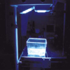
A prototype of high intensity gallium nitride blue LED phototherapy unit was developed and custom built by the Department of Pediatrics and Biomedical Engineering at the Samsung Medical Center, Sungkyunkwan University School of Medicine. High intensity blue LEDs (HLBB-L55B, Nissitronics Korea Inc., Seoul, Korea) had a dominant wavelength at 465-470 nm, and the overhead device had two focused arrays, each array equipped with 500 LEDs (Fig. 1). For conventional phototherapy, halogen quartz phototherapy device (Model: 6600-0084-900, Ohmeda Medical Co., MD, U.S.A.) was used. As halogen lamps could incur the risk of burn when positioned closer to the infant, phototherapy was administered 45 cm apart in accordance with the manufacturer's recommendation. At that distance, irradiance was measured with a phototherapy radiometer (BiliBlanket Meter II, Ohmeda Medical Co., MD, U.S.A.), with peak sensitivity at 450 nm, and illuminance was measured with an Illuminance Meter, TL-1 Minolta (Minolta Camera Co., Tokyo, Japan). The in vitro results do not necessarily reflect its in vivo efficacy due to confounding variables including poor skin penetration of blue light spectrum (14-16). We therefore conducted both in vitro and in vivo experiment to determine the bilirubin degradation efficacy of LED device in the present study.
In vitro experiment
Bilirubin (Sigma Chemical Co., MO, U.S.A.) was dissolved in a buffer solution containing 18.5 vol% 0.1 N NaOH, 44.5 vol% human albumin (5%) and 37 vol% 0.055 M Na2HPO4, with the final concentration and pH adjusted to 20 mg/dL and 7.4 respectively. In each group, ten microhematocrit tubes containing 100 µL of the bilirubin solution per tube, was placed horizontally on a black non-reflective background, and exposed to each phototherapy light at 45 cm distance for 5 hr at room temperature. In vitro photodegradation of bilirubin (%) was calculated as the difference between bilirubin concentrations at before and after exposure to the phototherapy divided by the bilirubin concentration at before exposure to the phototherapy times 100. Bilirubin concentration was measured with Bilirubin Tester (Wako Pure Chemical Industries Ltd., Osaka, Japan).
In vivo experiment
For in vivo experiment, Gunn rats were purchased from Harlan Sprague Dawley, Inc. (Indianapolis, IN, U.S.A.). Twenty of 8-day old jaundiced (jj) Gunn rats were randomly divided into two groups, and exposed to each phototherapy light at 45 cm distance for 5 hr at room temperature. In vivo bilirubin degradation (%) was also calculated as the difference between serum bilirubin concentrations of Gunn rats at before and after exposure to the phototherapy divided by the serum bilirubin concentration at before exposure to the phototherapy times 100.
RESULTS
Mean irradiance of blue LED and conventional phototherapy unit at 45 cm distance was 75 and 23 µW/cm2/nm, and illuminance was 680 and 1999 foot candle, respectively.
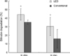
Bilirubin photodegradation of blue LED device was significantly higher than conventional phototherapy in both in vitro (44±7% vs. 35±2%) and in vivo (30±9% vs. 16±8%) experiment (Fig. 2).
DISCUSSION
In the present study, high intensity gallium nitride blue LED phototherapy was more effective than conventional halogen quartz phototherapy in both in vitro and in vivo bilirubin photodegradation. More rapid breakdown of bilirubin with the use of more efficient LED phototherapy devices would shorten the treatment times of neonatal jaundice (15, 16), and ultimately improve the prognosis of severe neonatal hyperbilirubinemia by reducing the need for exchange transfusion and minimizing the risk of bilirubin neurotoxicity (2, 4, 11).
The optimum phototherapy method with the least side effects has not been developed yet (1-3). The color (wavelength) and intensity (irradiance) of the light emitted during phototherapy are two major factors determining its efficacy (2, 4-6). Blue LEDs used for light sources of phototherapy in the present study would be more effective than day light because they emit a high intensity narrow band of blue light corresponding to the peak absorption wavelength at which bilirubin is broken down (14). Furthermore, as LEDs emit no significant ultraviolet or infrared light, they would reduce the potential risk of DNA damage (8) or fluid loss (7). However, two recent clinical trials of blue gallium nitride LED device using comparable light intensity were not found to be of higher efficacy than the conventional phototherapy (17, 18). In contrast, gallium nitride blue LED phototherapy was observed to be more effective than conventional phototherapy in both in vitro and in vivo bilirubin photodegradation in the present study. As the irradiance and illuminance of our LED device was nearly three times higher and three times less than the conventional phototherapy respectively, not the broad light intensity (illuminance) but the specific light intensity matching the bilirubin absorption spectrum (irradiance) seems to correlate with the magnitude of bilirubin photodegradation (6, 16). Taken together, these findings suggest that besides the wavelength, the intensity of light (irradiance) emitted during phototherapy is also very important for its best efficacy (2, 4-6, 16).
The dose of phototherapy light is the product of irradiance, surface area exposed, and duration of exposure (2, 4-6, 16). Irradiance depends not only on the power of the light source but also on the distance from the source (2, 4, 5). LEDs can deliver much higher doses of light than any of the conventional devices as they generate greater irradiance (14), and can be placed very close to the skin due to less heat production in contrast to halogen or fluorescent lights (1, 7). Furthermore, such direct contact LEDs in the form of mattresses, pads or jackets of any size and shape could cover a larger surface area than conventional devices, leading to more efficient and convenient phototherapy and abrogating the need for eye patches (2). Therefore, LED devices could markedly improve the efficacy and safety of phototherapy (14).
Tan (5, 19) demonstrated a dose response relationship with increased intensity of phototherapy resulting in greater responses. However, the rate of bilirubin decline progressively decreased with increasing irradiance until a saturation point of 30-40 µW/cm2/nm in the 425 to 475 nm range was reached, beyond which an increase in the irradiance produced no added efficacy. At the saturation point, a bilirubin decline of 40 to 50% in 24 hr was achieved (19). However, given that the conversion of bilirubin to excretable photoproducts is partly irreversible and follows first order kinetics (14, 15), it is not certain whether a saturation point suggested by Tan really exists. Our data of more effective bilirubin degradation with blue LEDs well above the saturation point of irradiance also contradict the concept of the saturation point. As LED phototherapy devices can provide significantly higher irradiance levels compared to all currently available conventional systems (14), further studies will be necessary to clarify this.
In addition to the features mentioned above, LEDs have several advantages not found in conventional devices such as light weight, compact size, high energy efficiency, use of low voltage battery power, extremely long life, the ability to be focused with a lens or through spatial orientation and no glass parts (2, 12-14). These features make the LED devices ideal light sources for phototherapy during transport or at home. Furthermore, as LEDs use direct current, the typical flickering glare that may be responsible for the untoward symptoms of headache, nausea and dizziness, reported with the use of conventional blue fluorescent lamps (11), is absent, thereby reducing nursing staff discomfort (17, 18).
In summary, a blue LED phototherapy unit with higher irradiance levels showed higher efficacy of both in vitro and in vivo bilirubin photodegradation compared to that of commercially used halogen quartz phototherapy device. As LED devices can provide much higher irradiance, and thus greater efficacy, they can be ideal light sources for the phototherapy of neonatal jaundice. Additional studies will be necessary to prove its clinical efficacy.
 XML Download