

 PDF
PDF ePub
ePub Citation
 Print
Print


Dear Editor,
Hydroa vacciniforme-like lymphoma (HVLL) is a disorder of childhood that is associated with Epstein–Barr virus (EBV). The characteristic clinical sign of HVLL is papulovesicular eruption and subsequent ulceration/scarring primarily on the face and upper extremities [1]. The neoplastic cells of HVLL are usually T cells and natural killer (NK) cells. Recently, increase of circulating EBV-infected γδ T cells has been demonstrated in patients with HVLL [234]. We report an unusual case of marked double-negative (CD4-negative/CD8-negative) γδ T cell lymphocytosis, wherein the morphological findings resembled those of larger granular lymphocytic leukemia (LGL).
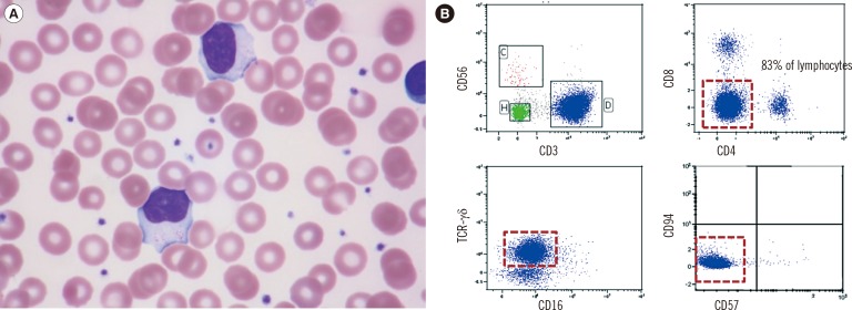
A 4-yr-old girl presented with multiple facial erythematous papules and crust/patches. She had a history of persistent lymphocytosis (for seven months). At the time of visit to our hospital, her white blood cell (WBC) count, hemoglobin, and platelet levels were 29.44×10⁹/L (lymphocytes: 76.6%, 22.55×10⁹/L), 12.2 g/dL, and 364×109/L, respectively. Serologic results for EBV infection were as follows: EBV-viral capsid antigen (EB-VCA) IgG, positive; EB-VCA IgM, negative; EBV-early antigen (EBV-EA), negative; EBV-nuclear antigen (EBNA) IgG, positive. The quantitative PCR result for EBV DNA was 186,620 copies/mL. Large granular lymphocytes and small lymphocytes without significant atypia increased in the peripheral blood (PB) (Fig. 1A). Immunophenotyping revealed increase of double-negative γδ T cells in the PB (83% of lymphocytes); surface CD3-positive/CD4-negative/CD8-negative/CD16-negative/CD56-negative/CD57-negative/T cell receptor (TCR)-γδ-positive (Fig. 1B). The flow cytometer, FACSCantoII, and monoclonal antibodies were purchased from Becton Dickinson (San Jose, CA, USA). Immunohistochemical staining results for the facial skin biopsies were as follows: CD3-positive, TCR-βF1-negative, TCR-CγM1-positive, CD4-negative, CD8-negative, CD20-negative, EBV in situ hybridization-positive, and Ki-67-positive (30%). To exclude T cell LGL (T-LGL), we analyzed STAT3 using PCR and sequencing after obtaining informed consent [5]. Primers for STAT3 were as follows: exon 19, Forward 5'-TTGGAACGAAGGGTAGGTTG-3' and Reverse 5'-TTTGCGAGTCTGAGTGAAACA-3'; exon 20, Forward 5'-CCCCTTCGAGGAAAGAAAAA-3' and Reverse 5'-CCAGGTTATTCAGGCATTTG-3'; exons 21-22, Forward 5'-GCAGATGGAGCTTTCCAGAC-3', Reverse 5'-TCCTACCATTCCGAGTGACC-3'. Sequencing was performed by using the BigDye Terminator Cycle Sequencing Ready Reaction Kit on the ABI Prism 3130 Genetic Analyzer (Applied Biosystems, Foster City, CA, USA). No mutations were found in STAT3. HVLL was diagnosed according to the 2008 WHO Classification [6].
Absolute lymphocytosis is unusual in hydroa vacciniforme-like lymphoproliferative disease [36]. Cytomorphological analysis of the neoplastic cells generally shows small-to-medium-sized cells without significant atypia. However, increase in large granular lymphocytes has been reported in some cases [7]. Large granular lymphocytosis is a prominent feature of T-LGL or chronic lymphoproliferative disorder of NK cells (CLPD-NK) [8]. Therefore, we analyzed the STAT3 mutation, a relatively specific molecular marker of T-LGL or CLPD-NK (20-40%) [5]. We also analyzed CD57 expression by flow cytometry (usually positive in T-LGL) [5]. We excluded T-LGL on the basis of negative CD57 expression, wild-type STAT3 expression, and skin biopsy results.
Recently, Kimura et al. [3] reported increase (>5% lymphocytes) of double-negative γδ T cells in hydroa vacciniforme-like lymphoproliferative diseases (10/11, 90.9%), with a mean value of 15.7%±2.9% (0.266±0.108×10⁹/L); the γδ T-cell fractions had higher EBV DNA concentrations than non-γδ T cell fractions. Similar results were observed in other studies [24]. The clinical and pathologic significance of circulating γδ T cells has not been fully understood until now. γδ T cells are generally predominant in the epithelium of the skin and mucosa. These cells play a role in innate and acquired immune regulatory functions. Considering the clonal T cell proliferation (confirmed by TCR rearrangement analysis) and clonal EBV proliferation (confirmed by terminal repeat analysis), γδ T cells may be important for the development of hydroa vacciniforme-like lymphoproliferative diseases [23].
HVLL is a rare disease and can remain undiagnosed or be misdiagnosed (e.g., cutaneous lupus and cellulitis) owing to prominent skin manifestations without an abnormal complete blood count [910]. Our patient remained undiagnosed for seven months although lymphocytosis with skin manifestations was persistent. Therefore, clinicians must rule out HVLL in children with multiple facial erythematous papules and crust/patches. Furthermore, complete blood count, peripheral blood smear, and serologic tests for EBV should be performed; subsequent flow cytometry and skin biopsy may help rule out lymphoproliferative diseases.
In conclusion, we report an unusual case of marked double-negative γδ T cell large granular lymphocytosis in a patient with HVLL. Although cytomorphological analysis of the neoplastic cells generally shows small-to-medium-sized cells without significant atypia, the findings for our case suggest that larger granular lymphocytes may be a prominent feature of HVLL, similar to T-LGL or CLPD-NK. Therefore, flow cytometry or STAT3 sequencing analysis may aid in differential diagnosis.
 XML Download