

 PDF
PDF ePub
ePub Citation
 Print
Print


Dear Editor,
Conjunctival pyogenic granuloma (PG) is a benign vascular proliferation of immature capillaries that usually denotes sequelae of incomplete surgical or traumatic wound healing [12]. It is most commonly seen at a traumatic wound site or near a suture line after surgery and other insults [3]. Herein, we report three cases of conjunctival PG with unknown etiology in children and a young adult.
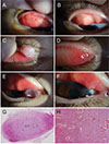
Case 1. An 8-year-old boy presented with a mass on his right eyelid that suddenly started to grow 2 months prior. A vascularized, broad-based, 7.0 mm × 4.0 mm sized, bright-red mass was observed on his right upper tarsal conjunctiva (Fig. 1A). He had no surgery or trauma history, and no other ocular abnormalities observed. After 4 weeks of using topical 0.5% loteprednol etabonate, the mass size had diminished by one-third. Two months after cessation of medication, the mass was completely regressed (Fig. 1B).
Case 2. A 10-year-old boy presented with an interpalpebral mass on his left eye. In his clinical history, we could not find any surgery performed on the eye or its adnexa nor any other trauma. Slit lamp examination revealed a 6.0 mm × 5.0 mm sized, pedunculated, pinkish mass on his left upper tarsal conjunctiva (Fig. 1C). Parts of the exposed tissue had necrotic debris. Under the assumption of PG with its morphology, excisional biopsy under general anesthesia was planned, but the mass fell off by itself after 2 weeks of observation (Fig. 1D).
Case 3. A 29-year-old man presented with a one-week history of bloody tears and discharges. He had no specific history except laser in situ keratomileusis 4 years prior. Examination revealed a pedunculated, dark-red, 6.0 mm × 4.0 mm, mobile mass emanating from his right upper tarsal conjunctiva (Fig. 1E). The mass was easily excised under slit lamp examination without extensive bleeding (Fig. 1F). Hematoxylin and eosin staining revealed an unusual vascular pattern of numerous blood-filled, capillary-sized channels with some neutrophils and lymphocytes (Fig. 1G, 1H), which are typical of PGs [4].
PGs are classically reported as a complication of ocular surgery or trauma and have a characteristic bright-red, lobular appearance located near a wound site [34]. In a previous review of ocular PGs, 87 out of 100 cases had predisposing factors, while 13 cases had unknown etiologies [1]. Similarly, our cases had no definite clinical history. These findings suggest that, even though specific etiologies do not exist, the diagnosis of PG should be considered with its typical clinical presentation of rapid growth and distinctive pink fleshy appearance.
Treatment options for PGs include surgical excision, cryotherapy, electrocautery, laser ablation, topical medical therapy, and observation [5]. Although surgery is a well-known method for definite diagnosis and treatment, this option requires local anesthesia at least and general anesthesia in children and poorly cooperative patients. Successful outcomes with topical steroids without adverse effects have been reported for conjunctival PGs at young age [4]. In case 1, the mass was diminished after 4 weeks of topical steroids and 2 months after cessation of the medication. Although this might be either the effect of topical steroids or the result of spontaneous regression, topical steroids can be prescribed in PGs before planning a surgical biopsy. Of course, intraocular pressure should be monitored routinely while using topical steroids [4]. Recently, small case studies, including 4 patients, reported that ocular surface PGs can be treated with topical timolol 0.5% [5]. This can be another treatment option after it is confirmed in further large studies. In cases of side effects after topical medication, regular follow-up without treatment could be an option considering the benign characteristics, as seen in our case 2 with a spontaneous resolution.
In summary, conjunctival PG should be considered when a lesion shows a vascularized mass protruding from the conjunctiva even with an undetermined etiology. We recommend prescribing topical steroids as an initial approach, especially in children and poorly cooperative patients, as they have a lower adverse-effect profile than conventional surgical therapies. Regarding non-responsiveness, repeated recurrence, or clinical uncertainty, we advise surgical biopsy and histological evaluation.
 XML Download