

 PDF
PDF ePub
ePub Citation
 Print
Print


Dear Editor,
The evaluation of retinal nerve fiber layer (RNFL) defects has long been regarded as one of the mainstays for the diagnosis of glaucoma [1]. Recently, optical coherence tomography (OCT) has become increasingly popular for RNFL evaluation [2]. However, red-free photography is still the gold standard for detecting RNFL defects. Although there are many limitations to this technique, such as interpretation subjectivity, invisibility in patients with high myopia or diffuse atrophy, and difficulty quantifying results [3], it plays an important role in diagnosing and monitoring glaucoma progression, especially in localized RNFL defects [4]. Here, we report three cases in which localized RNFL defects on later red-free photographs (VISUCAM 200; Carl Zeiss Meditec, Jena, Germany) looked improved than in previous ones, yet there was no evidence of RNFL defect improvement using other methods, such as OCT (Cirrus HD OCT; Carl Zeiss Meditec, Dublin, CA, USA) or visual field (28-2 SITA Standard Algorithm, Humphrey visual field analyzer; Carl Zeiss Meditec).
These three glaucoma patients were not diagnosed with other systemic disease and did not undergo any type of ophthalmologic surgeries except glaucoma operation. Intraocular pressure was measured by the Goldman applanation tonometer. The intraocular pressure of these patients has been stable.
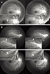
A 47-year-old man with primary open angle glaucoma in both eyes had been using latanoprost 0.005% (Xalatan; Pfizer, Tokyo, Japan) once daily since 2002. Fig. 1A shows superior temporal RNFL bundle defect change on red-free photographs of his right eye in May 2012 and July 2013 in order from left. The width of the RNFL bundle defect in July 2013 looked improved compared to that in May 2012.
A 12-year-old girl was diagnosed with juvenile-open angle glaucoma in 2008. She used latanoprost 0.005% (Xalatan, Pfizer) once daily in both eyes and brinzolamide 1% (Azopt; Alcon Laboratories, Fort Worth, TX, USA) twice daily in her left eye and had a history of trabeculectomy in both eyes in February 2011. Fig. 1B shows superior temporal RNFL bundle defect difference in red-free photographs of her right eye in June 2014 and August 2014 in order from left. In those red-free photographs, the width of the RNFL bundle defect in August 2014 seemed to be improved compared to that in June 2014.
A 38-year-old female primary open angle glaucoma patient was using bimatoprost 0.03% (Lumigan; Allergan, Irvine, CA, USA) once daily and fixed combination timolol maleate 0.5% and dorzolamide 2% (Cosopt; Merck & Co, Whitehouse Station, NJ, USA) twice daily in both eyes. Fig. 1C shows inferior temporal RNFL changes in her right eye in March 2013 and March 2014 in order from left. The RNFL bundle defect in 2013 was worse than in 2014.
All three patents in this case series showed improved RNFL defects on subsequent red-free photographs compared to earlier images. It is unlikely that these changes are reflective of true improvements, but instead are the result of a misidentification of RNFL defects. Considering the location of changes of RNFL defects, deformed parafoveal annular reflex is thought to be the one of the reasons for such erroneously identified change. Light reflection by the thickened retinal tissue causes a parafoveal annular reflex. In the normal retina, many nerve cells accumulate in the parafoveal area. The internal limiting membrane of the sloped and thickened parafoveal region is considered the cause of the parafoveal annular reflex. It is believed that these light reflections are affected by pathologic conditions of macular or optic disc under pathological conditions, and the annular reflex is altered, showing distortion, blunt borders, fragmentation, and irregular extension of the reflex area [5]. In addition, the angle and direction of incident light while taking red-free photographs might affect these changes. In conclusion, RNFL evaluation by red-free photography for determining glaucoma progression can be erroneous and should be analyzed repeatedly. Further study of the certain conditions under which this effect occurs should be performed.
 XML Download