Journal List > Korean J Ophthalmol > v.29(4) > 1025173

Jung, Park, and Woo: Iatrogenic Central Retinal Artery Occlusion Following Retrobulbar Anesthesia for Intraocular Surgery

Abstract

Purpose

To present clinical features of central retinal artery occlusion (CRAO) following retrobulbar anesthesia for intraocular surgery.

Methods

This observational case series describes 5 consecutive patients with acute CRAO following retrobulbar anesthesia for intraocular surgery. Data collected for this study included subject characteristics, retrobulbar anesthesia technique, treatment type, initial and final best-corrected visual acuity, and other ophthalmologic examinations.

Results

Mean subject age was 67.0 ± 8.2 years (range, 53 to 72 years). All patients had one or more vascular risk factors (e.g., hypertension, cerebral infarction, carotid artery stenosis) and presented with acute vision loss 1 day after uneventful intraocular surgery (cataract surgery in 2 eyes and vitrectomy in 3 eyes). All 5 patients received retrobulbar anesthesia during surgery, 4 of which involved the use of a sharp needle. No immediate complications were noted during intraocular surgery. Final visual prognosis was poor (from finger count to no light perception) although intraocular thrombolysis was attempted in 3 patients.

Conclusions

Iatrogenic CRAO is a potential complication of retrobulbar anesthesia for intraocular surgery in elderly patients with vascular risk factors. Unfortunately, this complication can lead to severe vision loss. We conclude that retrobulbar anesthesia for intraocular surgery should be performed with great care and special consideration for elderly patients with vascular risk factors.

Central retinal artery occlusion (CRAO) is an acute vascular event that causes painless and sudden visual loss in the affected eye [1]. It develops when the central retinal artery is occluded with emboli, which results in a retinal infarction (similar to a cerebral infarction) [1]. The incidence of CRAO is low, approximately 5 per 100,000 person-years in people over 50 years of age [2]. Individuals with cardiovascular disease are at a higher risk for developing CRAO [3]. Iatrogenic CRAO is rare but can occur during cosmetic facial filler injection, retrobulbar anesthesia, embolization treatment, and head and neck drug injection [4567]. Retrobulbar anesthesia has been used for years in intraocular surgery. Complications related to this procedure (retrobulbar hemorrhage, direct optic nerve trauma, scleral perforation, and CRAO) are rare, but may lead to dramatic and permanent vision loss if they do occur [89].
Despite the occurrence of numerous CRAOs following intraocular surgery with retrobulbar anesthesia [81011121314], clinical features of this condition, including detailed ophthalmologic evaluations, cerebrovascular imaging, and visual outcomes after treatment, are not well documented. We report a case series of 5 patients who developed CRAO after uneventful intraocular surgery with retrobulbar anesthesia.

Materials and Methods

This study was approved by the institutional review board of Seoul National University Bundang Hospital. Electronic medical records of consecutive patients who visited Seoul National University Bundang Hospital between November 2008 and August 2014 were reviewed for study eligibility. Inclusion criteria included acute non-arteritic CRAO that was newly diagnosed with fundus photography and/or fluorescein angiography within 1 day of intraocular surgery. Following the diagnosis of CRAO, some patients underwent super-selective intra-arterial thrombolysis with urokinase and tirofiban within 24 hours of symptom onset as a standard-of-care procedure. Informed consent was obtained from patients for this procedure.
Data collected included ocular and/or systemic disease presence, demographics, preoperative ocular abnormalities, anesthesia technique (i.e., anesthetic agent, anesthetic dose, needle type), treatment type, initial and final best-corrected visual acuity, and follow-up duration. Each patient underwent a complete ophthalmologic evaluation, including spectral domain optical coherence tomography, fluorescein angiography, and electroretinography. Systemic risk factors were also assessed and, in patients who underwent intra-arterial thrombolysis, cerebrovascular status was evaluated using transfemoral cerebral angiography.

Results

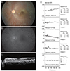
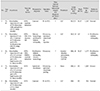
A total of 5 patients (2 men [40%] and 3 women [60%]) met all inclusion criteria (Figs. 1,2,3,4,5). Patient demographics and clinical characteristics are summarized in Table 1. A total of 3 (60%) patients had isolated CRAO and 2 (40%) patients had CRAO with ophthalmic artery occlusion. The mean patient age was 67.0 ± 8.2 years (range, 53 to 72 years). All patients had one or more vascular risk factor, including hypertension, diabetes, prior cerebral infarction, myocardial infarction, and/or carotid artery stenosis. All patients presented with acute visual disturbance noted when they removed their eye patches 1 day after uneventful intraocular surgery. There were 2 (40%) patients who underwent cataract surgery (phacoemulsification and posterior chamber intraocular lens implantation) and 3 (60%) patients who underwent vitrectomy (1 patient with air). Patients were managed with observation (1 case), anterior chamber paracentesis (1 case), or intra-arterial thrombolysis (3 cases). In the 3 patients who underwent intra-arterial thrombolysis, transfemoral cerebral angiography revealed ophthalmic artery stenosis in 1 case, internal carotid and ophthalmic artery stenosis in 1 case, and no steno-occlusive or remarkable lesions in 1 case. Mean follow-up duration was 1,139.4 ± 710.1 days (range, 4 to 1,807 days) and final visual outcome was poor in all patients. At the last follow-up visit, best-corrected visual acuity was no light perception in 2 eyes, light perception in 1 eye, and the ability to count fingers in 2 eyes.

Discussion

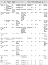
Despite various modifications in retrobulbar anesthesia methods, blindly inserting a needle into the retrobulbar space is still necessary and can result in vision-threatening complications [15]. Many previous articles have reported iatrogenic CRAO following retrobulbar anesthesia (Table 2) [5810111214161718]. In these reports, 10 of 17 cases occurred following intraocular surgery (cataract surgery or vitrectomy). Because no CRAOs were observed following intraocular surgery without retrobulbar anesthesia, the main cause of CRAO is thought to be retrobulbar anesthesia and not the intraocular surgery itself. As in the 5 cases reported here, the majority of previously reported cases had a poor visual outcome.
The mechanism of iatrogenic CRAO associated with retrobulbar anesthesia is not well understood. Klein et al. [5] suggested that direct central retinal artery trauma and pharmacologic and/or compressive anesthetic agent effects resulted in retinal vascular occlusions following retrobulbar anesthesia. Morgan et al. [13] also described mechanisms of vascular obstructions associated with retrobulbar anesthesia. They believe that trauma of and partial anesthetic injection into the central retinal artery resulted in embolization of the retinal circulation. Additionally, injection of anesthetic agents into the optic nerve sheath may occlude both the central retinal artery and vein [9121319]. Because central retinal vein occlusion was not observed in any of our patients, we consider optic nerve sheath hematoma to be an unlikely cause of CRAO in our patients.
Horven [20] and Vinerovsky et al. [21] documented that retrobulbar and peribulbar injection of a nesthetic agent causes vasoconstriction of the ophthalmic artery and a subsequent reduction in ocular blood flow. We recently suggested retrograde embolic propagation through collateral ophthalmic artery vessels as a mechanism for CRAO development following cosmetic facial filler injection [7]. Although injection agents and sites were different than those examined in this study, this phenomenon may still underlie CRAO development following retrobulbar anesthesia. Sharp needles were used in 4 of our 5 CRAO patients to deliver anesthetic, supporting the notion that direct injury to the ophthalmic artery or its collaterals can result in CRAO. In 4 out of the 17 previously reported cases (Table 2), retrobulbar hemorrhage or optic nerve sheath hematoma were observed. Although the cause of CRAO in these 4 cases remains unclear, and direct central retinal artery compression cannot be ruled out, injury to the central retinal artery or its branches may have caused the formation of vascular emboli and CRAO.
It is known that CRAO is associated with vascular risk factors, including hypertension, cardiac disease, carotid artery disease, and cerebral infarction [3]. Given that all of our patients (Table 1) and most previously reported patients (Table 2) had one or more vascular risk factor, we conclude that pre-existing cerebrovascular disease may increase the risk of developing iatrogenic CRAO following the administration of retrobulbar anesthesia.
This study is limited by the retrospective design and small sample size. However, to the best of our knowledge, the 5 cases included here make up the largest cohort of patients with retrobulbar anesthesia-associated CRAO in Korea. We hope that our detailed description of clinical features and visual outcomes will be helpful to ocular surgeons. In conclusion, iatrogenic CRAO is a vision-threatening complication associated with retrobulbar anesthesia administered for intraocular surgery. Patients who are elderly and have one or more vascular risk factor may be at an increased risk for this complication. Although rare, patients should be educated on iatrogenic CRAO prior to intraocular surgery with retrobulbar anesthesia. Precautionary measures (e.g., using a blunt needle) should be implemented to minimize the risk of iatrogenic CRAO, particularly in patients with vascular risk factors.

Figures and Tables

Fig. 1

Images of patient 1 who underwent cataract surgery (phacoemulsification and posterior chamber intraocular lens implant) with retrobulbar anesthesia. (A) Fundus photograph showing a cherry-red spot. (B) Fluorescein angiogram showing decreased choroidal perfusion and delayed filling of the retinal artery and vein. (C) Internal carotid angiogram showing stenosis of the right ophthalmic artery (arrow) and a tortuous proximal ophthalmic artery.

kjo-29-233-g001
Fig. 2

Images of patient 2 who underwent vitrectomy, endolaser, and intravitreal air injection for proliferative diabetic retinopathy vitreous hemorrhage. (A) Fundus photograph obtained 3 days after intraarterial thrombolysis. A cherry-red spot, retinal edema, and multiple retinal hemorrhages are apparent. (B) Spectral domain optical coherence tomography image obtained 3 days after intra-arterial thrombolysis. Increased reflectivity and inner retinal thickness (including the central macula), along with decreased outer retina reflectivity, are apparent. (C) Internal carotid angiogram showing severe stenosis of the right cervical internal carotid artery (arrow) and the ophthalmic artery.

kjo-29-233-g002
Fig. 3

Images of patient 3, who presented with severe visual decline resulting in no light perception after vitrectomy with retrobulbar anesthesia for proliferative diabetic retinopathy and vitreous hemorrhage. All images were taken 7 days after surgery. (A) A cherry-red spot and severe arterial narrowing and sclerosis are observed in the fundus photograph. (B) Early-phase (2 : 11) fluorescein angiogram showing poor arterial filling. (C) Increased reflectivity and thickness of the inner retina are apparent in spectral domain optical coherence tomography. (D) Standard electroretinogram (ERG) in the left eye showing decreases in both scotopic and photopic wave amplitude.

kjo-29-233-g003
Fig. 4

Images of patient 4, who underwent vitrectomy under retrobulbar anesthesia for treatment of epiretinal membrane. Preoperative best-corrected visual acuity was 20 / 32 in the right eye. One day following surgery, best-corrected visual acuity was at the level of "hand motion" in his right eye. (A) Fundus photograph 1 day after surgery, showing a cherry red spot and marked inner retinal edema in the macula. (B) Early-phase (20 seconds after fluorescein injection) fundus fluorescein angiography 13 days after surgery, showing a mild delay in arteriovenous transit time and multiple patchy capillary nonperfusion in the posterior pole. (C) Goldmann visual field test obtained 5 months after surgery, indicating the presence of a central scotoma.

kjo-29-233-g004
Fig. 5

Images of patient 5, who underwent cataract surgery (phacoemulsification and posterior chamber intraocular lens implant) under retrobulbar anesthesia and presented with visual decline 1 day after surgery. (A) Fundus photograph 1 day after surgery, showing a typical cherry red spot in the right eye. (B) Increased retinal reflectivity and internal retinal thickness are apparent in spectral domain optical coherence tomography. (C) Transfemoral cerebral angiogram performed 1 day after surgery, showing no definitive occlusion in the ophthalmic artery (arrow) or cerebral arteries.

kjo-29-233-g005
Table 1

Demographic and clinical characteristics of patients with CRAO following intraocular surgery

kjo-29-233-i001

CRAO = central retinal artery occlusion; BCVA = best-corrected visual acuity; IOP = intraocular pressure; HTN = hypertension; PE & PCL = phacoemulsification and posterior chamber intraocular lens implantation; IAT = intraarterial thrombolysis; NLP = no light perception; DM = diabetes mellitus; ICA = internal carotid artery; MI = myocardial infarction; LP = light perception; ESRD = end stage renal disease; Mx = medication; HM = handmotion; FC = finger count.

Table 2

Summary of previous reports of central retinal artery occlusion after retrobulbar anesthesia (total of 17 cases)

kjo-29-233-i002

VA = visual acuity; BCVA = best-corrected visual acuity; FU = follow-up duration; DM = diabetes mellitus; PDR = proliferative diabetic retinopathy; FC = finger count; SCR = sickle-cell retinopathy; NA = not available; HM = hand motion; OIS = ocular ischemic syndrome; AC = anterior chamber; PE & PCL = phacoemulsification and posterior chamber intraocular lens implantation; IOP = intraocular pressure; Mx = medication; CRVO = central retinal vein occlusion; IOL = intraocular lens; NLP = no light perception; LP = light perception; HTN = hypertension.

Acknowledgements

This work was partly supported by a grant from the Joint Research Project, Korea Research Council of Fundamental Science and Technology, Korea.

Notes

Conflict of Interest No potential conflict of interest relevant to this article was reported.

References

1. Hwang G, Woo SJ, Jung C, et al. Intra-arterial thrombolysis for central retinal artery occlusion: two cases report. J Korean Med Sci. 2010; 25:974–979.
2. Park SJ, Choi NK, Seo KH, et al. Nationwide incidence of clinically diagnosed central retinal artery occlusion in Korea, 2008 to 2011. Ophthalmology. 2014; 121:1933–1938.
3. Hazin R, Dixon JA, Bhatti MT. Thrombolytic therapy in central retinal artery occlusion: cutting edge therapy, standard of care therapy, or impractical therapy? Curr Opin Ophthalmol. 2009; 20:210–218.
4. Ashwin PT, Mirza S, Ajithkumar N, Tsaloumas MD. Iatrogenic central retinal artery occlusion during treatment for epistaxis. Br J Ophthalmol. 2007; 91:122–123.
5. Klein ML, Jampol LM, Condon PI, et al. Central retinal artery occlusion without retrobulbar hemorrhage after retrobulbar anesthesia. Am J Ophthalmol. 1982; 93:573–577.
6. Li W, Hu Z, Gao R. Iatrogenic central artery embolism. Yan Ke Xue Bao. 1990; 6:102–104.
7. Park SW, Woo SJ, Park KH, et al. Iatrogenic retinal artery occlusion caused by cosmetic facial filler injections. Am J Ophthalmol. 2012; 154:653–662.e1.
8. Tappeiner C, Garweg JG. Retinal vascular occlusion after vitrectomy with retrobulbar anesthesia-observational case series and survey of literature. Graefes Arch Clin Exp Ophthalmol. 2011; 249:1831–1835.
9. Feibel RM. Current concepts in retrobulbar anesthesia. Surv Ophthalmol. 1985; 30:102–110.
10. Torres RJ, Luchini A, Weis W, et al. Combined central retinal vein and artery occlusion after retrobulbar anesthesia: report of two cases. Arq Bras Oftalmol. 2005; 68:257–261.
11. Giuffre G, Vadala M, Manfre L. Retrobulbar anesthesia complicated by combined central retinal vein and artery occlusion and massive vitreoretinal f ibrosis. Retina. 1995; 15:439–441.
12. Sullivan KL, Brown GC, Forman AR, et al. Retrobulbar anesthesia and retinal vascular obstruction. Ophthalmology. 1983; 90:373–377.
13. Morgan CM, Schatz H, Vine AK, et al. Ocular complications associated with retrobulbar injections. Ophthalmology. 1988; 95:660–665.
14. Mieler WF, Bennett SR, Platt LW, Koenig SB. Localized retinal detachment with combined central retinal artery and vein occlusion after retrobulbar anesthesia. Retina. 1990; 10:278–283.
15. Jacobi PC, Dietlein TS, Jacobi FK. A comparative study of topical vs retrobulbar anesthesia in complicated cataract surgery. Arch Ophthalmol. 2000; 118:1037–1043.
16. Cowley M, Campochiaro PA, Newman SA, Fogle JA. Retinal vascular occlusion without retrobulbar or optic nerve sheath hemorrhage after retrobulbar injection of lidocaine. Ophthalmic Surg. 1988; 19:859–861.
17. Roth SE, Magargal LE, Kimmel AS, et al. Central retinal-artery occlusion in proliferative sickle-cell retinopathy after retrobulbar injection. Ann Ophthalmol. 1988; 20:221–224.
18. Mameletzi E, Pournaras JA, Ambresin A, Nguyen C. Retinal embolisation with localised retinal detachment following retrobulbar anaesthesia. Klin Monbl Augenheilkd. 2008; 225:476–478.
19. Stewart JM. Mechanism of ophthalmic artery occlusion following pars plana vitrectomy. Br J Ophthalmol. 2003; 87:376.
20. Horven I. Ophthalmic artery pressure during retrobulbar anaesthesia. Acta Ophthalmol (Copenh). 1978; 56:574–586.
21. Vinerovsky A, Rath EZ, Rehany U, Rumelt S. Central retinal artery occlusion after peribulbar anesthesia. J Cataract Refract Surg. 2004; 30:913–915.
TOOLS
Similar articles