

 PDF
PDF ePub
ePub Citation
 Print
Print


Dear Editor,
Recently, we experienced a case of pediatric orbital lymphangioma successfully treated with intralesional bleomycin injection. To our knowledge, this is the first such case report in Korea of this treatment used for this condition.
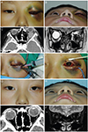
An 8-year-old male visited our clinic complaining of eyelid swelling and proptosis in his left eye for 5 days (Fig. 1A and 1B). He was diagnosed with an external stye and was administered antibiotics 4 days ago, but showed no improvement. The best-corrected visual acuity of the left eye was 20 / 30 and intraocular pressure was 32 mmHg. The patient complained of severe subconjunctival hemorrhage and pain. Initial computed tomography (CT) and magnetic resonance (MR) images showed a 22.2 × 25.4 mm-sized macrocystic lymphangioma in the left inferotemporal orbit (Fig. 1C and 1D).
An inferior lower eyelid incision was made to facilitate the approach to the macrocyst. An opening was made on the cystic wall and the internal wall of the cyst was exposed using Sewall retractors. Blood evacuation was performed with a suction catheter. Bleomycin (1 IU per milliliter saline) was injected into the cyst for exactly 5 minutes (Fig. 1E). Barovac (100 mL; Sewoon Medical, Seoul, Korea) was inserted into the cyst to provide continuous negative pressure to induce cyst shrinkage and reduce the risk of delayed or recurrent bleeding (Fig. 1F). On the third day after the operation, the visual acuity had returned to 20 / 20, and intraocular pressure remained at 20 mmHg. At this point, 10 mg of prednisolone was administered for 2 weeks with a tapering schedule. At 1 month of follow-up, the patient's symptoms and signs were completely resolved (Fig. 1G and 1H). CT and MR imaging results showed no residual macrocystic lymphangioma, and there was no sign of re-bleeding (Fig. 1I and 1J). There were also no signs of recurrence at 6 months of follow-up.
Lymphangioma is a congenital malformation of the lymphatic system that frequently occurs in the head and neck [1]. Orbital lymphangioma manifests in proptosis and restriction of ocular motility following minor trauma or upper respiratory tract infection. Its mass effect may lead to compressive optic neuropathy or exposure keratopathy. Surgery is considered when there are symptoms such as decreased visual acuity, amblyopia, and severe cosmetic disfigurement. However, complete resection of the lesion is difficult to achieve, increasing the risk of local recurrence and complications involving injury to surrounding structures. Multiple treatment modalities including radiotherapy, intralesional/oral corticosteroid and intralesional sclerosing therapy can be adopted in conjunction with surgery [1].
Intralesional sclerosing therapy is used to induce cyst collapse by destroying the epithelial lining of the cystic wall of macrocystic lymphangioma and by reducing lymphatic fluid production. OK-432 (Picibanil, Shuhei Ogita Fund, Japan), Sotradecol (sodium tetradecyl sulphate; Bioniche Pharma, Galway, Ireland), sodium morrhuate and bleomycin are commonly used as sclerosing agents [2,3]. Bleomycin is a chemotherapeutic agent that generates free radicals to induce DNA strand scissions. It is often used in patients with vascular malformation such as lymphangioma to cause sclerosis of vascular endothelium. Local injection of bleomycin shows a 72.3% success rate and complications such as erythema, edema and/or pain at the injection site have been reported [4]. Gooding and Meyer [5] injected bleomycin in 4 patients with orbital lymphangioma, all of whom presented favorable clinical outcomes without systemic or ophthalmic complications.
The present case is the first in Korea to report the use of local bleomycin injection as a successful treatment modality for orbital lymphangioma. Postoperative follow-up showed improved visual acuity, intraocular pressure, and proptosis without complications such as pain, erythema and swelling. No sign of recurrence was observed following a one-step surgery. The authors attribute the successful treatment to a number of factors that facilitated the surgery. For example, incision into the cystic wall enabled bleomycin injection directly into the macrocyst. Furthermore, minimal exposure of the surrounding tissue to the drug reduced its side effects. Also, control of hemorrhage increased the therapeutic potential of the drug without dilution. Finally, barovac insertion into the cyst to control additional bleeding and maintain negative pressure effectively induced cyst shrinkage.
It is imperative that surgeons confirm the precise location of the lesion, inject sclerosing agents for a specific period of time, and apply continuous negative pressure to ensure cystic shrinkage for effective surgical outcome. A longer follow-up period is required to investigate the rate of local recurrence.
 XML Download