

 PDF
PDF ePub
ePub Citation
 Print
Print


Dear Editor,
We report a unique case of visual recovery where intravitreal bevacizumab injection (IVB) completely resolved serous retinal detachment (SRD) secondary to posterior ciliary artery (PCA) occlusion after brain surgery.
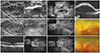
A 20-year-old male was referred to our clinic complaining of a sudden decrease in visual acuity in his left eye following neurosurgery. The patient had no other medical history except for meningioma in the left sphenoid ridge. According to the neurosurgeon, the removal of meningioma was uneventful and there were no complications during the entire surgical procedure. However, during the surgery, his scalp was compressed against the surgical bed for eight hours. His best-corrected visual acuity (BCVA) was 20 / 20 in the right eye and counting fingers at 30 cm in the left eye. Light reflex was intact and there was no relative afferent pupillary defect in either eye. Fundus examination revealed multiple patchy whitenings of the outer retina across the fundus. SRD in the distribution of a cilioretinal artery (CA) was noted (Fig. 1A and 1B). The right eye showed a completely normal fundus. Fluorescein angiography (FA) revealed delayed filling of the choroidal watershed zone and CA which persisted throughout the early phase, simultaneously with normal filling of arterial branches from the central retinal artery (Fig. 1C). Indocyanine green angiography (ICGA) revealed multiple patchy hypofluorescence and non-perfusion areas (Fig. 1D). The patient was observed during the following week, but because there were no signs of improvement in the degree of SRD (Fig. 1E and 1F), 1.25 mg of IVB was administered after patient consent was given despite the risk of aggravating the already ischemic retina. Three days later, his BCVA improved to 10 / 20 and SRD was markedly improved (Fig. 1G). Ten days later, BCVA improved to 20 / 20 and FA revealed improvement of leakage, but areas of hypofluorescence persisted (Fig. 1H). ICGA revealed that multiple patchy choroidal filling defects remained (Fig. 1I and 1J). Five weeks later, BCVA was stable at 20 / 20 and optical coherence tomography showed complete resolution of SRD. His BCVA remained stable at 20 / 20 without recurrence of any SRD at the one-year follow-up examination.
Occlusion of the choroidal vessels can manifest itself in many different ways, ranging from complete vessel obstruction to relative ischemia. In this case, irregular filling of choroidal vessels and CA, an enlarged watershed zone, and multiple geographic hypofluorescent areas with ischemic cloudy swelling of the retina extending along the length of the CA could be attributed to relative choroidal ischemia. Extensive retinal pigment epithelium (RPE) ischemic changes across the entire fundus along with resultant SRD, and multiple wedge-shaped choroidal filling defects suggested a disturbance in choroidal circulation, specifically in the PCA. Retinal circulation originating from the CA also appeared to be compromised, indicated by dilatation and staining of the CA. We speculate that the prolonged compression of the globe during brain surgery may have played a significant role in causing occlusion of the CA and PCA. PCA occlusion is known to result in RPE necrosis and subsequent blood retinal barrier (BRB) breakdown [1,2]. Multiple leakages on FA indicated a disturbance in the outer BRB. Late phase FA showed multiple stippled hyperfluorescence signals, most likely due to staining of the distressed RPE, which could be attributed to RPE ischemia resulting from PCA occlusion. Vascular endothelial growth factor (VEGF), released in response to choroidal ischemia, leads to a cascade of responses to ischemia, in which VEGF plays a crucial role in inducing disruption of vessel permeability [3]. It causes disturbances of the outer BRB, resulting in SRD [4]. Anti-VEGF can therefore be effective, by decreasing vascular permeability. The spontaneous resolution of choroidal ischemia has been reported previously [5], but in our case, because the patient showed no signs of any functional or anatomic improvement after observation for a week, we decided to treat the patient with IVB, which led to prompt visual and anatomical recovery.
This is the first case report of the occurrence of choroidal ischemia following brain tumor surgery and complete recovery of BCVA after IVB. In patients with severe visual loss and SRD associated with choroidal ischemia, IVB may be a viable treatment option.
 XML Download