

 PDF
PDF ePub
ePub Citation
 Print
Print


Recent advances have allowed us to understand corneal dystrophies on a basic molecular level, and several studies have reported specific mutations in the TGFBI gene showing various phenotypes [1]. Among these are lattice corneal dystrophy (LCD), granular corneal dystrophy, Thiel-Behnke corneal dystrophy, Reis-Bucklers corneal dystrophy, and Cogan's dystrophy [2,3].
LCD is one of the most common corneal disorders and is characterized by lineal stromal amyloid depositions that usually present bilaterally and symmetrically. Among the LCD subtypes, type IV is extremely rare, and only a few cases of this subtype have been discovered in the Japanese [4] and Indian populations [3], and one case in the Italian population [2]. We recently discovered a Korean LCD type IV patient, and therefore we are hereby reporting the first Korean case of LCD type IV, confirmed by DNA sequencing and clinical examination.
Case Report
An 87-year-old woman visited our clinic for a scheduled cataract surgery. At the time of preoperative evaluation, mild localized semitransparent opacity was noted in both corneas. The opacity showed lattice-shaped, granular deposits with asymmetrical patterns in the stroma (Fig. 1). The right eye was more severely affected, showing nodulolinear amyloid deposits located mainly in the anterior stroma; the left eye showed less linear and macular opacity. The patient demonstrated a good visual outcome after the surgery, with a best-corrected visual acuity of 0.8 in both eyes.
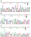
Genomic DNA samples from the patient were extracted from peripheral leukocytes using the Easy-DNA Kit (Invitrogen, Carlsbad, CA, USA). Polymerase chain reaction was performed on this genomic DNA with specific primers for the TGFBI gene (Table 1); the results showed a Leu527Arg mutation in the TGFBI gene (Fig. 2A). In addition to the Leu527Arg mutation, two more single-nucleotide polymorphisms (SNPs) were also identified: a T>C variation at cDNA position 1620 resulting in Phe540Phe and a G>A variation at cDNA position 1678+23 (Fig. 2B and 2C).
The patient's only family members, her son and granddaughter, did not show any corneal stromal manifestations. They also did not have any known mutations in the TGFBI gene specific for LCD type IV, though both showed the T>C variation at cDNA position 1620 and the G>A variation at cDNA position 1678+23, just as in the present case.
Discussion
LCD type IV was first reported in 1998 [4]. The LCD type IV patients with L527R are characterized by asymmetric corneal opacities, sporadic occurrences, and late onset [5]. In the present case, because visual impairment was not directly related to the cornea, visual acuity was improved after cataract surgery. We could not identify the etiology of the condition because the patient's lesion did not cause her visual symptoms.
Similar to previous LCD type IV cases, the patient in this report also showed only mild clinical manifestation without subjective symptoms. We assert that the novelty of this case is two-fold. Firstly, we found two novel SNP mutations in addition to the well-known L527R mutation; these were also found in the patient's family members. Since the effects of these SNP mutations on the development and progression of LCD type IV remain uncertain, the role of these mutations should be investigated in future studies.
Secondly, since the initial report of the L527R mutation in the BIGH3 gene [4], all LCD type IV patients with an L527R mutation have been found exclusively in the Japanese population. This has led Fukuoka et al. [5] to report that the L527R SNP mutation might have been caused by a founder mutation in a single Japanese ancestor. To our knowledge, present case is the first report of an L527R mutation outside Japan, and it could challenge the idea that L527R is the product of a Japanese founder effect [5].
Lastly, because the clinical manifestations of LCD type IV are sporadic and extremely mild with subtle subjective symptoms, only a few cases have been detected and reported.
In summary, the patient in this report showed typical asymmetric nodulolinear stromal opacity in both corneas, and an L527R mutation in the TGFBI gene with two novel SNP mutations. This is the first report of the L527R mutation in Korea, as well as outside Japan, challenging the idea that L527R was caused by a founder mutation in a single Japanese ancestor.


 XML Download